Vol. 22 No. 1, p 1-7 ISSN: 1978-3019
http://journal.ipb.ac.id/index.php/hayati DOI: 10.4308/hjb.22.1.
_________________
∗Corresponding author. Phone/Fax: +62-251-8622833,
E-mail: [email protected]
Characterization of Bacteriophage Specific to
Bacillus pumilus
from Ciapus River in Bogor, West Java, Indonesia
ANIK KUSMIATUN, IMAN RUSMANA, SRI BUDIARTI*
Department of Biology, Faculty of Mathemathics and Natural Sciences, Bogor Agricultural University, Darmaga Campus, Bogor 16680, Indonesia
Received August 11, 2014/Accepted December 5, 2014
Bacillus pumilus is a spore-forming bacteria that is rod-shaped, gram positive, and aerobic. B. pumilus produced pumilacidins, known to have toxic effects on epithelial cells. Antibiotics were usually used to treat the disease caused by bacteria. Antibiotic typing test of B. pumilus indigenous from sewage water showed that this isolate was resistant to ampicillin and clindamycin. An alternative way was by application of bacteriophages as biocontrol agents to reduce
B. pumilus in environment. The aim of this study were to isolate and characterize B. pumilus bacteriophage isolated from Ciapus River in Bogor, West Java. Bacteriophages infecting B. pumilus were isolated from river water using the
double agar overlay method. Phages were defined by plaque morphology, structure, host range, and characteristic of molecular weight protein phage. Phage FBa1, FBa2, and FBa3 had narrow host range and they were specific for
infecting B. pumilus. Electron microscope observation showed that phage FBa1 had icosahedral head without tail (166.67 nm in diameter), so it is called phage-like particles. Characterization of phage FBa1 by SDS-PAGE showed
five proteins band. Molecular weight of FBa1 proteins was 70.9, 54.9, 33.8, 28.3, and 21.4 kDa, respectively.
Key words: Bacillus pumilus, bacteriophage, food poisoning
___________________________________________________________________________
INTRODUCTION
Bacillus pumilus is a spore-forming bacteria that is commonly associated with foodborne illness caused by intoxication. From et al. (2007) reported a food poisoning outbreak caused by B. pumilus. In this case, B. pumilus was found as contaminant from reheated rice at a Chinese restaurant. Large numbers of B. pumilus strain was produce a complex of lipopeptides known as pumilacidins, a substance toxic to mammalian cells (From et al. 2007). This toxin can destroy cell membranes through production of selective cationic channels (Sheppard et al. 1991). The structural of pumilacidin is similar to surfactin (Naruse et al.1990). Bacillus pumilus could grow well at low temperatures (10-15 oC) and produce
large amounts of a toxic metabolite. Food poisoning incident by B. pumilus was occurred after ingestion of foods containing large numbers of B. pumilus
(105-107 cfu/g) followed by stomach cramps and
diarrhea (Suominen et al. 2001). Parvathi et al. (2009) reported that B. pumilus had cesA and cesB genes encoded cereulide synthetase. Cereulide is a stable cyclic dodecadepsipeptide produced by some strains of B. cereus. This toxin has high toxicity to humans (Agata et al. 1995; Yokohama et al. 1999).
Multidrug resistant bacteria are a serious health problem these days. Antibiotics are not bacteria specific and thus can result in disruption in the
balance of normal flora (Giuliano et al. 1987). Phage therapy seems to be a good option for this problem.
Phage therapy is use of a specific bacteriophage to
a bacterial cell. The introduction of phage therapy was seen in the early 1930s (Levin & Bull 2004). Bacteriophages (phages) are viruses that infect bacteria. They have the absolute requirement to infect host cells in order to multiply and survive. Phages are viruses that have a protein coat that encloses a nucleid acid (DNA or RNA). There are two types life cycle of phages, lytic and lysogenic phages. Lytic phage kill bacteria through a multiple-step process. The bacteria are destroyed through lysis, resulting in release many particles of phages. Lytic phage provides opportunity to control bacteria. Lysogenic phage do not immediately replicate in the bacterium they infect, but coexist within the bacterium as a prophage. When lysogenic bacteria are stress, the prophage can become activated and the virus killing the bacteria through lysis.
as a biocontrol to prevent foodborne disease in Indonesia (Budiarti et al. 2011). Phage FGCSSa1
isolated from sewage had potential for biocontrol of
Salmonella spp. in foods (Carey-Smith et al. 2006). Oral application of phage FR38 in rat diet to reduce
Salmonella P38 had no effecton body weight, blood chemistry, kidney and liver functions in rat (Sartika
et al. 2012). Bacteriophage cocktail as a biocontrol agent of Salmonella enterica serovar Typhimurium and S. enterica serovar Enteritidis were used in food matrices and their production (Spicigo et al. 2013). A cocktail of phages isolated from cattle feces reduced
Escherichia coli O157:H7 populations in the gut of sheep (Callaway et al. 2008). Application of phage
P100 on meat could reduced Listeria monocytogenes
(Soni & Nannapaneni 2010). Cocktail of phages application in milk decreased contamination of S. aureus (Martínez et al. 2008). Many phages infecting
Bacillus spp. have been isolated such as phages
FWLBc1 and FWLBc2 which infected B. cereus (Lee
et al. 2011), Tsamsa phage isolated from B. anthracis
(Ganz et al. 2014), SPP1 phage infecting B. subtilis
(Jakutytėet al. 2011), and phiAGATE phage infecting B. pumilus (Barylski et al. 2014). Nevertheless, B. pumilus phages have not been reported in Indonesia. The objectives of this research were to isolate and characterize B. pumilus bacteriophage isolated from Ciapus river in Bogor, West Java, Indonesia.
MATERIALS AND METHODS
Identification of Host Bacterium. Host bacterium was isolated from sewage water located in Cigudeg, Bogor, Indonesia. Haemolitic activity of this indigenous bacterium was determined using
blood agar plates. Identification was performed using
API 50CHB (bioMérieux, Marcy I`Etoile, France). The isolate was maintained aerobically on Tryptic Soy Agar (Difco), at 37 oC for 24 h. Antibiotics test
with disk diffusion method was used to identify the susceptibility of the bacterium to ampicillin,
amoxicillin, ciprofloxacin, and clindamycin (CLSI
2012). Antibiotic test was assessed by measuring the diameter (in millimeters) of the inhibition zone surrounding the disks.
Isolation and Purification of Phage. Isolation of
phage from water samples was collected in Ciapus river, Bogor, West Java, Indonesia. Water sample of 4.5 mL was incubated overnight with 0.5 mL of Nutrient broth (Difco) and 0.5 mL of host bacteria culture (previously grown 24 h in Nutrient broth medium). An enrichment culture was used to multiply phages. The culture was centrifugated at 5000 rpm for
25 min and filtered through a 0.22 µm sterile filter.
Stock solution of phage were stored in sterile tube at 4 oC. Soft agar (0.8%) was prepared and autoclaved.
As much as 7 mL of soft agar was warmed in water bath at 42 oC before used as an agar overlay. 100
µL of phage stock solution (10-5 to 10-8 dilutions)
was mixed with 100 µL B. pumilus culture which contain 108 CFU/mL, and incubated at 37 oC for
30 min. After incubation, it was mixed with 7 mL soft agar, and poured as an overlay onto the top of a solid Nutrient Agar (Difco) base plates. Plates were incubated at 37 oC for 24 h.
Plaques were purified according to methods in Goodridge et al. (2001). Plaques were purified by
single-plaque isolation based on plaque morphology.
Purified phage was diluted in saline-magnesium (SM)
buffer and stored at 4 oC. SM buffer is a buffer used
for storage of phage stocks (5 M NaCl, 1 M MgSO4, 1 M Tris-HCl pH 7.5, 1% gelatin in distilled water). The suspension was mixed using vortex for 30 sec and then incubated at room temperature for 1-2 h. The suspension was centrifugated at 5000 rpm for 20 min. After centrifugation, the supernatant was
filtered through a 0.22 µm sterile filter and transferred to sterile tubes. The stock solution of purified phage
was stored at 4 oC.
Phage Quantification. Quantification of phage
solution was measured by counting the number of Plaque Forming Unit (PFU) on the top agar. Phage
quantification (plaque assay) was examined using
double layer method as mentioned earlier. Stock
solution of purified phage was serially diluted (10-1
to 10-8) in 0.85% NaCl. Each dilution was subjected
to plaque assay. Plaques were counted on the plate that contain 30-300 plaques and expressed as plaque forming unit per milliliter (PFU mL-1).
Phage titer (PFU mL-1) = number of plaques x 10
x reciprocal of counted dilution
Host Range Determination. Exponential phase cultures of the host bacteria (B. pumilus, Proteus mirabilis, Photobacterium damselae, Salmonella sp., EPEC K1.1) were prepared, and agar overlays were
inoculated with a host (100 µL) and phage stocks (100 µL). The plates were incubated at 37 oC for 24
h and the plaques were observed for each host.
Transmission Electron Microscopy (TEM). Stock solution phage (5 µL) was dropped in to grid for 30 sec, and then dried up with filter paper. Uranyl acetate 2% solution (5 µL) was also dropped in to grid for 1 min, and then dried up with filter paper for 60
min. The grid placed in holder and left for perfect dry. Specimen was observed using transmission electron microscope (TEM JEOL JEM-1010 model) at 80 kV, and phages were examined at 20000-80000 times
Structural Analysis of Phage Proteins. Stock of buffer sample (2 mL mercaptoethanol, 4 mL glycerol, 0.3 g Tris, 2 mL bromfenol blue 0.1%, pH 6.8) was prepared and added with 0.92 g Sodium Dodecyl Sulfate (SDS), homogenized with addition of aquades until the volume of buffer reached 20 mL. Stock phage were mixed with buffer sample (4:1),
and boiled for 5-10 min. Sixty µL phage protein samples were loaded onto a SDS-Polyacrylamide Gel Electrophoresis (SDS-PAGE) gel (12% acrilamide)
at 20 mA, 50 volt, for 3.5 h. Silver staining was used for visualization of the result.
RESULTS
Identification of Host Bacterium. The bacterium
was isolated from sewage water in Cigudeg, Bogor, West Java, Indonesia. Several tests were used to identify species of the bacterial isolate (Table 1). Our result showed that the isolate were Bacillus gram positive, and showed a clear zone around the colonies on blood agar. It was an indication that
the isolate exhibited β-haemolytic. The isolate was identified as B. pumilus with 99.9% this biochemical
identification was matched with identification noted
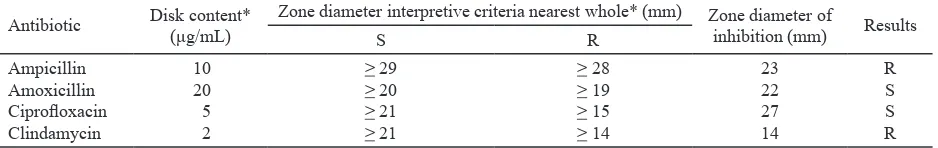
on API 50CHB. The antibiotic test of B. pumilus
showed that the isolate was resistant to ampicillin and clindamycin (Table 2).
Isolation and Purification of Phage. The phage
isolate of indigenous B. pumilus was found in a river water sample collected from Ciapus river, Bogor, West Java, Indonesia. Samples were collected from river water because of phage is naturally part of
environmental ecosystem. Treatment of B. pumilus
with phage resulted in complete lysis of the bacterial cells. The lytic activity formed two types of plaque, haloing plaque and plaque without halo. Average plaque diameter was 0.5 to 2 mm after 18 h incubation at 37 oC (Figure 1).
Phage FBa1 produced one 2 mm diameter translucent plaque, FBa2 1 mm diameter translucent plaque, and FBa3 0.5 mm diameter translucent plaque. Three phages were successfully isolated and they had differences on plaque morphology (Table 3). FBa1 and FBa2 phage were formed plaque with halo, and FBa3 phage was formed plaque without
halo. Purified phages were test on B. pumilus for three times. Plaques morphology were the same as plaques morphology of isolation step, but with differences in plaque size or diameter.
Phage Quantification. Natural phage number in
water samples was too low to produce a quantifiable
titer. Enrichment method was used to isolate phages
and to produce quantifiable titer. Plaque count was
showed in 10-6 phage dilution. The titer of FBa1
Table 1. Identification of host bacterium isolated from water in Cigudeg, Bogor
Identification test Results
Gram staining
Hemolytic activity API 50 CHB
Gram-positive, rod shaped cells β-hemolytic
Positive: glycerol, L-arabinose, D-ribose, D-glucose, D-fructose, D-mannose, D-mannitol, amygdalin, arbutin, esculin ferric citrate, salicin, D-cellobiose, D-saccharose (sucrose), D-trehalose, D-tagatose
Negative: erythritol, D-arabinose, D-xylose, L-xylose, D-adonitol, D-galactose, L-sorbose,
L-rhamnose, dulcitol, inositol, D-sorbitol, mannopyranoside,
methyl-αD-glucopyranoside, N-acetylglucosamine, D-maltose, D-lactose, D-melibiose, inulin,
D-melezitose, D-raffinose, amidon, glycogen, xylitol, gentiobiose, D-turanose,
D-lyxose, D-fucose, L-fucose, D-arabitol, L-arabitol, potassium gluconate, potassium 2-ketogluconate, potassium 5-ketogluconate
Table 2. Results of antibiotic susceptibility test of Bacillus pumilus
Antibiotic Disk content*
(μg/mL)
Zone diameter interpretive criteria nearest whole* (mm) Zone diameter of
inhibition (mm) Results
S R
Ampicillin Amoxicillin
Ciprofloxacin
Clindamycin
10 20 5 2
> 29 > 20 > 21 > 21
> 28 > 19 > 15 > 14
23 22 27 14
R S S R
*Standards by CLSI (2012), S: sensitive, R: resistant.
phage was 10.2 x 10⁸ PFU/mL, FBa2 phage was 5.9
x 108 PFU/mL, and FBa3 phage was 8.5 x 10⁸ PFU/
mL (Table 4). High concentration of B. pumilus -phage in Ciapus river indicated that this river was contaminated with B. pumilus.
Host Range Determination. The host range of
phage was defined by types of bacterial cells which
it can lyse. The phages of FBa1, FBa2, and FBa3 were tested on exponential-phase cultures of other pathogenic bacteria i.e. Photobacterium damselae,
Proteus mirabilis, Salmonella sp., and EPEC K1.1. FBa1, FBa2, and FBa3 phage isolates were lytic on
B. pumilus but did not infect the other tested bacterial isolates (Table 5).
Transmission Electron Microscopy (TEM).
Phage FBa1 was selected for characterization according to the morphological feature. Phage FBa1 was assigned to phage-like particles with 166.67
nm in diameter (Figure 2). It was a phage with icosahedral capsids without tail.
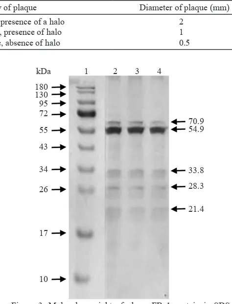
Structural Analysis of Phage Proteins. Capsids of many phage are constructed from multiple copies of the same protein(s). Analysis of phage FBa1
proteins by SDS-PAGE showed five protein bands
(Figure 3). Molecular weight of phage FBa1 proteins was 70.9, 54.9, 33.8, 28.3, and 21.4 kDa.
DISCUSSION
Indigenous B. pumilus was isolated from sewage water in Cigudeg, Bogor, West Java, Indonesia. It indicated that water of their drainage system in the area was contaminated with B. pumilus. The B. pumilus isolate produced a toxin that lysed red blood cells in culture media. Hoult and Tuxford (1991) reported that B. pumilus produced hemolytic activity. Hemolysin toxin produced by B. pumilus caused diarrhea in human. Hemolysin is a toxin having dermonecrotic and vascular permeability activities
and causing fluid accumulation. It implicated in
diarrheal illness.
Most studies on bacterial antibiotic resistance have focused on pathogenic microorganisms. Very
Table 3. Phage isolates from Ciapus river water, Bogor
Phage isolate Morphology of plaque Diameter of plaque (mm)
FBa1 FBa2 FBa3
Translucent plaque, presence of a halo Translucent plaque, presence of halo
Translucent plaque, absence of halo
2 1 0.5
Table 4. Phage quantification of FBa1, FBa2, and FBa3
Phage isolate Plaque count Titer (PFU/mL)
FBa1
Table 5. Host range of phage FBa1, FBa2, FBa3
Host bacteria Phage isolate
+: clear plaques; -: no plaque.
200 nm
Figure 2. TEM images of phages negatively stained with 2% uranyl acetate. Phage FBa1 morphology is circular as phage-like particles. Phage diameter 166.67 nm.
PAGE gel. Lane 1, molecular size marker (PageRuler
limited information on antimicrobial susceptibility profiles of B. pumilus is available. In India, B. pumilus isolates were identified resistant to penicillin
(Parvathi et al. 2009). In Turkey, Ozkocaman (2006) reported that B. pumilus was resistant to amikacin, tobramycin, gentamycin, penicillin, cefepim, cefotaxime, ampicillin-sulbactam, ceftazidime, piperacillin, imipenem, meropenem, aztreonam, cefoperazone-sulbactam, piperacillin-tazobactam, and tetracycline. Four antibiotics (ampicillin,
amoxicillin, ciprofloxacin, and clindamycin) which
had different mode of action were used to treat B. pumilus isolate in our study. Ampicillin acts as a competitive inhibitor of the enzyme transpeptidase, and amoxicillin had same potency but much better
than ampicillin. Ciprofloxacin acts by binding to
complexes DNA and gyrase or topoisomerase IV. Clindamycin acts as inhibitor in bacterial protein synthesis by inhibiting ribosomal translocation. Our results showed that B. pumilus isolate was resistant to ampicillin and clindamycin. Resistance to antibiotic would be a problem. This could solved by an alternative solution such as bacteriophage therapy.
Isolation of bacteriophage specific to B. pumilus
was reported by Grilione and Carr (1960), but it
has not been yet reported in Indonesia. Our study found phage isolate that can infect and kill B. pumilus through lysis. The lytic activity by phage formed plaque on the medium. The different of plaque size in this research may be caused by delay in adsorption. Delay in adsorption makes a lower adsorption rate and resulted in a smaller plaque size (Abedon et al. 2001). Plaque size was influenced
by numerous factors, such as addition of sodium azide accompanied by an extended incubation period, reducing the agar concentration, condition of incubation, and log phase cells of host bacteria (Clokie & Kropinski 2009). In this study, log phase of host cell was used for optimum phage replication and produced large burst size.
The titer of FBa1 phage was higher than that of FBa2 phage and FBa3 phage. It means that FBa1 phage population in environment was higher than the other phages. Titer values could indicated phage generation time. The phage generation time was divided into three period, (i) diffusion of phage progeny to new host cells; (ii) the phage eclipse period; (iii) a period of progeny maturation (Abedon
et al. 2001). The phage generation was controlled by
the phage lysis time. The phage lysis time was defined
by the infective phage particles released from the host bacteria. The longer lysis time resulted in larger burst size (Wang 2006). The FBa1 phage with larger burst size associated with longer lysis time. In the longer
lysis time, FBa1 phage could continue to accumulate progeny virions before lysis. Larger burst size formed larger plaque size.
Our study showed that phage FBa1, FBa2, and
FBa3 had specific activity to B. pumilus. It cannot infect Photobacterium damselae, Proteus mirabilis,
Salmonella sp., and EPEC K1.1. A specific phage
is known to be able to infect a narrow host range.
Narrow host specificity means that non target bacteria will not be killed by them (Goodridge & Abedon 2003). Specificity interaction of phage with bacterial cell is determined by specificity of adsorption and
dependent on the structural of reseptors on bacterial cell surface (Braun & Hantke 1997). The nature of
reseptors contacting phages is largely defined by
composition of host cell wall and surface structures. The outer membrane of gram-positive bacteria differs in structure from the inner membrane and from the plasma membrane of gram-negative bacteria. Many
phages are attracted to bacterial pili, flagella, capsular
and slime polysaccharides as receptors. Among
phages adsorbing to flagella, several agents have been reported including phage χ infecting Salmonella,
Serratia, and E. coli, phage PBS7 specific to B. pumilus (Shade et al. 1967; Lovett 1972).
Phage FBa1 was assigned to phage-like particles. The phage-like particles are phage without tail and were more abundant than tailed phage (Ashelford
et al. 2003). Phage FBa1 did not have tail to attach to B. pumilus. Phage FBa1 may be have the same
mechanism of adsorption as phage PBP7 that specific
to B. pumilus as discribed by Lovett (1972). Phage FBa1 in our result were different from previously
discovered of phiAGATE. Phage phiAGATE
infecting B. pumilus by virions phiAGATE had tail
morphology and assumed that phage was a member of Myoviridae family (Barylski et al. 2014). This
different morphology between FBa1 and phiAGATE
might caused by different water sample collection.
Phage phiAGATE was isolated from water sample
in litoral and pelagic zone of lake, and phage FBa1 was isolated from river water. Ashelford et al. (2003) reported that different sample types might harbor different population of bacteriophage.
Molecular weight of phage proteins was analyzed
using SDS-PAGE. Five bands were observed at SDS-PAGE gel, they may correlated to structural
and functional protein of phage FBa1. Three bands were observed at 70.9, 54.9, and 33.8 kDa; they may correlated to structural proteins of phage FBa1. Capsid proteins of phage SPP1 infecting B. subtilis
was reported between 47.5 and 32.5 kDa (Vinga et al.
2006). While 28.3 kDa protein was similar to purified
from the B. cereus-infecting bacteriophage B4 (Son
et al. 2012), and 21.4 kDa protein may correlated to endolysin of phage. Molecular weight of endolysin of phage BtCS33 infecting B. thuringiensis was 24 and 11 kDa (Yuan et al. 2012).
Lytic phage produced holin and endolysin to degradate bacterial cell wall (Hanlon 2007). The holin-endolysin is essential for host lysis (Young 2002). Holin forms a hole in the cell membrane, and endolysin passes through the hole and destroys the peptidoglycan structure. Two gene products were determined to be the possible lysin, an N-acetylmuramoyl-L-alanine amidases and/or L-alanoyl-D-glutamate peptidase. The endolysin have two domains connected by a short linker: the N-terminal catalytic domain is responsible for cell lytic activity and the C-terminal cell wall binding
domain that recognizes and binds a specific substrate
in the cell wall of target bacteria (Fischetti 2008). Several advantages of using phage as a biocontrol
agent is its high specificity to target host bacterium,
self-replication because phage will multiply as long as there is still a host present, low toxicity because phage consist mostly of nucleic acids and proteins, phage are relatively cheap and easy to isolate, and phage can add on food processing (Sillankorva et al. 2012). Indigenous phage FBa1, FBa2, FBa3 isolated from Ciapus river water, Bogor were capable to
specifically lyse B. pumilus. Our study showed that these phage isolates had potency for biocontrol of B. pumilus in the environment.
REFERENCES
Abedon ST, Herschler TD, Stopar D. 2001. Bacteriophage latent-period evolution as a response to resource availability. Appl Environ Microbiol 67:4233-4241.
Agata N, Ohta M, Mori M, Isobe M. 1995. A novel dodecadepsipeptide, cereulide, is an emetic toxin of Bacillus cereus. FEMS Microbiol Lett 129:17-20.
Ashelford KE, Day MJ, Fry JC. 2003. Elevated abundance of bacteriophage infecting bacteria in soil. Appl Environ Microbiol 69:285-289.
Barylski J, Nowicki G, Gozdzicka-Jozefiak A. 2014. The
discovery of phiAGATE, a novel phage infecting Bacillus
pumilus, leads to new insights into the phylogeny of the subfamily Spounavirinae. PLOS ONE 9:1-14.
Braun V, Hantke K. 1977. Bacterial receptors for phages and colicins as constituents of specific transport systems.
Microbial Interactions. Receptor and Recognition 3:101-137. Budiarti S, Hidayati R, Rusmana I. 2011. Infectivity of lytic
phage to Enteropathogenic Escherichia coli from diarrheal patients in Indonesia. J US-China Med Sci 8:273-282. Callaway TR, Edrington TS, Brabban AD, Anderson RC,
Rossman ML, Engler MJ, Carr MA, Genovese KJ, Keen
JE, Looper ML, Kutter EM, Nisbet DJ. 2008. Bacteriophage isolated from feedlot cattle can reduce Escherichia coli
O157:H7 populations in ruminant gastrointestinal tracts.
Foodborne Pathogens and Diseases 5:183-191.
Carey-Smith GV, Billington C, Cornelius AJ, Hudson A,
Heinemann JA. 2006. Isolation and characterization of bacteriophages infecting Salmonella spp. J FEMSLE 217:1-5. Clokie MRJ, Kropinski AM. 2009. Bacteriophages Methods
and Protocols, Volume 1: Isolation, Characterization, and Interactions. New York: Humana Pr.
[CLSI] Clinical and Laboratory Standards Institute. 2012. Performance Standards for Antimicrobial Susceptibility Testing: Twenty-Second Informational Supplement. Pennsylvania (USA): CLSI.
Fischetti VA. 2008. Bacteriophage lysins as effective antibacterials. Curr Opin Microbiol 11:393-400.
From C, Hormazabal V, Granum PE. 2007. Food poisoning
associated with pumilacidin-producing Bacillus pumilus in rice. Int J Food Microbiol 115:319-324.
Ganz HH, Law C, Schmuki M, Eichenseher F, Calendar R, Loessner MJ, Getz WM, Korlach J, Beyer W, Klumpp J.
2014. Novel giant siphovirus from Bacillus anthracis features unusual genome characteristics. PLOS ONE 9:1-9.
Giuliano M, Barza M, Jacobus NV, Gorbach SL. 1987. Effect
of broad spectrum parenteral antibiotics on compotition of intestinal microflora in humans. Antimicrob Agents Chemother 31:202-206.
Goodridge L, Gallaccio A, Griffiths WM. 2001. Morphological,
host range, and genetic characterization of two coliphages.
Appl Environ Microbiol 69:5364-5371.
Goodridge L, Abedon ST. 2003. Bacteriophage biocontrol and
bioprocessing application of phage therapy to industry. SIM News 53:254-262.
Grilione PL, Carr JH. 1960. Isolation and study of a bacteriophage
for Bacillus pumilus. J Bacteriol 80:47-50.
Hanlon GW. 2007. Bacteriophages: an appraisal of their role in
the treatment of bacterial infections. Int J Antimicrob Agents
30:118-128.
Hoult B, Tuxford AF. 1991. Toxin production by Bacillus pumilus. J Clin Pathol 44:455-458.
Jakutytė L, Baptista C, São-José C, Daugelavičius R,
Carballido-López R, Tavares P. 2011. Bacteriophage infection in rod-shaped gram-positive bacteria: evidence for preferential polar route for phage SPP1 entry in Bacillus subtilis. J Bacteriol
193:4893-4903.
Lee WJ, Billington C, Hudson JA, Heinemann JA. 2011. Isolation and characterization of phages infecting Bacillus cereus. Lett Appl Microbiol 52:456-464.
Levin BR, Bull JJ. 2004. Population and evolutionary dynamics of phage therapy. Microbiology: Nature Reviews 2:166-173. Lovett PS. 1972. PBP1: a flagella specific bacteriophage
mediating transduction in Bacillus pumilus. Virology
47:743-752.
Martínez B, Obeso JM, Rodríguez A, García P. 2008.
Nisin-bacteriophage crossresistance in Staphylococcus aureus. Int J Food Microbiol 122:253-258.
Naruse N, Tenmyo O, Kobaru S, Kamei H, Miyaki T, Konishi M, Oki T. 1990. Pumilacidin, a complex of new antiviral antibiotics. J Antibiot 43:267-280.
Ozkocaman V, Ozcelik T, Ali R, Ozkalemkas F, Ozkan A, Ozakin C, Akalin H, Ursavas A, Coscun F, Erner B, Tunali A. 2006. Bacillus spp. among hospitalized patients with haemotological malignancies: clinical features, epidemics and outcomes. J Hosp Infect 64:169-176.
Parvathi A, Krishna K, Jose J, Joseph N, Nair S. 2009. Biochemical and molecular characterization of Bacillus pumilus isolated from coastal environment in Cochin, India.
Rakhuba DV, Kolomiets EI, Szwajcer Dey E, Novik GI. 2010.
Bacteriophage reseptors, mechanisms of phage adsorbtion and penetration into host cell. Pol J Microbiol 59:145-155. Sartika D, Budiarti S, Sudarwanto M. 2012. Phage FR38
treatment on sprague dawley rat inferred from blood parameters and organ systems. Hayati J Biosci 19:131-136.
Shade SZ, Adler J, Ris H. 1967. How bacteriophage χ attack
motile bacteria. J Virol 1:599-609.
Sheppard JD, Jumarie C, Cooper DG, Leprade R. 1991. Ionic
channels induced by surfactin in planar lipid bilayer membranes. Biochimia et Biophysica Acta 1064:13-23. Sillankorva SM, Oliveira H, Azeredo J. 2012. Bacteriophages and
their role in food safety. Int J Microbiol 2012:1-13. Son B, Yun J, Lim J-A, Shin H, Heu S, Ryu S. 2012.
Characterization of LysB4, an endolysin from the Bacillus cereus-infecting bacteriophage B4. BMC Microbiol 12:1-9. Soni KA, Nannapaneni R. 2010. Removal of Listeria monocytogenes biofilms with bacteriophage P100. J Food Protection 73:1519-1524.
Spricigo DA, Bardina C, Cortes P, Llagostera M. 2013. Use of bacteriophage cocktail to control Salmonella in food and the food industry. Int J Food Microbiol 165:169-174.
Suominen I, Andersson MA, Andersson MC, Hallaksela AM, Kampfer P, Rainey FA, Salkinoja-Salonen M. 2001. Toxic
Bacillus pumilus from indoor air, recycled paper pulp, Norway spruce, food poisoning outbreaks and clinical samples. Syst Appl Microbiol 24:267-276.
Vinga I, Dröge A, Stiege AC, Lurz R, Santor MA, Daugelavicius R, Tavares P. 2006. The minor capsid protein gp7 of
bacteriophage SPP1 is required for efficient infection of
Bacillus subtilis. Mol Microbiol 61:1609-1621.
Wang I-N. 2006. Lysis timing and bacteriophage fitness. Genetics
172:17-26.
Yokohama K, Ito M, Agata N, Isobe M, Shibayama K, Horii T, Ohta M. 1999. Pathological effect of synthetic cereulide, an emetic toxin of Bacillus cereus, is reversible in mice. FEMS Immunol Med Microbiol 24:115-120.
Young R. 2002. Bacteriophage holins: deadly diversity. J Mol Microbiol Biotechnol 4:21-36.
Yuan Y, Peng Q, Gao M. 2012. Characteristics of a broad