Nanosecond Pulsed Electric Fields (nsPEFs) Menginduksi Ekspresi Gen Socs1 dan Socs3 namun
bukan pada Sel HeLa
1 2 3
Martina Kurnia R , Diana Lyrawati , Kenichi Yano
1Master in Biomedical Science Faculty of Medicine University of Brawijaya Indonesia
2Departement of Pharmacology Faculty of Medicine University of Brawijaya Indonesia
3Bioelectric Laboratory Institute of Pulse Power Science Kumamoto University Japan
ABSTRACT
Nanosecond Pulsed Electric Fields (nsPEFs) is one of bioelectric technologies applied widely in a number of sciences. nsPEFs cause some biological responses and known to play a role as a novel cancer therapy. However, the nsPEFs molecular mechanisms have not been fully elucidated. This study determines the effects of nsPEFs in socs (Suppressor of Cytokine Signaling) genes which are target genes of JAK/STAT signaling pathway. Through a negative feedback mechanism, SOCS proteins can suppress both cytokine signal transduction and overgrowth factor, so the cell growth is controlled. In cervix cancer, the presence of E6 and E7 HPV's oncoprotein is associated with methylation and inactivation of socs1 and socs3 genes. This mechanism is related to the increase of STAT expression and cancer prognostic. In this research, nsPEFs as much as 20 kV/cm for 80 ns was exposed over HeLa S3 cells in 4 mm cuvette. Socs1, socs2 and socs3 gene expressions were analyzed using real time PCR SYBR green and reverse transcription PCR (RT-PCR). This study shows that at 20 and 30 shots, nsPEFs significantly increase socs1 and socs3 but not socs2 gene expression. Effect of nsPEFs on socs1 and socs3 gene expression pattern is influenced by duration of post exposure incubation and each cell activity on internal cell condition. This research provides a new cancer therapy target for nsPEFs.
Keywords: Bioelectric, gene expression, nsPEFs, shot, socs gene
ABSTRAK
Nanosecond Pulsed Electric Fields (nsPEFs) merupakan salah satu teknologi bioelektrik yang secara luas diaplikasikan pada sejumlah bidang sains. NsPEFs dapat menyebabkan sejumlah respon biologis dan diketahui berperan sebagai salah satu novel terapi kanker. Meski demikian, mekanisme molekular nsPEFs belum banyak diketahui. Penelitian ini bertujuan untuk mengetahui efek nsPEFs pada gen socs (Suppressor of Cytokine Signaling) yang merupakan salah satu gen target jalur sinyal JAK/STAT. Melalui aktivitas umpan balik negative, protein SOCS dapat menekan transduksi sinyal sitokin dan faktor pertumbuhan yang berlebih sehingga dapat mengontrol pertumbuhan sel. Pada kanker serviks, kehadiran onkoprotein HPV E6 dan E7 diasosiasikan dengan metilasi dan inaktivasi gen socs1 dan socs3. Mekanisme tersebut juga berkaitan dengan peningkatan ekspresi STAT dan peningkatan prognosivitas kanker. Pada penelitian ini, nsPEFs sebesar 20 kV/cm selama 80 ns dipaparkan pada sel HeLa S3 di dalam kuvet 4 mm. Ekspresi gen socs1, socs2 dan socs3 dianalisis menggunakan real time PCR SYBR green dan reverse transcription PCR (RT-PCR). Penelitian ini menunjukkan bahwa pada 20 dan 30 kali tembakan, nsPEFs mampu meningkatkan ekspresi gen socs1 dan socs3, namun bukan socs2. Efek nsPEFs pada kedua gen tersebut juga dipengaruhi lamanya waktu inkubasi pasca paparan dan aktivitas masing-masing gen pada kondisi internal sel. Penelitian ini memberikan target terapi kanker baru bagi nsPEFs.
Kata Kunci: Bioelektrik, ekspresi gen, gen socs, nsPEFs, tembakan
have not been fully elucidated. In this research, we will INTRODUCTION
investigate the effect of nsPEFs on socs1, socs2 and socs3
Nanosecond pulsed electric fields (nsPEFs) are one of gene expressions. This research will provide a new bioelectric application that developed from conventional possibility role of nsPEFs on cancer therapy.
electroporations. Generally, electroporations are used to deliver plasmid, gene, antibody or other molecules to cell
METHOD for some purposes especially for therapy (1).Unlike
electroporations, nsPEFs are exposurewith high electric Cell Culture and Sample Preparation fields (1-100 kV/cm) in very short duration (1-300
HeLa S3 cells (ATCC) were cultured in α-minimum essential nanoseconds). It causes low energy density and non
medium (α-MEM)(WAKO) supplemented with 10% fetal thermal effect (2). In cell membrane, nsPEFs cause
b o v i n e s e r u m ( F B S ) ( E q u i t e c h B i o ) a n d nanoporeformation (1-1,5 nm) without disturb the
penicillin/streptomycin (WAKO). Cells were maintained in membrane integrity (3). NsPEFs also activate
voltage-2+ 2+ a humidified atmosphere of 5% CO at 37 C. The cells were
gated Ca channels and increases Ca influx into 2 6
counted using cell counter coulter Z.1. The 2x10 cell cytoplasm (4).
nsPEFs cells were suspended in 400 µl α-MEM-10%FBS Unlike conventional electroporations, nsPEFs raise some lacking antibiotics.
intracellular responses. In the high intensity, nsPEFs reach
Application of NsPEFs
subcellular level and can be active in cellular system (5). In
mitochondria, nsPEFs increase mitochondrial membrane The cell suspensions (400 µl) were placed in an permeability (mPTP) andcause cytochrome c release (6,7). electroporation cuvette that contained a pair of parallel
2+
NsPEFs also cause Ca influx into mitochondria and induce aluminium electrodes with a 4 mm-gap (#5540, MβP, dissipation of membrane potential (ΔΨm) that implicate Thermo Fisher Scientific). A train of nsPEFs was generated
2+
to cell viability (7,8). NsPEFs also cause Ca release from by a pulsed power modulator and applied to the cell ER (9), involve in some signaling pathways (10-12), and suspensions in the cuvette at 1 Hz. Voltage waveform of induce stress responses (13). In the nucleus, nsPEFs electric pulses were monitored by a high-voltage probe increase some endogen gene expressions (14). In the high (P6015A, Tektronix) in the oscilloscope. NsPEFs were intensity (>60kV/cm), nsPEFs can cause actin filament exposured at 20 kV/cm with pulses of 80 ns duration (was depolymerization, disturb telomere attachment to the standarderized in Bioelectric Laboratory, Institute of Pulse nucleus and disturb DNA stability (15). Power Science, Kumamoto University, Japan). After exposure, the cells were immediately diluted 4-folds Recently, application of electric fields in an extremly short
αMEM-10%FBS lacking antibiotics and incubated in 5% CO duration with high intensity is widely used in various fields 2
incubator at 37 C during 1 and 4 hours (hr). of the life science including for cancer therapy. NsPEFs can
induce apoptosis via intrinsic and extrinsic pathways (16- RNA Isolation 18). NsPEFs also decrease cell proliferation and metastasis
Total RNA was isolated from all samples using RNAiso Plus via suppression of NFκB and Wnt/β-catenin signaling (12),
reagent (Takara Bio). Aliquots of the total RNA were decrease viability of cancer cell (19), decrease tumor mass
subjected to real time PCR SYBR Green and RT-PCR. The and size (12)(20), haveantiangiogenic activity (21,22) and
total RNA concentration and purity were confirmed using increase the immune system (23).
Biophotometer plus (eppendorf). Suppressor of Cytokine Signaling (SOCS) proteins
Real Time PCR Analysis
areidentified as tumor suppressor proteins andone of the
cancer targeted therapies. In normal condition, the active Quantitative mRNA analysis was conducted by quantative SOCS have important role in negative feedback real time PCR using iScript One-Step RT-PCR kit with SYBR mechanism of JAK/STAT signaling pathway and inhibit Green (Biorad) on an MJ Mini Thermal Cycler equipped cytokine and growth factor signal transduction (24). In with a Mini Opticon Real Time PCR system (BioRad). Real some cancers, overexpressions and dysregulations of time PCR data were calculated by measuring the average JAK/STAT signaling pathway are associated with high cycle threshold (Ct) number for the mRNA of target, which proliferation and low apoptosis. However, SOCS are was normalized to the values of the mRNA for methylated and silenced in many cancers (25-32).
glyceraldehydes-3-phosphate dehydrogenase (GAPDH). Dysregulations of SOCS are also associated with
phosphorylation defect and mutation (33,34). SOCS1, Reverse Transcription PCR (RT-PCR)
SOCS2 and SOCS3 are reported as the three most active
Semiquantitative mRNA analysis was conducted by RT-PCR SOCS protein with some tumor suppression roles.
using OneStep RT-PCR Kit (Qiagen) on RT-PCR system. PCR Cervical cancer is one of gynecological cancer that caused products were separated by agarose gel electrophoresis by HPV infection. Two oncoproteins of HPV, E6 and E7, can and visualized by UV transluminator. Intensity of band was trigger carcinogenesis and genetic instability (35). The quantified by ImageJ software.
presences of HPV's genomes are associated with
Primer Optimization
overexpression of STAT3 and STAT5, and are also
implicateto prognosivity of cancer (36,37). STAT3 and The primer pairs for each gene (socs1, socs2 and socs3) STAT5 overexpressions are also assoaciated with that used in this research were optimized based on primer epigenetic control as well as methylation at promoter of dimmer presence and consistency of melt curve in the real
socs1 gene (38). E6 and E7 can induce methylation in some time PCR (Supplementary data). The primer pairsthat used tumor suppressor genes including socs1 and socs3 (39). as the result from the optimizationof the methods as Wherease, the activity of socs2 has not found in cervical f o l l o w s ( 3 2 ) ( 1 0 ) ,s o c s 1 ( 2 0 2 b p ) , F : 5 ' -cancer. However, the overexpression of STAT3 and STAT5 A G A C C C C T T C T C A C C T C T T G 3 ' ; R : 5 ' -as the degradation target of socs2 is one reason to CTGCACAGCAGAAAAATAAAGC-3'; socs2 (244 bp), F: 5'-investigate socs2 gene expression (40). G G ATG G TA C TG G G G A A G TATG A C TG 3 ' ; R : 5 ' -The role and molecular mechanisms of nsPEFs in cancer AGTCGATCAGATGAACCACACTGTC-3'; socs3 (107 bp), F:
5'-°
at 1 hr and 4 hr were observed using different analysis T C C C C C C A G A A G A G C C T A T T A C 3 ' ; R : 5 '
-respectively using real time PCR and RT-PCR. Based on the TCCGACAGAGATGCTGAAGAGTG-3'; GAPDH (452 bp), F:
simple t-test, the expression of socs1, socs2 and socs3
5 ' A C C A C A G T C C A T G C C A T C A C 3 ' , R : 5 '
-were significantly different between at 1 hr and 4 hr in 0, TCCACCACCCTGTTGCTGTA-3'.
20, and 30 shots nsPEFs respectively socs1 (p<0,000;
Statistical Analysis p<0,014, p< 0,010), socs2 (p<0,015; p<0,024; p<0,047),
and socs3 (p<0,044; p<0,002; p<0,006). The present data were expressed as mean ± SD. The
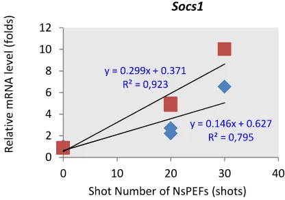
distribution of data was evaluated using Kolmogorov- Shot Number of NsPEFs Correlated with Socs1 and Socs3 Smirnov analysis. The data were analyzed using one way but not Socs2 Gene Expressions
analysis of variance (ANOVA) with HSD Tukey post hoc
Based on the correlation-regression analysis, we suggest test, simple t-test, and correlation-regression test.
that different shot number of nsPEFs correlated with socs1
Statistic significance was set at P value < 0,05.
and socs3 but not socs2 gene expressions as we show at the Figure 2.
RESULTS
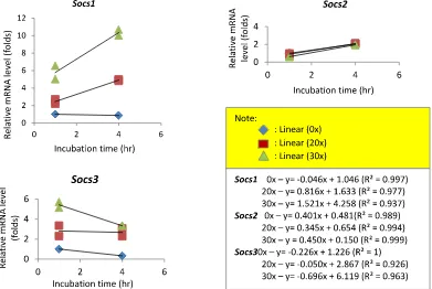
NsPEFs IncreasedSocs1 and Socs3 but not Socs2 Gene Expressions
Previous study reported that 20 kV/cm during 80 ns duration at 20 and 30 shots treated into HeLa S3 cell decreased 15% and 30% cell viability and cell growth (41). Our results showed that these numbers of nsPEFs increased socs1 (Figure 1A) and socs3 (Figure 1C) but not
socs2 (Figure 1B) gene expressions.
Based on the one way ANOVA, we suggest that nsPEFs significantly increased socs1 and socs3 but not socs2 gene expressions. At 1 hr, 20 and 30 shots nsPEFs increased
socs1 geneexpression approximately 2,45 and 5,78 folds.
Socs1 gene expression increased up to 4,90 and 10,34 folds at 4 hr. NsPEFs also increased socs3 gene expression. At 1 hr, 20 and 30 shots nsPEFs increased socs3 gene
expressions approximately 3,19 and 5,42 folds. At 4 hr, 20 Figure 2. Correlations between shot number of NsPEFs with and 30 shots nsPEFs increased socs3 gene expression Socs1, Socs2 and Socs3 gene expressions
approximately 2,28 and 3,33 folds. NsPEFs did not increasesocs2 gene expression.
m;
Effect of nsPEFs on socs1, socs2 and socs3 gen expressions
Note: The correlation between shot number and socs1 and socs3 gene expressions were indicated with positive correlation, but negative correlation for socs2. 1 jam; 4 ja
Figure 1. Effect of NsPEFs on Socs gene expressions
treatment group. Gene expression pattern of socs2 in control group and treatment group increased, but these expressions were not significantly different. These data showed that nsPEFs resulted in changes gene expressions pattern in socs1 and socs3, but not in socs2.
DISCUSSION
NsPEFs Probably Decrease Proliferation and Induce Apoptosis via Increasing of Socs1 and Socs3 Gene Expressions
In the cervical cancer, HPV infections cause socs1 and socs3
methylations (36, 37, 39). These methylations implicate to STAT3 and 5 overexpressions (38). Generally, methylation of socs1 and socs3 implicate to high proliferation and low apoptosis in some cancers (36-39). Wherease, the rolesof
socs2 have not been known in this cancer. Our results show that 20 and 30 shots of 20 kV/cm nsPEFs in 80 ns duration increased socs1 and socs3 but not socs2 gene expressions. From the control group of our results indicate that there were not any changes on socs2 gene expressions.
Figure 2. Correlations between shot number of NsPEFs with Socs1, socs2
and socs3 are involved in negative feedback
Socs1, Socs2 and Socs3 gene expressions
mechanism of JAK/STAT signaling pathway (41). By this
Note:
mechanism, increasing of socs1, socs2, socs3 decrease JAK/STAT target genes that regulate cell proliferation and apoptosis, such as c-myc and bcl-xl (24). According to these theories, we propose that nsPEFs may decrease proliferation and induce apoptosis via increasing of socs1 Different Pattern of Socs1 and Socs3 Gene Expressions
and socs3 gene expressions. Inducing socs1 and socs3 gene
Followed NsPEFs
expressions by nsPEF probably decrease JAK/STAT signal Gene expression pattern was determined to know the transduction and implicate to decreasing of c-myc and bcl-dynamic of nsPEFs effect on socs1, socs2, and socs3 gene xl. Decreasing of c-myc (induce proliferation) and anti expressions during a certain time (see Figure 3). Gene apoptotic bcl-xl can decrease cell proliferation and induce expression pattern of socs1due to 20 and 30 shots nsPEFs apoptosis. Increasing of socs1 by nsPEFs probably causes increased from 1 hr to 4 hrs. In contrary, the control group decreasing of c-myc and bcl-xl directly by E7 degradations. was decreased during 4 hrs. Unlike socs1, socs3 gene
expression initially increased at 1 hr but subsequently However, we need further investigations to prove this decreased after 1 hr up to 4 hr. The pattern of control hypothesis. Our results and hypothesis are described on group of socs3 decreased too, but still lower than Figure 4.
Methylations on promoter of some genes in cervical cancer such as socs1 and socs3 are associated with HPV infections and presence of E6 and E7 oncoproteins (38-39). E6 and E7 increase DNA methylatransferase 1 (DNMT1) that catalizes attachment of methyl group to cytosine in CpG Island. Binding E7 to pRb (E7/pRb) cause the release of E2F, favoring the expression of DNMT1. Binding of E2F to DNMT1 (E7/DNMT1) induced a conformational change in DNMT1 and exposed its DNA binding site and promoting DNA binding (39).
Difference Gene Expression between Socs1 and Socs3 are Determined by Time of NsPEFs Post Exposure and Activity of Each Gene
Based on gene expression pattern analysis, we know that
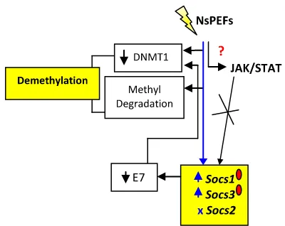
Figure 4 . A schematic diagrams of effect of NsPEFs on socs1,
nsPEFs caused different gene expression pattern between
socs2 and socs3 gene expressions in HeLa S3 cells.
socs1 and socs3 genes. We can see this different expression in Figure 6.
Possibility Mechanismof NsPEFs to Induce Socs1 and Socs3 Gene Expressions
This research did not provide any data to explain the mechanism of nsPEFs to inducesocs1 and socs3 gene expressions. The high intensity of nsPEFs enter cell and nucleus and induce some endogen genes (14). Possibly, mechanism of nsPEFs to induce socs1 and socs3 gene expressions is demethylation.
In general, JAK/STAT signal transduction determines all
socs genes as a target of this pathway. However, methylation of socs1 and socs3 gene expressions in cervical cancer makes this signal can not raise socs1 and
Figure 6. A schematic diagrams of socs1 and socs3 gene
socs3 genes. Therefore, we think that methylation process
expressions pattern due to nsPEFs exposure.
is more important to determine socs1 and socs3 gene expressions than JAK/STAT signal transductions. Our results show that nsPEFs only induced socsmethylated genes, socs1 and socs3 but not socs2. These results suggest that nsPEFs may induce socs1 and socs3 by demethylation process. We also propose that nsPEFs does not induce socs1 and socs3 by JAK/STAT signal
transduction. Our results and hypothesis are describes in At 1 hr, nsPEFs induced socs1 and socs3 gene expressions. Figure 5. Probably, increasing of these gene expressions are induced by directly effect of nsPEFs on socs1 and socs3
demethylations that involve decreasing of DNMT1 and methyl degradation. However, there are different gene expressions at 4 hrs between socs1 and socs3. Socs1 gene expressions increased from 1 hr up to 4 hrs, but socs3
decreased.
The high expressions of socs1 and socs3 at 1 hr on time of nsPEFs post exposure probably were caused by directly effect of nsPEFs on degradation of DNMT1 or methyl group (demethylation process). However, different expression on
socs1 and socs3 probably depend on activity of each gene after socs1 and socs3 were demethylated. SOCS1 can interact to other signaling pathway NFκβ. Not only by JAK/STAT signal transduction, socs1 is also induced by NFκβ, but socs3 is not (44).
The high expressions of socs3 at 1 hr probably caused negative feedback mechanism on JAK/STAT and decreased the target gene such as c-mycandbcl-xl. From this
Figure 5. A schematic diagrams of possibility mechanism of
mechanism, socs3 expression also decreased at 4 hrs but
NsPEFs to increase socs1 and socs3 gene expressions
still higher that control (cancer cell). Unlike socs3, socs1
gene expression increased at 4 hrs. The high expressiob of
socs1 gene probably also caused by induction of NFκβ. In
Note: Blue sign show our result - ( )= gene expressions inductions, (x)= no effect; green sign show our hypothesis – ( / ) = increasing/decreasing of expressions; = gene methylation; ?: unknown effect)
Note: 1: primary demethylation effect by nsPEFs; 2a: secondary demethylation effect by E7 degradation; 2b: decreasing of demethylation effect; ?: The process between 1 hr to 4 hrs, it may be associated with negative feedback mechanism of SOCS on JAK/STAT
cervical cancer, E6 and E7 induce NFκβ signaling pathway activity of these gene to decrease proliferation in HeLa S3 and increase expression of the target genes including cell. This result is suitable with the previous research (42).
cyclin, cdk, bcl-xl, and socs1. However, socs1 was High Shot Number do not always Associate with High Gene methylated in HPV infection (39) and cannot decrease 065 Expressions
subunit (45). Therefore, proliferation increase and
apoptosis decrease. Demethylated socs1 by nsPEFs Shot number is one of characteristic that also determine probably invlove in JAK/STAT and NFκβ. High expression of the electric fields. Different shot number is proven NFκβ by E6 and E7 may induce socs1 gene expression associated with different cell viability (41) and expression without methylated at 4 hrs. of some gene (13). Our result show that shot number is positively correlates with socs1 and socs3 gene SOCS1 has many activities in cervical cancer than SOCS3. expressions. Higher shot number caused higher socs1 and SOCS1 is better to decrease proliferation in HeLa S3 than socs3 gene expression, but not in socs2. We suggest that SOCS3 (42). This study also showed that SOCS1 is adaptor high shot number do not always assoaciate with high gene of E3 that specific to degrade E7. E7 degradation is expressions.
important to repress genetic instability in cervical cancer.
Finally, we conclude that nsPEFs increase socs1 and socs3
Low level of E7 is implicated to DNMT1 repression and
but not socs2 gene expressions. The shot number demethylation some tumor supressor genes such as socs1
correlates positively with the socs1 and socs3 gene and socs3 (39). Low level of E7 also implicated to inhibition
expressions. High shot number do not always correlate E2F and repression of some genes such as c-jun and c-fos
with high gene expression. Effect of nsPEFs on socs1 and (46). SOCS1 can degrade p65 subunit of NFκβ and
socs3 gene expression pattern depends on time of nsPEFs decrease cyclin, cdk, and bcl-xl (45). SOCS1 but not SOCS3
post exposure and type of gene. Because socs1 and socs3
can increase p53 and p21 expressions and implicated to
have some roles in tumor suppressor, nsPEFs possibly have low proliferation and apoptosis induction (47).
potentially as anti cancer. According to the discussion, nsPEFs probably directly
induce demethylation and activation of socs1 and socs3. ACKNOWLEDGEMENTS However, after activation, the expressions of socs1 and
socs3 are determined by activity of each gene in cellular We thank JASSO (Japan Student Services Organization) for mechanism. From this result we know that the higher providing Martina scholarships in IJEP short-term
exchange program, Kumamoto University, Japan. expression of socs1 than socs3 probably determine the
REFERENCES Transient Feature in Nanosecond Pulsed Electric Fields Differentially Modulate Mitochondria and Viability.
1. Chen C, Smye SW, Robinson MP, and Evan JA. PLoS One. 2012;7(12): e51349
Membrane Electroporation Theories: A Review.
Medical & Biological Engineering & Computing. 2006; 9. Vernier PT, Sun Y, Mercu L, Salemi S, Craft SM, and 44(1-2): 5-14. Gundersen MA. Calcium Bursts Induced by Nanosecond Electric Pulses. Biochemical and 2. Beebe SJ and Schoenbach KH. Nanosecond Pulsed Biophysical Research Communications. 2003; 310(2):
Electric Fields: A New Stimulus to Activate 286-295. Intracellular Signaling. Journal of Biomedicine and
Biotechnology. 2005; 2005(4): 297-300. 10. Morotomi-Yano K, Akiyama H, and Yano K.
Nanosecond Pulsed Electric Fields Activate MAPK
3. Deng J, Schoenbach KH, Buescher ES, Hair P, Fox PM, Pathways in Human Cells. Biochemical and Biophysical and Beebe SJ. The Effect of Intense Submicrosecond Research Communications. 2011; 515(1-2): 99-106. Electrical Pulses on cells. Biophysical Journal. 2003;
84(4): 2709-2714. 11. Morotomi-Yano K, Akiyama H, and Yano K.
Nanosecond Pulsed Electric Fields Activate
AMP-4. Crasivo GL, Choe S, Chatterjee P, Chatterjee I, and Activated Protein Kinase: Implications for Calcium-Vernier PT. Nanosecond Electric Pulses: A Novel
2+ Mediated Activation of Cellular Signaling. Biochemical
Stimulus for Triggering Ca Influx into Chromaffin
2+ and Biophysical Research Communications. 2012; Cells Via Voltage-Gated Ca Channels. Cellular and 428(3): 371-375.
Molecular Neurobiology. 2010; 30(8): 1259-1265.
12. Ren Z, Chen X, Cui G, et al.Nanosecond Pulsed Electric
5. Weaver JC, Smith KC, Esser AT, Son RS, and Field Inhibits cancer Growth Followed by Alteration in Gowrishanker TR. A Brief Overview of Electroporation Expression of NF-kB and Wnt/β-catenin Signaling Pulse Strength-Duration Space: A Region Where Molecules. PLoS One. 2013; 8(9): e74322.
Additional Intracellular Effects are expected.
Bioelectrochemistry. 2012; 87: 236-243. 13. Morotomi-Yano K, Oyadomari S, Akiyama H, and Yano K. Nanosecond Pulsed Electric Act as a Novel Cellular
6. Napotnik TB, Wu Y, Gundersen MA, Miklavcic D, and Stress That Induces Translational Suppression Vernier PT. Nanosecond Electric Pulses Cause Accompanied by elF2a Phosphorylation and 4E-BP1 Mitochondrial Membrane Permeabilization in Jurkat Dephosphorylation. Experimental Cell Research. Cells. Bioelectromagnetic. 2012; 33(3): 257-264. 2012; 318: 1733-1744.
7. Beebe SJ, Sain NM, and Ren W. Induction of Cell Death 14. Beebe SJ, Blackmore PF, White J, Joshi RP, and Mechanisms and Apoptosis by Nanosecond Pulsed Schoenbach KH. Nanosecond Pulsed Electric Fields Electric Fields (nsPEFs). Cells. 2013; 2(1): 136-162. Modulate Cell Function through Intracellular Signal
Measurement. 2004; 25(4): 1077-1093. 21. Nuccitelli R, Pliquett U, Chen X, et al. Nanosecond Pulsed Electric Fields Cause Melanomas to
Self-15. Stacey M, Fox P, Buescher S, and Kolb J. Nanosecond
Destruct. Biochemical and Biophysical Research
Pulsed Electric Field Induced Cytoskeleton, Nuclear
Communications. 2006; 343(2): 351-360.
Membrane and Telomere Damage Adversely Impact
Cell Survical. Bioelectrochemistry. 2011; 82(2): 131- 22. Wu S, Wang Y, Guo J, Chen Q, Zhang J, and Fang J.
134. Nanosecond Pulsed Electric fields as a Novel Drug Free
Therapy for Breast Cancer: An In Vivo Study. Cancer 16. Beebe SJ, Fox PM, Rec LJ, Willis LK, and Schoenbach
Letters. 2014; 343(2): 268-274. KH. Nanosecond, High-Intensity Pulsed Electric Fields
Induce Apoptosis in Human Cell. The Journal of the 23. Beebe SJ. Bioelectrics in Basic Science and Medicine: Federation of American Societies for Experimental Impact of Electric Fields on Cellular Structures and Biology. 2003; 17(11): 1493-1495. Functions. Nanomedicine and Nanotechnology. 2013; 17. Ren W and Beebe SJ. An Apoptosis Targeted Stimulus 4(2): 1-8.
with Nanosecond Pulsed Electric Fields (NsPEFs) in E4 24. Rawlings JS, Rosler KM, and Horrison DA. The JAK/STAT Squamous Cell Carcinoma. Apoptosis. 2011; 16(4): Signaling Pathway. Journal of Cell Science. 2004; 117:
382-393. 1281-1283.
18. Ford WE, Ren W, Blackmore PF, Schoenback KH, and 25. Elliott J, Hookham MB, and Johnston JA. The Beebe SJ. Nanosecond Pulsed Electric Fields Stimulate Suppressors of Cytokine Signalling E3 Ligases Behaves Apoptosis without Release of Pro-Apoptototic Factors
as Tumor Suppressors. Biochemical Society
from Mitochondria in B16f10 Melanoma. Archieves of
Transactions. 2008; 36(Pt3): 464-468. Bioechemistry and Biophysics.2010; 497(1-2): 82-89.
26. Weniger MA, Melzner I, Menz CK, et al.Mutations of
19. Stacey M, Stickley J, Fox P, et al. Differential Effects in
the Tumor Suppressor Gene SOCS1 in Classical Hodgkin Cells Exposed to Ultra-Short, High Intensity Electric
Lymphoma are Frequent and Associated with Nuclear Fields: Cell Survival, DNA Damage, and Cell Cycle
Phospho-STAT5 Accumulation. Oncogene. 2006;
Analysis. Mutation Research. 2003; 542(1-2): 65-75.
25(18): 2679–2684. 20. Nagahama M, Shimomura N, Nakagawa A, Teranishi K,
27. Nagai H, Naka T, Terada Y, et al. Hypermethylation
Uto Y, and Hori H. In Vivo Experimental Study of
Associated with Inactivation of the SOCS-1 Gene, a Nanosecond Pulsed Electric Field Effects on Solid
JAK/STAT Inhibitor, in Human Hepatoblastomas. Tumors. IEEE Transactions on Dielectrics and Electrical