Wayne C. Drevets
Neuroimaging studies of major depression have identified neurophysiologic abnormalities in multiple areas of the orbital and medial prefrontal cortex, the amygdala, and related parts of the striatum and thalamus. Some of these abnormalities appear mood state– dependent and are lo-cated in regions where cerebral blood flow increases during normal and other pathologic emotional states. These neurophysiologic differences between depressives and control subjects may thus implicate areas where physiologic activity changes to mediate or respond to the emotional, behavioral, and cognitive manifestations of major depressive episodes. Other abnormalities persist following symptom remission, and are found in orbital and medial prefrontal cortex areas where postmortem studies demonstrate reductions in cortex volume and histopatho-logic changes in primary mood disorders. These areas appear to modulate emotional behavior and stress re-sponses, based upon evidence from brain mapping, lesion analysis, and electrophysiologic studies of humans and/or experimental animals. Dysfunction involving these regions is thus hypothesized to play a role in the pathogenesis of depressive symptoms. Taken together, these findings im-plicate interconnected neural circuits in which pathologic patterns of neurotransmission may result in the emotional, motivational, cognitive, and behavioral manifestations of primary and secondary affective disorders. Biol Psychi-atry 2000;48:813– 829 © 2000 Society of Biological Psychiatry
Key Words:Positron emission tomography (PET), mag-netic resonance imaging (MRI), major depression, pre-frontal cortex, anterior cingulate, amygdala
Introduction
N
euroimaging technology provides unprecedented op-portunities for elucidating the anatomic correlates of affective disease. Functional imaging tools such as positron emission tomography (PET), single photon emis-sion computed tomography, and functional magnetic res-onance imaging (fMRI) have enabled in vivocharacter-ization of neurophysiologic and/or receptor pharmacologic correlates of normal and pathologic emotional states, treatment response and resistance, and chronic or recurrent illness. Structural MRI allows assessment of neuromor-phology and neuromorphometry in primary mood disor-ders, and localization of pathology in major depressive episodes (MDEs) arising secondary to cerebral lesions. In vivo neuroimaging data are beginning to guide postmor-tem studies of mood disorders by delimiting areas where gray matter volume is abnormal and characterizing the clinical conditions under which such abnormalities are evident. Future studies will combine neuroimaging and genetic approaches to facilitate investigations of geno-types that may underlie the vulnerability to mood disor-ders or to specific neuroimaging abnormalities. This arti-cle reviews the psychiatric imaging literature and integrates its major findings with data from electrophysi-ologic and lesion analysis studies to develop hypotheses regarding the neural substrates of major depression.
Neuroimaging Approaches to Understanding
the Functional Anatomy of Depression
A constellation of cerebral blood flow (CBF) and glucose metabolic abnormalities are found in limbic and prefrontal cortex (PFC) structures by PET studies of major depres-sive disorder (MDD). Some abnormalities reverse during symptom remission and likely reflect areas where physi-ologic activity changes to mediate or respond to the emotional, behavioral, and cognitive manifestations of MDEs. Others persist despite symptom remission, and have in some cases been linked to anatomic differences between depressives and control subjects.
The interpretation of neurophysiologic differences be-tween depressives and control subjects is complex. Be-cause neural activity, CBF, and glucose metabolism are coupled, physiologic images can dynamically reflect the functional anatomic correlates of cognitive, emotional, or behavioral processes as changes in regional CBF or metabolism during mental activity. These changes com-prise summations of the chemical and hemodynamic processes involved in neural activity, which is dominated by the energy utilization associated with terminal field synaptic transmission (DiRocco et al 1989; Magistretti et al 1995; Raichle 1987). When CBF or glucose metabolism From the Departments of Psychiatry and Radiology, University of Pittsburgh,
Pittsburgh, Pennsylvania.
Address reprint requests to Wayne C. Drevets, M.D., University of Pittsburgh Medical Center, PET Facility Room B-938 PUH, 200 Lothrop Street, Pitts-burgh PA 15213.
Received February 16, 2000; revised June 19, 2000; accepted August 8, 2000.
© 2000 Society of Biological Psychiatry 0006-3223/00/$20.00
images acquired during performance of a neuropsycho-logic task are compared with images obtained in the same subject during a control condition, regional increases in CBF or metabolism generally signify increasing afferent synaptic transmission from local or distal structures in the experimental condition, relative to the control condition, while reductions in these parameters reflect decreasing afferent transmission (DiRocco et al 1989; Raichle 1987). Differences in regional CBF or metabolism between de-pressives and control subjects may thus reflect changes in neurotransmission associated with depressive symptoms; however, CBF and metabolism are also affected by changes in neurotransmitter/neuroreceptor function, neu-ropathologic alterations in the number of cells and synap-tic contacts, and disease processes affecting cerebrovascu-lar or thyroid function (Chimowitz et al 1992; Drevets et
al 1999b; Fazekas 1989; Wooten and Collins 1981). Since such factors may be abnormal in some mood-disordered subtypes, interpreting differences between depressives and control subjects requires thoughtful research design and information obtained using complementary approaches.
Critical Assessment of the Psychiatric Imaging Literature
The literature is in disagreement regarding the specific locations and the direction of neurophysiologic abnormal-ities in MDD. Critical review of design and data analysis resolves some inconsistencies across studies. Design is-sues that relate to technical aspects of image acquisition are discussed elsewhere (Drevets and Botteron 1997; Raichle 1987). Issues involving subject selection and
image analysis that are particularly relevant to the inter-pretation of imaging studies of depression are addressed here.
Image analysis techniques differ widely in their sensi-tivity for detecting the relatively subtle abnormalities extant in mood disorders. Region of interest (ROI) ap-proaches involving coregistration of high resolution PET and anatomic MRI images have the greatest sensitivity for detecting abnormalities when an affected region can be delimited in MRI scans; however, ROI placement may undersample or dilute focal abnormalities of radiotracer uptake when anatomic boundaries are unknown.
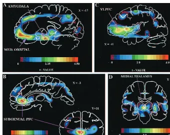
To address this type II error source, omnibus image analysis strategies are used to survey brain images voxel by voxel (e.g., Figure 1; Drevets et al 1992; Friston et al 1991). Primary images are spatially transformed into a standardized stereotaxic space so that tomographic data can be averaged across subjects. Since current spatial transformation algorithms do not precisely align the vari-able, complex three-dimensional structure of images from different brains, images are blurred before analysis to
minimize the impact of misalignment error (Poline et al 1997); however, the loss of spatial resolution from blur-ring and the error in overlaying brain structure across subjects decrease sensitivity for detecting abnormalities in small structures (e.g., amygdala, subgenual PFC) or areas characterized by high anatomic variability (e.g., orbital cortex). Voxel-by-voxel analyses may also increase type I error because they require thousands of statistical compar-isons, and thep values obtained in published studies of MDD have generally not been appropriately corrected for multiple comparisons (Poline et al 1997). Nevertheless, when constrained by appropriate statistical considerations and followed by replication using targeted, MRI-based, ROI analyses in independent samples, approaches involv-ing omnibus statistical mappinvolv-ing techniques prove invalu-able for delineating abnormalities in MDD (e.g., Drevets et al 1992, 1995c, 1997).
The sensitivity for identifying abnormalities is also affected by sample size, medication effects, and biological heterogeneity. Antidepressant and antianxiety treatments reportedly decrease CBF and metabolism in frontal,
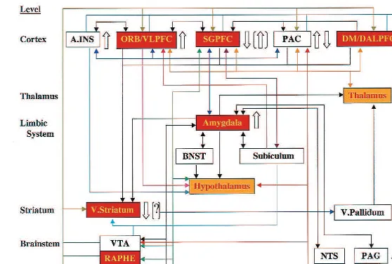
pari-Figure 2. Anatomic circuits implicated by neuroimaging and neuropathologic studies of familial mood disorders. The regional abnormalities summarized are specifically hypothesized to contribute to the genesis of pathologic emotional behavior. Regions in red have neuromorpho-metric and/or histopathologic abnormalities in primary major depressive disorder and/or bipolar disorder (BD; see text). Regions in yellow have not been microscopically examined in mood disorders, but are areas where structural abnormalities aresuspectedbased upon the finding of third ventricle enlargement in children and adults with BD. Open arrows to the right of each region indicate the direction of abnormalities in cerebral blood flow (CBF) and metabolism reported in depressives relative to control subjects (?indicates where experimental data await replication). Parenthetical open arrows indicate the direction of metabolic abnormalities after correcting the positron emission tomography (PET) measures for partial volume effects of reduced gray matter volume [(?)indicates where decreased gray matter is suspected as the explanation for reductions in CBF and metabolism, but partial volume-corrected PET results have not been reported]. Solid lines indicate
etal, and temporal lobe regions, and studies including images from subjects taking such agents usually fail to detect the areas of hypermetabolism identified in unmedi-cated depressives, and instead report areas of reduced CBF or metabolism not evident in unmedicated samples (for a review, see Drevets et al 1999b). Moreover, MDD and bipolar disorder (BD) likely encompass groups of disor-ders that are heterogenous with respect to etiology and pathophysiology, and “enrichment” of samples for the likelihood of having psychobiological markers appears necessary to replicate some imaging findings.
In some studies, biological variability was reduced by selecting subjects with familial mood disorders. Subjects with primary, familial MDD (i.e., familial pure depressive disease [FPDD]) were previously shown to be more likely to have abnormalities of hypothalamic–pituitary–adrenal (HPA) axis function, platelet [3H]imipramine binding, and sleep electroencephalography relative to MDD subjects who either lacked mood-disordered relatives or had alco-holic relatives (Kupfer et al 1992; Lewis et al 1983; Lewis and McChesney 1985; Winokur 1982). Depressed samples with familial MDD or BD have also been more likely to show elevated CBF/metabolism in the amygdala, orbital cortex, and medial thalamus and decreased metabolism and cortex volume in the subgenual PFC than nonfamilial cases or cases with secondary depressive syndromes (Dre-vets et al 1992, 1995c, 1999b; Hirayasu et al 1999; Kegeles et al 1999; O¨ ngu¨r et al 1998). Studies using alternative means for selecting enriched MDD samples, such as melancholic subtype criteria or responsiveness to sleep deprivation or phototherapy, have also found ele-vated metabolism in the amygdala and orbital cortex during MDEs (Cohen et al 1992; Ebert et al 1991; Nofzinger et al 1999; Wu et al 1992).
The mood-disordered subtype for which a distinct set of neuroimaging abnormalities has been best characterized is comprised of elderly depressives with a late age at illness onset. Depressives with onset of first MDE after age;55 are more likely than both age-matched, healthy control subjects and age-matched depressives with an earlier age of onset to have large “patches” of MR signal hyperinten-sity (in T2-weighted images) in the periventricular and deep white matter and lacunae in the cortex and striatum (Drevets et al 1999b; Krishnan et al 1993). Tissue acquired postmortem from brain areas showing patches of MR signal hyperintensity reveals arteriosclerosis, gliosis, white matter necrosis, and axon loss in the affected areas, but not in the surrounding tissue where the MRI signal appears normal (Awad et al 1986; Chimowitz et al 1992). Functional imaging studies confirm that CBF is decreased in areas where white matter hyperintensities are evident in MR images (Chimowitz et al 1992; Fazekas 1989). Late-onset depressives also commonly have widened cortical
sulci, ventricular enlargement, and reduced frontal lobe and basal ganglia volumes, which are thought to reflect tissue atrophy secondary to ischemia (for a review, see Drevets et al 1999b). Because cerebrovascular disease alters CBF, radiotracer delivery, and relationships between neuronal activity, CBF, metabolism, and oxygen extrac-tion (Derdeyn et al 1998), the funcextrac-tional imaging corre-lates of late-onset MDD profoundly differ from those of depressives with early or midlife illness onset (Drevets et al 1999b). Nevertheless, most imaging studies of MDD that included elderly subjects failed to distinguish or exclude such subjects, confounding interpretation of their results.
The MRI and clinical correlates of late-onset depression suggest this mood-disordered subgroup develops MDEs secondary to cerebrovascular disease (Krishnan et al 1993). The regions most commonly affected by white matter hyperintensities or lacunae in late-onset MDD are also the regions where infarction increases the risk for developing MDEs following stroke—namely, the left frontal lobe and striatum (for reviews, see Drevets et al 1999b; Starkstein and Robinson 1989). These regions contain neuropathologic abnormalities in familial MDD and BD subjects studied early in life, as well, which are of a distinct, idiopathic nature (see below). Thus, dysfunction involving an overlapping set of brain structures may confer vulnerability to MDEs in both early- and late-onset mood disorders.
Functional Anatomic Correlates of Major
Depression
The Subgenual Anterior Cingulate Cortex
The anterior cingulate cortex situated anterior and ventral to the genu of the corpus callosum (termedpregenualand subgenual,respectively) has been implicated by numerous studies of MDD and BD (for reviews, see Drevets 1999; Drevets and Raichle 1998). In the subgenual PFC, CBF and metabolism are decreased in unipolar and bipolar depressives relative to healthy control subjects (Figure 1B; Buchsbaum et al 1997; Drevets et al 1997; Kegeles et al 1999). This abnormality appears to be accounted for by a left-lateralized, volumetric reduction of the corresponding cortex, initially demonstrated by MRI-based morphomet-ric measures (Drevets et al 1997; Hirayasu et al 1999) and later by postmortem neuropathologic studies of familial BD and MDD (O¨ ngu¨r et al 1998). This reduction in volume exists early in the illness in familial BD (Hirayasu et al 1999) and MDD (Botteron et al 1999), but appears to follow illness onset, based upon preliminary evidence in twins discordant for MDD (Botteron et al 1999).
simulations that correct PET data for the partial volume effect of reduced gray matter volume conclude the “actu-al” metabolic activity in the remaining subgenual PFC tissue is increased in depressives relative to control subjects (Figure 2; Drevets 1999). This hypothesis appears compatible with findings that effective antidepressant drug (AD) treatments result in adecreasein metabolic activity in this region in MDD (Buchsbaum et al 1997; Drevets 1999; Mayberg et al 1999). Computer simulations of posttreatment images find that actual (partial volume corrected) metabolism decreases to normative levels dur-ing effective treatment (Drevets 2000). This mood state dependency of subgenual PFC metabolism would appear consistent with PET studies showing that flow increases in the subgenual PFC of healthy, nondepressed humans during experimentally induced sadness (Damasio et al 1998; George et al 1995; Mayberg et al 1999).
Although effective treatment with selective serotonin reuptake inhibitor (SSRI) ADs did not alter the subgenual PFC volume in MDD (Drevets et al 1997), this cortex was significantly larger in BD subjects chronically medicated with lithium or divalproex than in BD subjects who were unmedicated or were medicated with other agents (W.C. Drevets et al, unpublished data). Chronic administration of these mood stabilizers markedly increases levels of the neuroprotective protein Bcl-2 in the frontal cortex, stria-tum, and hippocampus of experimental animals (Manji et al 1999). Bcl-2 administration increases neurite sprouting and protects against glutamate-mediated excitoxicity, rais-ing the possibility that this difference in subgenual PFC volume reflects a neuroprotective/neurotrophic effect of mood-stabilizing medications (Manji et al 1999 [this reference also considers alternative explanations for lithi-um’s effects on cortical volume, however]).
Postmortem assessment of the subgenual PFC (agranu-lar cortex on the prelimbic anterior cingulate gyrus [part of Brodmann’s area (BA) 24]) demonstrated this abnormal reduction in gray matter was associated with areductionin glia, no equivalent loss of neurons, and increased neuronal density in MDD and BD relative to healthy and schizo-phrenic control samples (O¨ ngu¨r et al 1998). These findings suggest the volumetric abnormality in the subgenual PFC may specifically reflect a reduction of neuropil, the fibrous layers comprised of dendrites and axons that occupy most of the cortex volume (McEwen 1999). Glia are dividing cells that support neurons and synaptic transmission, so the reduction in glia may conceivably occur secondary to the reduction of synapses expected from a reduction in neuropil (Magistretti et al 1995; McEwen 1999). Never-theless, because glia maintain potassium homeostasis, transport glutamate and g-aminobutyric acid (GABA) from the extracellular fluid, and provide trophic factors and energy substrates to neurons, glial hypofunction may
also disturb synaptic transmission within the affected cortex (Azmitia 1999; Magistretti et al 1995).
The subgenual PFC has extensive reciprocal connec-tions with the orbital cortex, hypothalamus, amygdala, accumbens, ventral tegmental area (VTA), substantia nigra, raphe, locus coeruleus, periaqueductal gray (PAG), and nucleus tractus solitarius (Figure 2; Carmichael and Price 1995; Leichnetz and Astruc 1976). Humans with lesions that include the subgenual PFC show abnormal autonomic responses to emotionally provocative stimuli, inability to experience emotion related to concepts that ordinarily evoke emotion, and inability to use information regarding the likelihood of punishment and reward in guiding social behavior (Damasio et al 1990). In rats, bilateral or right-lateralized lesions of the prelimbic (the apparent homologue of the primate subgenual PFC) and infralimbic cortices attenuatesympathetic autonomic re-sponses, stress-induced corticosterone secretion, and gas-tric stress pathology during restraint stress or exposure to fear-conditioned stimuli (Frysztak and Neafsey 1994; Morgan and LeDoux 1995; Sullivan and Gratton 1999). In contrast, left-sided lesions of this area increase sympa-thetic autonomic arousal and corticosterone responses to restraint stress (Sullivan and Gratton 1999). Although these latter data await replication, they suggest the hypoth-esis that the right subgenual PFC facilitates expression of visceral responses during emotional processing, whereas the left subgenual PFC modulates such responses (Sulli-van and Gratton 1999). This hypothesis is noteworthy in light of the left-lateralized nature of the volumetric reduc-tion of the subgenual PFC in MDD and BD, and of PET data showing thatrightsubgenual PFC metabolism corre-lates positively with depression severity in MDD (rated by the Hamilton Depression Rating Scale [HDRS];r5 .76, p , .01; Drevets 2000). It is thus conceivable that dysfunction of the left subgenual PFC contributes to the altered neuroendocrine and autonomic function in depres-sion (Carney et al 1988; Dioro et al 1993; Holsboer 1995; Veith et al 1994).
conceivably contribute to disturbances of hedonic percep-tion and motivated behavior in mood disorders. In this regard, the magnitude of abnormal metabolic activity in the subgenual PFC may relate to switches between depres-sion and mania, as, even in the presence of reduced volume, apparent subgenual PFC activity appears abnor-mally increased in small samples of manic subjects (e.g., Drevets et al 1997).
The Pregenual Anterior Cingulate Cortex
The psychiatric imaging literature is less consistent with respect to the pregenual anterior cingulate cortex. This region consistently shows elevated CBF during various emotional conditions elicited in healthy or anxiety-disor-dered humans (for a review, see Drevets and Raichle 1998). Electrical stimulation of this region elicits fear, panic, or a sense of foreboding in humans, and vocaliza-tion in experimental animals (for a review, see Price et al 1996).
Most imaging studies find that CBF and metabolism are increased in this area during MDEs (for a review, see Drevets 1999); however, Mayberg et al (1997) reported that, although metabolism in this area was abnormally increased in depressives who subsequently showed a good response to ADs, metabolism was abnormally decreased in depressives who later had poor or incomplete treatment responses. In contrast, other groups found inverse corre-lations between basal metabolism and subsequent antide-pressant response in MDD, with lower baseline pregenual anterior cingulate metabolism predicting superior respon-siveness to ADs (Brody et al 1999; Ketter et al 1999). The effects of treatment on pregenual anterior cingulate CBF and metabolism have also differed across studies, with activity decreasing in some but increasing in others in posttreatment scans, relative to pretreatment scans (for a review, see Drevets 1999). The extent to which these discrepancies are explained by differential effects in sub-regions of this area remains unclear.
The Orbital and Anterior Insular Cortices
In the left and right posterior orbital cortices, the left ventrolateral PFC (VLPFC), and the anterior (agranular) insula, CBF and metabolism are abnormallyincreasedin unmedicated subjects with primary MDD (Figure 1, A and C; e.g., Baxter et al 1987, Table 3; Biver et al 1994; Cohen et al 1992; Drevets et al 1992, 1995c; Ebert et al 1991). Flow and metabolism also increase in the these areas during experimentally induced sadness and anxiety in healthy subjects and during induced anxiety and obses-sional states in subjects with obsessive– compulsive, post-traumatic stress, simple phobic, and panic disorders (for a review, see Drevets and Raichle 1998). The elevation of
physiologic activity in these areas during MDEs appears mood state dependent, as a variety of effective, somatic antidepressant therapies result in decreases in CBF and metabolism in the remitted phase of MDD relative to the depressed phase (e.g., Brody et al 1999; Drevets 1999; Drevets et al 1992; Mayberg et al 1999; Nobler et al 1994). A complex relationship exists between depression se-verity and metabolic activity in the orbital cortex and VLPFC. Although CBF and metabolism are elevated in these areas in the depressed phase of MDD relative to the remitted phase, these measures correlate inversely with ratings of depression severity and depressive ideation (Drevets et al 1992, 1995c). Compatible with these data, although metabolic activity is abnormally increased in these areas in outpatient, treatment-responsive, unipolar and bipolar depressives, more severely ill or treatment-refractory BD samples and inpatient MDD samples have shown mean CBF and metabolic values that either did not significantly differ or were decreased relative to control samples (Drevets et al 1997; Ketter et al 1999; Mayberg et al 1997). Similarly, PET studies of depression secondary to Parkinson’s disease, cerebrovascular disease, or com-plex partial seizures have shown that flow is either unchanged or abnormally decreased in these conditions relative to nondepressed control subjects with the same neurologic conditions (Bromfield et al 1992; Lesser et al 1994; Mayberg et al 1990; Ring et al 1994). Elevated orbital activity is, therefore, not essential to the production of depressive symptoms.
Instead, these relationships between orbital metabolism and depression severity are consistent with evidence from imaging, lesion analysis, and electrophysiologic studies that the posterior orbital cortex participates in modulating behavioral, visceral, and cognitive responses associated with defensive, fear, and reward-directed behavior as reinforcement contingencies change. Posterior orbital cor-tex CBF increases in subjects with obsessive– compulsive disorder or animal phobias during exposure to phobic stimuli and in healthy subjects during induced sadness (Drevets et al 1995b; Rauch et al 1994; Schneider et al 1995). In each of these cases, DCBF in the posterior orbital cortex correlates inversely with concomitant changes in obsessive thinking, anxiety, and sadness, re-spectively. In animal phobic subjects, serial PET images acquired during repeated exposures to phobic stimuli revealed that the orbital CBF was unchanged during initial exposures when the fear response was greatest, but as subjects habituated to the stimuli during subsequent expo-sures, posterior orbital CBF progressively increased, and theDCBF was inversely correlated with changes in anxi-ety ratings and heart rate (Drevets et al 1995b).
stimulus and response, and the patterns of posttrial activ-ity, which relate to the presence or absence of reward, suggest these cells play a role in extinguishing unrein-forced responses to appetitive and aversive stimuli (Rolls 1995). This role may be partly mediated by interactions with the amygdala and other limbic structures (Mogenson et al 1993; Price et al 1996). The orbital cortex and amygdala send direct projections to each other and over-lapping projections to the striatum, hypothalamus, and PAG through which they modulate each other’s transmis-sion (Figure 2; Carmichael and Price 1995; Mogenson et al 1993; Price 1999). Such interactions are evidenced by findings that defensive behaviors and cardiovascular re-sponses evoked by electrical stimulation of the amygdala are attenuated or ablated by concomitant stimulation of orbital cortex sites, which when stimulated alone produce no autonomic changes (Timms 1977). Compatible with the hypothesis that orbital cortex activity modulates amyg-dalar function during MDEs, glucose metabolisms in the orbital cortex and amygdala are inversely correlated in depressed humans (Drevets 2000).
Although tumors and cerebrovascular lesions involv-ing the frontal lobe increase the risk for developinvolv-ing MDEs, the specific PFC regions where dysfunction confers this risk have not been established (Mayeux 1982; Starkstein and Robinson 1989). Humans with lesions of the orbital cortex show impaired performance on tasks requiring application of information related to reward or punishment, exhibit difficulty shifting intel-lectual strategies in response to changing demands, and perseverate in strategies that become inappropriate (Bechara et al 1998; Rolls 1995). Lesion analysis studies in monkeys and functional imaging studies in humans show that the lateral orbital cortex/VLPFC in particular is involved when responses to stimuli require the suppression of previously rewarded responses (El-liott et al 2000; Iversen and Mishkin 1970).
During MDEs orbital cortex activation may reflect endogenous attempts to attenuate emotional expression or interrupt perseverative patterns of aversive, nonrewarding thought and emotion. Nevertheless, evidence that neuro-pathologic changes exist in the orbital cortex in primary mood disorders raises the possibility that impairment of these orbital functions predisposes to MDEs. Postmortem studies of MDD and BD report abnormal reductions of gray matter, glia, and neuronal size, but no decrement in neuronal number, in the posterior orbital cortex and VLPFC (Bowen et al 1989; Rajkowska et al 1997, 1999). Like the histopathologic changes found in the subgenual PFC, these data are most consistent with a reduction in neuropil. If such abnormalities are associated with dis-turbed synaptic interactions between the orbital cortex and amygdala, striatum, hypothalamus, or PAG, then orbital
dysfunction may conceivably contribute to the develop-ment of excessive emotional responses to stressors and ruminative ideation.
The abnormalities of serotonergic and catecholaminer-gic neurotransmitter function reported in mood disorders suggest other mechanisms by which orbital activity may be impaired in depression. In healthy subjects serotonin (5-HT) depletion (via tryptophan-free diet) produced per-formance deficits on decision-making tasks involving risk/reward probabilities that were similar to those seen in subjects with orbital cortex lesions (Rogers et al 1999). Moreover, depressive relapse occurring in remitted MDD subjects scanned during serotonin depletion was associ-ated with reductions in metabolism in the orbital cortex and VLPFC (Bremner et al 1997 [the area termedmiddle frontalby Bremner is part of the VLPFC area described herein]; Smith et al 1999). Finally, orbital cortex metab-olism is decreased in depressed subjects with Parkinson’s disease, relative to nondepressed subjects, suggesting that dopamine depletion may impair orbital cortex function (Mayberg et al 1990; Ring et al 1994).
The effects of antidepressant treatment on neurophysi-ologic activity in the orbital cortex and VLPFC are noteworthy in this regard. The highly replicated finding that CBF and metabolism decrease in the orbital/insular cortex and VLPFC during effective AD treatment may indicate that this cortex can “relax,” as such treatments directly inhibit pathologic activity in limbic structures such as the amygdala (for a review, see Drevets 1999). In contrast, nonpharmacologic treatments such as cognitive behavioral therapy and repetitive transcranial magnetic stimulation reportedlyincreasemetabolism in the VLPFC and posterior orbital cortex, respectively (Brody et al 1999; Teneback et al 1999), suggesting the hypothesis that their therapeutic mechanisms depend upon enhancing the function of PFC mechanisms for attenuating emotional expression.
The Dorsomedial/Dorsal Anterolateral Prefrontal Cortex
MDD, Rajkowska et al (1999) observed abnormal reduc-tions in the density and size of neurons and glia in the supra- and infragranular layers of the DALPFC (rostral BA 9), a finding that may relate to the reduction in metabolic activity in this area in MDD.
In brain mapping studies of healthy humans, CBF in the DMPFC increases during performance of tasks that elicit emotional responses or require emotional evaluations (Dolan et al 1996; Drevets et al 1994; Reiman et al 1997). In healthy humans imaged during anticipation of an electrical shock, the relationship between DCBF and emotion ratings suggested that while this region activated during anxiety it exerted a modulatory influence on emotional expression, such that the change in anxiety ratings and heart rate correlated inversely with DCBF (Drevets et al 1994). In rats, lesions of the area that appears homologous with the human DMPFC result in exaggerated heart rate responses to fear-conditioned stim-uli, and electrical and chemical stimulation of these sites attenuates defensive behavior and cardiovascular re-sponses evoked by amygdala stimulation (for a review, see Frysztak and Neafsey 1994). The DMPFC sends efferent projections to the PAG through which it may modulate cardiovascular responses associated with emotional be-havior or stress (Price 1999). If the histopathologic changes found in this area in MDD by Rajkowska et al (1999) are associated with functional impairment, they may conceivably contribute to the pathologic stress re-sponses and elevated resting heart rate seen in MDD.
Dorsolateral PFC and Dorsal Anterior Cingulate Cortex
Abnormal reductions of CBF and metabolism have also been reported in MDD in areas of the lateral and dorso-lateral PFC (L/DLPFC) and the dorsal anterior cingulate cortex situated posterior to the areas described in the preceding section (for reviews, see Drevets et al 1999b; Drevets and Raichle 1998). These abnormalities appear mood state dependent, reversing during symptom remis-sion (Bench et al 1995; Mayberg et al 1999). Initial studies assessing neuromorphometry or histology have not found abnormalities in these areas in MDD or BD (Bowen et al 1989; Drevets et al 1997; Rajkowska et al 1997).
Human PET and fMRI studies show that hemodynamic responses in the anterior cingulate cortex dorsal and posterior to the genu of the corpus callosum consistently increase during tasks requiring discriminative attention and selection for action (for a review, see Drevets and Raichle 1998). Attentional demand, target frequency, and/or frequency of being incited to action modulate the magnitude of DCBF in this region (for a review, see Drevets and Raichle 1998). In contrast, CBF isdecreased
in this area in healthy subjects during experimentally induced anxiety and depressed subjects with MDD, rela-tive to control conditions (Bench et al 1992; Drevets and Raichle 1998). Moreover, the normal activation of this region during a verbal fluency task was attenuated during the depressed mood associated with acute tryptophan depletion (Smith et al 1999).
Multiple L/DLPFC areas activate when verbal or visuo-spatial information is maintained in working memory and processed in some way (for a review, see Drevets and Raichle 1998). In the vicinity of these areas, CBF and metabolism are reportedly decreased in the depressed phase of MDD, as compared with the remitted phase, in depressives relative to healthy control subjects, and during experimentally induced sadness in healthy subjects (Bench et al 1992; Biver et al 1994; Mayberg et al 1999). These DLPFC areas do not appear activated during emotional processing, and lesions of this region do not impair performance on tasks involving the application of infor-mation related to reward or punishment (e.g., Bechara et al 1998). In one left DLPFC area, Dolan et al (1993) reported that the reduction in CBF correlated with impoverishment of speech (thought to reflect slowed cognitive processing) in MDD and schizophrenia, although this finding awaits replication.
Interpreting reversible decreases in CBF and metabo-lism found during MDEs in areas that have not been implicated in emotional processing requires an under-standing of the regulation of neurophysiologic activity across anatomic systems. Brain mapping studies show that, whereas CBF increases in brain regions putatively activated to perform an experimental task, CBF concom-itantlydecreasesin some other neural systems that appear nonessential to task performance (Drevets and Raichle 1998). Such “deactivated” areas are thought to reflect attention-related processes in which signal processing is enhanced via suppression of neural transmission convey-ing competconvey-ing, unattended information (for a review, see Drevets et al 1995a).
Drevets et al 1995a). The reduced afferent synaptic trans-mission into the portions of the SI representing unattended skin sites presumably accounts for the reduction in CBF in these areas in PET images (DiRocco et al 1989; Drevets et al 1995a). Similar interactions appear to occur across sensory modalities (for a review, see Drevets and Raichle 1998), as exemplified by findings that CBF decreases in the primary auditory cortex, auditory association cortex, somatosensory cortex, and posterior cingulate and midcin-gulate cortices as subjects process complex visual stimuli (Haxby et al 1994).
Areas specialized for emotional versus higher cognitive functions may also engage in such cross-modal relation-ships. In the amygdala, posterior orbital cortex, and ventromedial PFC sites where CBF increases during emo-tion-related tasks, flowdecreases during performance of attentionallydemanding, cognitive tasks (Drevets and Raichle 1998; Shulman et al 1997). Conversely, in dorsal anterior cingulate and DLPFC areas where flow increases while attentionally demanding cognitive tasks are per-formed, CBF decreases during some emotional states (Drevets and Raichle 1998; Mayberg et al 1999).
These reciprocal patterns of neural activity hold intrigu-ing implications for interactions between emotion and cognition (Drevets and Raichle 1998). The reduction of CBF in dorsal anterior cingulate and DLPFC areas spe-cialized for verbal, attentional, visuospatial, and mne-monic processing during depression may reflect a suppres-sion of afferent activity to those regions as dysphoric emotions or thoughts are processed. This suppression may relate to the subtle impairments of attention, memory, and
visuospatial function that accompany MDEs (Drevets and Raichle 1998).
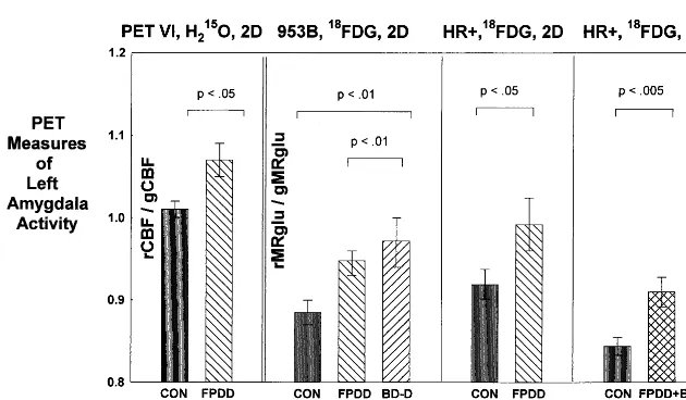
The Amygdala
Resting CBF and glucose metabolism are abnormally elevated in the amygdala in depressives with FPDD, type II BD or nonpsychotic, type I BD (Figures 1 and 3; Drevets et al 1992, 1995b; Wu et al 1992). In contrast, this abnormality is not evident in more severe, psychotic, type I BD subjects (Drevets 1995), and the extent to which this severity-based difference between BD samples may reflect differential magnitudes of partial volume effects associ-ated with abnormalities of amygdala structure in BD remains unclear (Bowley et al, in press; Pearlson et al 1997). Abnormalities of resting amygdalar CBF/metabo-lism have also not been found in MDD samples meeting Winokur (1982) criteria for depression spectrum disease (Drevets et al 1995c), obsessive– compulsive disorder, panic disorder, phobic disorders, or schizophrenia, sug-gesting this abnormality may be specific to some primary mood disorder subtypes (for a review, see Drevets and Botteron 1997).
In FPDD the magnitude of this abnormality as measured by PET is about 5% to 7% (Figure 3). When corrected for spatial resolution effects, this difference would reflect an increase in the actual CBF and metabolism of about 50 –70% (Drevets et al 1992; Links et al 1996). These magnitudes are in the physiologic range, as CBF increases ;50% in the rat amygdala during exposure to fear-Figure 3. Mean physiologic activity (6
conditioned stimuli as measured by tissue autoradiography (LeDoux et al 1983).
Amygdalar CBF and metabolism correlate positively with depression severity (Abercrombie et al 1996; Drevets et al 1992, 1995b). During AD treatment that both induces and maintains symptom remission, amygdala metabolism decreases to normative levels, compatible with preclinical evidence that chronic AD administration has inhibitory effects on amygdala function (for a review, see Drevets 1999). Nevertheless, CBF and metabolism in the left amygdala appear abnormally increased (although to a lesser extent) during the unmedicated, remitted phase of FPDD (Drevets et al 1992), and AD-medicated, remitted MDD subjects who relapse in response to serotonin depletion (via tryptophan-free diet) have higher amygdala metabolism before depletion than those who do not relapse (Bremner et al 1997). Abnormal amygdala activity may thus relate to both the severity of MDEs and the suscep-tibility to MDE recurrence. The extent to which these relationships reflect differentiable components (e.g., re-lated to involvement of distinct amygdalar nuclei) remains unclear due to the limited spatial resolution of PET.
The positive correlation between amygdala metabolism and depression severity rated by HDRS scores may reflect the amygdala’s role in organizing multiple aspects of emotional/stress responses (Davis 1992). In humans, elec-trical stimulation of the amygdala can produce anxiety, fear, dysphoria, recollection of emotionally provocative events, and increased cortisol secretion (for a review, see Drevets 1999). Moreover, an excessive amygdalar drive on the PAG (LeDoux 1996) may conceivably contribute to depressive signs such as inactivity, panic attacks, and reduced pain sensitivity, since in experimental animals stimulation of ventrolateral PAG produces social with-drawal, behavioral quiescence, and hypoalgesia, whereas stimulation of lateral PAG produces defensive behaviors, sympathetic autonomic arousal, and hypoalgesia (for a review, see Price 1999). Excessive efferent amygdala transmission to the lateral hypothalamus and locus coer-uleus could also potentially contribute to the elevated sympathetic tone, behavioral arousal, and insomnia seen in MDD (Carney et al 1988; Davis 1992; Veith et al 1994). In addition, activation of the amygdalar projections to the ventral striatum arrests goal-directed behavior in experi-mental animals (Mogenson et al 1993), suggesting a possible neural mechanism for the cessation of motivated or reward-directed behavior during MDEs.
The amygdala also facilitates stress-related cortico-tropin-releasing hormone (CRH) release via both intrinsic CRH-containing neurons and bisynaptic (double GABAergic) anatomic projections to the paraventricular nucleus of the hypothalamus (Herman and Cullinan 1997). These data thus suggest a pathway through which
exces-sive amygdala activity may contribute to the abnormal elevation of CRH secretion in MDD, which has been thought to be partly mediated by increased limbic drive (for a review, see Holsboer 1995). Corticotropin-releasing hormone administration is reported to induce appetite suppression, decreased sexual behavior, sleep disturbance, and anxiety in rats, suggesting another mechanism by which amygdalar hyperactivity may yield depressive symptoms (Musselman and Nemeroff 1993).
Finally, amygdala dysfunction may conceivably alter the interpretation of social or emotionally valenced stimuli in mood disorders. Neuroimaging, electrophysiologic, and lesion analysis studies demonstrate that the amygdala is involved in the acquisition and expression of emotional/ arousing memories (e.g., aversive conditioning; Canli et al 2000; LeDoux 1996; Phelps and Anderson 1997). For example, single-trial fMRI studies show that the human amygdala activates during initial exposures to fear-condi-tioned stimuli, but becomes deactivated during repeated exposures to the same stimulus (Bu¨chel et al 1998; LaBar et al 1998). The amygdala is also involved in interpreting the emotional significance of social cues. In humans, blood flow increases in the amygdala as subjects view faces expressing fear or sadness (Blair et al 1999; Morris et al 1996), and amygdala lesions impair the ability to recognize fear or sadness in facial expression (Adolphs et al 1994; Anderson and Phelps 1997) and fear and anger in spoken language (Scott et al 1997). Preliminary studies employing such neuropsychologic tasks in depressed hu-mans suggest that the pattern of hemodynamic responses to facially expressed emotion is altered in primary mood disorders (Casey et al 2000; Drevets 1999).
The preliminary finding that amygdala metabolism remains abnormally increased in MDD during sleep ar-gues that it is unlikely that the abnormal elevation in resting metabolism is simply accounted for by a height-ened response to the stressful stimuli related to scanning. Nofzinger et al (1999) reported that, although amygdala metabolism is increased in depressives relative to control subjects during wakefulness, the normal increase in me-tabolism that occurs during rapid eye movement sleep is also greater in depressives than in control subjects.
Abnormalities in the Striatum and Other Brain Areas
abnormally decreased in the caudate in MDD (Baxter et al 1985; Drevets et al 1992). The volumes of the caudate head and ventral striatum appear abnormally decreased in MRI and postmortem studies of MDD (Baumann et al 1999; Krishnan et al 1992). The partial volume effects associated with this abnormality may thus contribute to the CBF and metabolic reductions in the caudate in MDD. Nevertheless, depressive relapse during acute tryptophan depletion results in a corresponding reduction in caudate blood flow relative to baseline, suggesting that dynamic aspects of caudate function are also involved in mood disorders (Smith et al 1999).
Regional CBF and metabolic abnormalities in other structures have been less consistently replicated. Abnor-mally increased CBF has been reported in the posterior cingulate cortex and medial cerebellum in MDD (Bench et al 1992; Buchsbaum et al 1997). Medial cerebellar CBF also increases during experimentally induced anxiety or sadness in healthy or anxiety-disordered subjects (for reviews, see Drevets and Botteron 1997; George et al 1995). Some studies report reduced CBF and metabolism in sensory association areas in the lateral temporal and inferior parietal areas in MDEs (e.g., Biver et al 1994; Cohen et al 1992; Drevets et al 1992). Deactivation of these areas during MDE may reflect phenomena like those discussed above for the L/DLPFC (Drevets and Raichle 1998).
Anatomic Circuits Implicated in MDD
The abnormalities of function and structure in mood disorders implicate limbic–thalamic– cortical (LTC) cir-cuits, involving the amygdala, medial thalamus, and or-bital and medial PFCs, and limbic– cortical–striatal–palli-dal–thalamic (LCSPT) circuits, involving the components of the LTC circuit along with related parts of the striatum and pallidum (Figure 2; Drevets et al 1992). The amygdala and PFC are interconnected by excitatory projections with each other and with the MD (Carmichael and Price 1995; Price et al 1996). Through these connections the amygdala is in a position to directly activate the PFC and to modulate the reciprocal interaction between the PFC and MD (Drevets et al 1992).
Pathologically increased amygdala activity could also produce abnormal activity in the PFC and MD through the striatum and pallidum. The amygdala and PFC send excitatory projections to overlapping parts of the ventro-medial caudate and nucleus accumbens (Carmichael and Price 1995; Price et al 1996). This part of the striatum sends an inhibitory projection to the ventral pallidum, which in turn sends GABAergic, inhibitory fibers to the MD (Graybiel 1990; Kuroda and Price 1991). Because the pallidal neurons have relatively high spontaneous firing
rates (DeLong 1972), activity in the amygdala or PFC that activates the striatum and in turn inhibits the ventral pallidum may release the MD from an inhibitory pallidal influence.
The putative functions of the orbital and medial PFCs in modulating emotional and stress responses discussed above could potentially be impaired by dysfunction arising within these PFC regions themselves or within their efferent terminal projection fields in the striatum. Consis-tent with this hypothesis, lesions involving either the PFC or the striatum (e.g., strokes or tumors) and degenerative diseases affecting the striatum (e.g., Parkinson’s and Huntington’s diseases) are associated with higher rates of secondary major depression than similarly debilitating conditions that spare these regions (Folstein et al 1991; Mayeux 1982; Starkstein and Robinson 1989). Because these conditions disturb the LCSPT and LTC circuitry in different ways, imbalances within these circuits, rather than overall increased or decreased synaptic activity in a particular structure, may increase the risk for developing MDEs (Drevets et al 1992). It is nevertheless noteworthy that surgical lesions that interrupt projections from the orbital cortex into the striatum do not result in depression if the amygdalar projections into the striatum or anterior cingulate are also severed, as would occur during neuro-surgical interventions for intractable depression (e.g., sub-caudate tractotomy, prefrontal/limbic leukectomy; Ballan-tine et al 1987; Corsellis and Jack 1973; Knight 1965; Nauta 1973; Newcombe 1975). Therefore, neural mecha-nisms of depression may more specifically require dys-function of the orbitomedial PFC and/or other brain systems that results in disinhibition of the amygdala and other limbic structures involved in mediating emotional responses.
Implications of Histopathologic Findings in the LTC and LCSPT Circuits
receive projections from the orbital and medial PFCs may also be affected (Carmichael and Price 1995). In contrast, the volumes of the whole brain, entire PFC, dorsal anterior cingulate, somatosensory cortex, lateral temporal cortex, and other control regions have not differed between midlife or early-onset MDD or BD subjects and healthy control subjects (e.g., Drevets et al 1997; Pearlson et al 1997). Morphometric data in the hippocampus have been less clear, as subtle reductions of hippocampal volume were found by some but not most MRI studies of MDD or BD, and histopathologic assessments of the hippocampus have thus far been negative (for reviews, see Drevets et al 1999a, 1999b).
Although the etiology and time course of the neuro-pathologic abnormalities in mood disorders are unknown, the histopathology and the apparent specificity for areas implicated in the modulation of emotional behavior sug-gest clues regarding their pathogenesis. The finding that the gray matter volumetric deficit is accompanied by a reduction in glia with no equivalent loss of neurons does not support neurodegenerative hypotheses, and instead implies that the neuropil volume is decreased in primary mood disorders. The neuropil volume can be modulated in some regions of the adult brain by exposure to increased concentrations of excitatory amino acid neurotransmitters or cortisol, and by decreased function of neurotrophins, 5-HT1A receptors, estrogen receptors, and other factors that maintain the cytoskeleton (for reviews, see Azmitia 1999; McEwen 1999). Glia are dividing cells that support neurons and synaptic transmission (Magistretti et al 1995), so the reduction in glia may conceivably arise secondary to a reduction of synapses associated with retraction of the neuropil.
In primary mood disorders, abnormally elevated cortisol concentrations and reduced 5-HT1Areceptor function may comprise risk factors for developing reductions in neuropil that could affect widespread areas of the brain (e.g., Drevets et al 1999a; Musselman and Nemeroff 1993; Sargent et al 2000; Young et al 1993); however, the targeted nature of the gray matter volume reductions to specific areas of the LTC and LCSPT circuits (e.g., the left subgenual PFC, but not the right; Drevets et al 1997; Hirayasu et al 1999; Figure 2) suggests that glutamatergic neurotransmission also plays a role in inducing neuropil alterations in primary mood disorders (McEwen 1999). The finding that during MDEs metabolic activity is ele-vated in the LTC pathway, which is formed by predomi-nantly glutamatergic projections, suggests a potential source for chronic glutamate exposure (Drevets et al 1992). Glutamate is predominantly removed from the extracellular fluid by astrocyte-based transporter sites situated adjacent to synaptic clefts (Magistretti et al 1995). If the reduction of astroglia found in mood disorders
(Rajkowska 2000) impairs the efficiency of glutamate transport, it is conceivable that glutamate concentrations may increase. The excitotoxic effects of elevated gluta-mate concentrations may be facilitated in affective illness by increased release of cortisol (Sapolsky 1996).
Currently the only evidence that glutamate transport may be insufficient in depression is that high-affinity N-methyl-D-aspartate (NMDA) glutamatergic receptors are desensitized in the PFC of suicide victims, compatible with antemortem exposure to elevated glutamate concen-trations (Nowak et al 1995). Nevertheless, antidepressant treatments may compensate for impaired glutamate trans-port, as repeated electroconvulsive shock and chronic AD administration desensitize NMDA-glutamatergic receptors in the rat frontal cortex (Paul et al 1994). Moreover, some anticonvulsant agents that are effective in BD reduce glutamatergic transmission (Sporn and Sachs 1997). The recently discovered neurotrophic and neuroprotective ef-fects of chronic AD and mood-stabilizing treatments may also play roles in ameliorating the neuromorphometric changes in primary mood disorders (Duman et al 1997; Manji et al 1999).
Finally, the putative effect of chronic AD treatment of increasing serotonin transmission, tonically activating postsynaptic 5-HT1A receptors, and enhancing negative feedback inhibition of cortisol release suggests other mechanisms through which such agents may protect against or reverse neuropil reduction (Chaput et al 1991; Duman et al 1997; Haddjeri et al 1998; Magarinos et al 1999; McEwen 1999). Stimulation of neuron-based 5-HT1A receptors inhibits disassociation of the tubulin polymers that form the dendritic cytoskeleton, and stimu-lation of astroglial-based 5-HT1Areceptors induces release of the neurotrophic factor S100b, which promotes tubulin polymerization and inhibits microtubule breakdown (Azmitia 1999). Conversely, administration of serotonin-depleting agents, 5-HT1Areceptor antagonists, or antibod-ies to S100b all produce similar losses of dendrites, spines, and/or synapses in adult and developing animals, effects that are blocked by administration of 5-HT1A receptor agonists or SSRIs (Azmitia 1999).
sub-groups with FPDD or BD are reportedly more likely to have neuroendocrine evidence of elevated limbic–HPA axis activity (e.g., Lewis et al 1983; Winokur 1982). Finally, the 5-HT1A receptor imaging studies using PET and [carbonyl-11C]WAY-100635 (Drevets et al 1999a; Sargent et al 2000) converge with in vitro studies acquired postmortem (Bowen et al 1989; Lo´pez et al 1998) or antemortem (Francis et al 1989) to indicate that 5-HT1A receptor binding is abnormally decreased in primary MDD and BD. In contrast, the 5-HT1Areceptor data from suicide victims who may have secondary mood disorders or neuropsychiatric conditions other than mood disorders have been highly variable (for a review, see Drevets et al 1999b).
Directions for Future Studies
Among the questions raised by the neuroimaging and neuropathologic data is the critical problem of understand-ing cause and effect. The extent to which the abnormalities discussed above reflect primary pathophysiology that produces affective disease as opposed to secondary re-sponses to alterations in behavior, adaptations to chronic illness, or drug treatment remains unclear. Future neuro-imaging studies may elucidate these issues in studies of healthy subjects at high familial risk for developing mood disorders and studies of the relationships between genetic markers and neuroimaging correlates of illness and illness vulnerability.
The author thanks collaborators Joseph L. Price, Ph.D., and Marcus E. Raichle, M.D., for seminal scientific discussions that synthesized many of the concepts presented herein.
Aspects of this work were presented at the conference “Depression in the Twenty-First Century: New Insights into Drug Development and Neurobiology,” February 21–22, 2000, Dana Point, California. The conference was sponsored by the Society of Biological Psychiatry through an unrestricted educational grant provided jointly by Pharmacia & Upjohn and Janssen Pharmaceutica.
References
Abercrombie HC, Larson CL, Ward RT, Schaefer SM, Holden JE, Perlman SB, et al (1996): Metabolic rate in the amygdala predicts negative affect and depression severity in depressed patients: An FDG-PET study.Neuroimage3:S217.
Adolphs R, Tranel D, Damasio H, Damasio A (1994): Impaired recognition of emotion in facial expressions following bilat-eral damage to the human amygdala.Nature372:669 – 672. Anderson AK, Phelps EA (1997): Production of facial emotion
following unilateral temporal lobectomy.Soc Neurosci Abstr
23:2113.
Awad IA, Johnson PC, Spetzler RJ, Awad CA, Carey R (1986): Incidental subcortical lesions identified on magnetic
reso-nance imaging in the elderly, II: Postmortem pathological correlations.Stroke17:1090 –1097.
Azmitia EC (1999): Serotonin neurons, neuroplasticity, and homeostasis of neural tissue. Neuropsychopharmacology
21(suppl 2):33S– 45S.
Ballantine HT Jr, Bouckoms AJ, Thomas EK, Giriunas IE (1987): Treatment of psychiatric illness by stereotactic cin-gulotomy.Biol Psychiatry22:807– 819.
Baumann B, Danos P, Krell D, Diekmann S, Leschinger A, Stauch R, et al (1999): Reduced volume of limbic system-affiliated basal ganglia in mood disorders: Preliminary data from a post mortem study.J Neuropsychiatry Clin Neurosci11:71–78. Baxter LR, Phelps ME, Mazziotta JC, Guze BH, Schwartz JM,
Selin CE (1987): Local cerebral glucose metabolic rates in obsessive-compulsive disorder—a comparison with rates in unipolar depression and in normal controls.Arch Gen Psy-chiatry44:211–218.
Baxter LR, Phelps ME, Mazziotta JC, Schwartz JM, Gerner RH, Selin CE, Sumida RM (1985): Cerebral metabolic rates for glucose in mood disorders.Arch Gen Psychiatry42:441– 447. Baxter LR, Schwartz JM, Phelps ME, Mazziota JC, Guze BH, Selin CE, et al (1989): Reduction of prefrontal cortex glucose metabolism common to three types of depression.Arch Gen Psychiatry46:243–250.
Bechara A, Damasio H, Tranel D, Anderson SW (1998): Disso-ciation of working memory from decision-making within the human prefrontal cortex.J Neurosci18:428 – 437.
Bench CJ, Frackowiak RSJ, Dolan RJ (1995): Changes in regional cerebral blood flow on recovery from depression.
Psychol Med25:247–251.
Bench CJ, Friston KJ, Brown RG, Scott LC, Frackowiak RSJ, Dolan RJ (1992): The anatomy of melancholia—focal abnor-malities of cerebral blood flow in major depression.Psychol Med22:607– 615.
Biver F, Goldman S, Delvenne V, Luxen A, DeMaertelaer V, Hubain P, et al (1994): Frontal and parietal metabolic distur-bances in unipolar depression.Biol Psychiatry36:381–388. Blair RJR, Morris JS, Frith CD, Perrett DI, Dolan RJ (1999):
Neural responses to sad and angry expressions.Brain 122: 883– 893.
Botteron KN, Raichle ME, Heath AC, Price JL, Sternhell KE, Singer TM, Todd RD (1999): An epidemiological twin study of prefrontal neuromorphometry in early onset depression.
Biol Psychiatry45:59S.
Bowen DM, Najlerahim A, Procter AW, Francis PT, Murphy E (1989): Circumscribed changes of the cerebral cortex in neuropsychiatric disorders of later life.Proc Natl Acad Sci U S A86:9504 –9508.
Bowley MP, Drevets WC, O¨ ngu¨r D, Price JL (in press): Glial changes in the amygdala and entorhinal cortex in mood disorders.Biol Psychiatry.
Bremner JD, Innis RB, Salomon RM, Staib LH, Ng CK, Miller HL, et al (1997): Positron emission tomography measurement of cerebral metabolic correlates of tryptophan depletion-induced depressive relapse.Arch Gen Psychiatry54:346 –374. Brody AL, Saxena S, Silverman DHS, Alborzian S, Fairbanks
Bromfield EB, Altshuler L, Leiderman DB, Balish M, Ketter TA, Devinsky O, et al (1992): Cerebral metabolism and depres-sion in patients with complex partial seizures.Arch Neurol
49:617– 623.
Bu¨chel C, Morris J, Dolan RJ, Friston KJ (1998): Brain systems mediating aversive conditioning: An event related fMRI study.Neuron20:947–957.
Buchsbaum MS, Wu J, Siegel BV, Hackett E, Trenary M, Abel L, Reynolds C (1997): Effect of sertraline on regional metabolic rate in patients with affective disorder.Biol Psy-chiatry41:15–22.
Canli T, Zhao Z, Brewer J, Gabrieli JDE, Cahill L (2000): Event-related activation in the human amygdala associates with later memory for individual emotional experience. J Neurosci20:RC99.
Carmichael ST, Price JL (1995): Limbic connections of the orbital and medial prefrontal cortex in Macaque monkeys.
J Comp Neurol363:615– 641.
Carney RM, Rich MW, teVelde A, Saini J, Clark K, Freedland KE (1988): The relationship between heart rate, heart rate variability and depression in patients with coronary artery disease.J Psychosom Res32:159 –164.
Casey BJ, Thomas KM, Eccard CH, Drevets WC, Dahl RE, Whalen PJ, et al (2000): Functional responsivity of the amygdala in children with disorders of anxiety and major depression.Neuroimage11(suppl):249.
Chaput Y, deMontigny C, Blier P (1991): Presynaptic and postsynaptic modifications of the serotonin system by long-term administration of antidepressant treatments. An in vivo electrophysiologic study in the rat. Neuropsychopharmacol-ogy5:219 –229.
Chimowitz MI, Estes ML, Furlan AJ, Awad IA (1992): Further observations on the pathology of subcortical lesions identified on magnetic resonance imaging.Arch Neurol49:747–752. Cohen RM, Gross M, Nordahl TE, Semple WE, Oren DA,
Rosenthal N (1992): Preliminary data on the metabolic brain pattern of patients with winter seasonal affective disorder.
Arch Gen Psychiatry49:545–552.
Corsellis J, Jack AB (1973): Neuropathological observations on yttrium implants and on undercutting in the orbito-frontal areas of the brain. In: Laitinen LV, Livingston KE, editors.
Surgical Approaches in Psychiatry.Lancaster, UK: Medical and Technical Publishing, 90 –95.
Damasio AR, Grabowski TJ, Bechara A, Damasio H, Ponto LLB, Hichwa RD (1998): Neural correlates of the experience of emotions.Soc Neurosci Abstr24:258.
Damasio AR, Tranel D, Damasio H (1990): Individuals with sociopathic behavior caused by frontal damage fail to respond autonomically to social stimuli.Behav Brain Res41:81–94. Davis M (1992): The role of the amygdala in conditioned fear.
In: Aggleton JP, editor. The Amygdala: Neurobiological Aspects of Emotion.New York: Wiley-Liss, 255–305. DeLong MR (1972): Activity of basal ganglia neurons during
movement.Brain Res40:127–135.
Derdeyn CP, Yundt KD, Videen TO, Carpenter DA, Grubb RL Jr, Powers WJ (1998): Increased oxygen extraction fraction is associated with prior ischemic events in patients with carotid occlusion.Stroke29:754 –758.
Dioro D, Viau V, Meaney MJ (1993): The role of the medial
prefrontal cortex (cingulate gyrus) in the regulation of hypo-thalamic-pituitary-adrenal responses to stress. J Neurosci
3:3839 –3847.
DiRocco RJ, Kageyama GH, Wong-Riley MT (1989): The relationship between CNS metabolism and cytoarchitecture: A review of 14C-deoxyglucose studies with correlation to
cytochrome oxidase histochemistry. Comput Med Imag Graph13:81–92.
Dolan RJ, Bench CJ, Liddle PF (1993): Dorsolateral prefrontal cortex dysfunction in the major psychoses: Symptom or disease specificity?J Neurol Neurosurg Psychiatry56:1290 –1294. Dolan RJ, Fletcher P, Morris J, Kapur N, Deakin JF, Frith CD
(1996): Neural activation during covert processing of positive emotional expressions.Neuroimage4:194 –200.
Drevets WC (1999): Prefrontal cortical-amygdalar metabolism in major depression.Ann N Y Acad Sci877:614 – 637. Drevets WC (2000): Functional anatomical abnormalities in
limbic and prefrontal cortical structures in major depression.
Prog Brain Res126:413– 431.
Drevets WC (1995): PET and the functional anatomy of major depression. In: Nakajima T, Ono T, editors.Emotion, Memory and Behavior-Study of Human and Nonhuman Primates.
Tokyo: Japan Scientific Societies Press, 43– 62.
Drevets WC, Botteron K (1997): Neuroimaging in psychiatry. In: Guze SB, editor.Adult Psychiatry.St. Louis: Mosby, 53– 81. Drevets WC, Burton H, Simpson JR, Videen TO, Snyder AZ, Raichle ME (1995a): Blood flow changes in human somato-sensory cortex during anticipated stimulation. Nature 373: 249 –252.
Drevets WC, Frank E, Price JC, Kupfer DJ, Holt D, Greer PJ, et al (1999a): PET imaging of serotonin 1A receptor binding in depression.Biol Psychiatry46:1375–1387.
Drevets WC, Gadde K, Krishnan R (1999b): Neuroimaging studies of depression. In: Charney DS, Nestler EJ, Bunney BJ, editors. Neurobiology of Mental Illness. New York: Oxford University Press, 394 – 418.
Drevets WC, Price JL, Simpson JR, Todd RD, Reich T, Vannier M, Raichle ME (1997): Subgenual prefrontal cortex abnor-malities in mood disorders.Nature386:824 – 827.
Drevets WC, Raichle ME (1998): Reciprocal suppression of regional cerebral blood flow during emotional versus higher cognitive processes: Implications for interactions between emotion and cognition.Cogn Emotion12:353–385. Drevets WC, Simpson JR, Raichle ME (1995b): Regional blood
flow changes in response to phobic anxiety and habituation.
J Cereb Blood Flow Metab15:S856.
Drevets WC, Spitznagel E, Raichle ME (1995c): Functional anatomical differences between major depressive subtypes.
J Cereb Blood Flow Metab15:S93.
Drevets WC, Todd RD (1997): Depression, mania and related disorders. In: Guze SB, editors.Adult Psychiatry.St. Louis: Mosby, 99 –141.
Drevets WC, Videen TO, Price JL, Preskorn SH, Carmichael ST, Raichle ME (1992): A functional anatomical study of unipo-lar depression.J Neurosci12:3628 –3641.
Drevets WC, Videen TO, Snyder AZ, MacLeod AK, Raichle ME (1994): Regional cerebral blood flow changes during antici-patory anxiety.Abstr Soc Neurosci20:368.
Ebert D, Feistel H, Barocka A (1991): Effects of sleep depriva-tion on the limbic system and the frontal lobes in affective disorders: A study with Tc-99 m-HMPAO SPECT. Psychia-try Res Neuroimaging40:247–251.
Elliott R, Dolan RJ, Frith CD (2000): Dissociable functions in the medial and lateral orbitofrontal cortex: Evidence from human neuroimaging studies.Cereb Cortex10:308 –317. Fazekas F (1989): Magnetic resonance signal abnormalities in
asymptomatic individuals: Their incidence and functional correlates.Eur Neurol29:164 –168.
Folstein SE, Peyser CE, Starkstein SE, Folstein MF (1991): Subcortical triad of Huntington’s disease—a model for a neuropathology of depression, dementia, and dyskinesia. In: Carrol BJ, Barrett JE, editors. Psychopathology and the Brain.New York: Raven, 65–75.
Francis PT, Poynton A, Lowe SL, Najlerahim A, Bridges PK, Bartlett JR, et al (1989): Brain amino acid concentrations and Ca21-dependent release in intractable depression assessed
antemortem.Brain Res494:314 –324.
Friston KJ, Frith CD, Liddle PF, Frackowiak RSJ (1991): Comparing functional (PET) images: The assessment of significant change.J Cereb Blood Flow Metab11:690 – 699. Frysztak RJ, Neafsey EJ (1994): The effect of medial frontal cortex lesions on cardiovascular conditioned emotional re-sponses in the rat.Brain Res643:181–193.
George MS, Ketter TA, Parekh PI, Horwitz B, Herscovitch P, Post RM (1995): Brain activity during transient sadness and happiness in healthy women.Am J Psychiatry152:341–351. Graybiel AM (1990): Neurotransmitters and neuromodulators in
the basal ganglia.Trends Neurosci13:244 –254.
Haddjeri N, Blier P, de Montigny C (1998): Long-term antide-pressant treatments result in tonic activation of forebrain 5-HT1A receptors.J Neurosci18:10150 –10156.
Haxby JV, Horwitz B, Ungerleider LG, Maisog JM, Pietrini P, Grady CL (1994): The functional organization of human extrastriate cortex: A PET-rCBF study of selective attention to faces and locations.J Neurosci14:6336 – 6353.
Herman JP, Cullinan WE (1997): Neurocircuitry of stress: Central control of the hypothalamo-pituitary-adrenocortical axis.Trends Neurosci20:78 – 84.
Hirayasu Y, Shenton ME, Salisbury DF, Kwon JS, Wible CG, Fischer IA, et al (1999): Subgenual cingulate cortex volume in first-episode psychosis.Am J Psychiatry156:1091–1093. Holsboer F (1995): Neuroendocrinology of mood disorders. In: Bloom FE, Kupfer DJ, editors. Psychopharmacology: The Fourth Generation of Progress.New York: Raven, 957–969. Iversen SD, Mishkin M (1970): Perseverative interference in monkeys following selective lesions of the inferior prefrontal convexity.Exp Brain Res11:376 –386.
Kegeles LS, Malone KM, Slifstein M, Anjilvel S, Xanthopoulos C, Campell M, et al (1999): Response of cortical metabolic deficits to serotonergic challenges in mood disorders. Biol Psychiatry45:76S.
Ketter T, Kimbrell TA, Little JT, George MS, Sachs N, Winsberg ME, et al (1999, December): Differences and commonalties in cerebral function in bipolar compared to unipolar depres-sion. Presented at the 38th annual meeting of the American College of Neuropsychopharmacology, Acapulco, Mexico. Knight G (1965): Stereotactic tractotomy in the surgical
treat-ment of treat-mental illness.J Neurol Neurosurg Psychiatry28:30.
Krishnan KRR, McDonald WM, Doraiswamy PM, Tupler LA, Hussain M, Boyko OB, et al (1993): Neuroanatomical sub-strates of depression in the elderly. Eur Arch Psychiatry Neurosci243:41– 46.
Krishnan KRR, McDonald WM, Escalona PR, Doraiswamy PM, Na C, Husain MM, et al (1992): Magnetic resonanace imaging of the caudate nuclei in depression: Preliminary observations.Arch Gen Psychiatry49:553–557.
Kupfer DJ, Targ E, Stack J (1992): Electroencephalographic sleep in unipolar depressive subtypes. Support for a biological and familial classification.J Nerv Ment Dis170:494 – 498. Kuroda M, Price JL (1991): Synaptic organization of projections
from basal forebrain structures to the mediodorsal nucleus of the rat.J Comp Neurol303:513.
LaBar KS, Gatenby JC, Gore JC, LeDoux JE, Phelps EA (1998): Human amygdala activation during conditioned fear acquisi-tion and extincacquisi-tion: A mixed trial fMRI study. Neuron
20:937–945.
LeDoux J (1996):The Emotional Brain.New York: Simon and Schuster.
LeDoux JE, Thompson ME, Iadecola C, Tucker LW, Reis DJ (1983): Local cerebral blood flow increases during auditory and emotional processing in the conscious rat.Science 221: 576 –578.
Leichnetz GR, Astruc J (1976): The efferent projections of the medial prefrontal cortex in the squirrel monkey (saimiri sciureus).Brain Res109:455– 472.
Lesser IM, Mena I, Boone KB, Miller BL, Mehringer CM, Mohl M (1994): Reduction of cerebral blood flow in older de-pressed patients.Arch Gen Psychiatry51:677– 686. Lewis DA, Kathol RG, Sherman BM, Winokur G, Schlesser MA
(1983): Differentiation of depressive subtypes by insulin subsensitivity in the recovered phase. Arch Gen Psychiatry
40:167–170.
Lewis DA, McChesney C (1985): Tritiated imipramine binding distinguishes among subtypes of depression.Arch Gen Psy-chiatry42:485– 488.
Links JM, Zubieta JK, Meltzer CC, Stumpf MJ, Frost JJ (1996): Influence of spatially heterogenous background activity on “hot object” quantitation in brain emission computed tomog-raphy.J Comput Assist Tomogr20:680 – 687.
Lo´pez JF, Chalmers DT, Little KY, Watson SJ (1998): Regula-tion of serotonin1A, glucocorticoid, and mineralocorticoid
receptor in rat and human hippocampus: Implications for the neurobiology of depression.Biol Psychiatry43:547–573. Magarinos AM, Deslandes A, McEwen BS (1999): Effects of
antidepressant and benzodiazepine treatments on the dendritic structure of CA3 pyramidal neurons after chronic stress.Eur J Pharmacol371:113–122.
Magistretti PJ, Pellerin L, Martin JL (1995): Brain energy metabolism: An integrated cellular perspective. In: Bloom FE, Kupfer DJ, editors. Psychopharmacology: The Fourth Generation of Progress.New York: Raven, 921–932. Manji HK, Moore GJ, Chen G (1999): Lithium at 50: Have the
neuroprotective effects of this unique cation been over-looked?Biol Psychiatry46:929 –940.
Mayberg HS, Brannan SK, Mahurin RK, Jerabek PA, Brickman JS, Tekel JL, et al. (1997): Cingulate function in depression: A potential predictor of treatment response. Neuroreport