COMMON MISCONCEPTIONS THAT ARISE IN THE
FIRST-Y EAR MEDICAL PHY SIOLOGY CURRICULUM
CONCERNING HEART FAILURE
Thomas H. Hintze and Gong Zhao
Depa rtm ent of Physiology, New York Medica l College, Va lha lla , New York 10595
T
here are a number of misconceptions that first-year medical students have concerning the pathophysiology of heart failure. These stem from 1) a poor definition of heart failure,2) a lack of care in distinguishing between similar but distinct concepts, and3) the inability to recognize the relationship between the various stages of heart failure and the clinical manifestation of the disease. In this paper we provide a list of some of the misconceptions that we have encountered, some explanations of the distinctions to be made, and some of the rationale behind current surgical procedures and drug treatment. The misconceptions include failing to differenti-ate between the Frank-Starling mechanism and cardiac dilation as well as not grasping the significance that changes in cardiac b-receptor function have in limiting the positive inotropic actions of circulating catecholamines. Finally, we review some of the altered neurohumoral mechanisms in heart failure and explain the basis for some common therapeutic approaches, including the use of angiotensin-converting enzyme inhibitors, in this disease.AM. J. PHYSIOL. 277 (ADV. PHYSIOL. EDUC. 22): S260–S267, 1999.
Key words:myocardial failure; circulatory failure; cardiac dilation; cardiac decompensation; neurohumoral mechanisms in heart failure; peripheral edema
Over the past several years, we have encountered several misconceptions commonly voiced by our first-year medical students regarding the altered hemo-dynamics and clinical manifestations of heart failure. This article reviews many of the underlying concepts by stating the misconception and then addressing the problem. First, some background meant to avert some of the problems is given.
The term ‘‘failure’’ is often loosely used in medicine and can refer to any number of clinical entities. As defined by Braunwald (2), heart failure is quite simply a ‘‘pathophysiologic state in which an abnormality in cardiac function is responsible for the failure of the heart to pump blood at a rate commensurate with the peripheral needs.’’ This can be subdivided into
these cases, whether it be tamponade, fibrosis, or effusion, the major change is not in the myocyte but rather the impaired filling of the cardiac chambers. When excess fluid is removed from the pericardial space, arterial pressure returns to normal almost instantaneously, as most of the students will recognize from evening television medical dramas.
Myocyte and cardiac failure should be contrasted with another type of failure—circulatory failure—in which some defect in the circulation leads to reduced perfusion. An example of this is hemorrhage. In this state, the myocyte and the valves function normally, yet cardiac output is insufficient to meet peripheral demands because of reduced blood volume. Obvi-ously, the way to correct this is to replace the lost blood volume.
Another misconception is that all forms of heart failure are ‘‘congestive.’’ Heart failure often occurs without congestion, unless the underlying defect is large and/or long lasting. Congestion is defined as the abnormal accumulation of fluid in the periphery as edema or, in the lung, most evident as rales. The mechanism leading to edema is altered Starling forces across the capillary bed, resulting in a net efflux of fluid into the tissue.
MISCONCEPTIONS CONCERNING THE CONSEQUENCES OF HEART FAILURE
The clinical manifestations of heart failure include low arterial pressure, tachycardia, exercise intolerance, difficulty breathing (dyspnea) in the supine position, swelling in the extremities, and cachexia. All of these are manifestations of altered physiological control mechanisms and the inability of various compensatory mechanisms to restore some level of normal function.
Low arterial pressure and tachycardia are manifesta-tions of the low cardiac output. With the use of the equation mean arterial blood pressure 5 cardiac output3total peripheral resistance, it is immediately obvious that a reduction in cardiac output with other variables constant results in lower arterial pressure. The accompanying tachycardia is, in part, a barorecep-tor reflex-mediated attempt to increase cardiac out-put. The tachycardia of the failing heart may also be caused by atrial stretch, eliciting a Bainbridge reflex.
The Bainbridge reflex is caused by vagal withdrawal, resulting in only a moderate tachycardia, similar to the tachycardia of heart failure. A common misconception that occurs at this point is that the tachycardia of heart failure is sympathetic in origin (2). Mild tachycardias, up to heart rates of ,120 beats/min in humans are
caused by the withdrawal of vagal tone and not by recruitment of sympathetic activity. This misconcep-tion results from the fact that students may have heard that there is a rise in circulating catecholamines with heart failure, and some well-informed students may even know that the best predictor of mortality in heart failure is the extent to which plasma norepinephrine increases. At this point, it is helpful to discuss the mechanism leading to tachycardia by using the trans-planted human heart as an example. The transtrans-planted (denervated) human heart beats at ,110–120 beats/
min, but the denervated heart has no vagal and no sympathetic tone. Because heart rate at rest in an innervated heart is,70 beats/min, some mechanism
must lower heart rate from the intrinsic 110 beats/min (seen in the denervated heart) to 70 beats/min in the intact heart. This mechanism is, of course, tonic parasympathetic drive to the sinoatrial (SA) node. Sympathetic drive to the SA node is evident if heart rate is.110 or 120 beats/min, and this only occurs late in the heart failure process.
capacity. Patients in the early stages of heart failure, like the patient in Fig. 1, will be able to perform moderate levels of exercise. It is their maximum capacity that is first affected. This example serves two functions, to reinforce the concept of a dynamic definition of heart failure and to illustrate the use of a ‘‘treadmill stress test,’’ which the students have all heard about. Finally, this allows for a discussion of Guyton’s concept of the permissive heart, i.e., that the heart will just pump out all of the blood that is returned to it by the circulation (4).
Another concept that is often misinterpreted by stu-dents is that the extended jugular vein, the prominent jugular venous pressure pulse, the difficulty breathing in the supine position, and the swollen feet that everyone associates with heart failure are directly related to the reduced output of the heart. The misconception to be addressed is that the reduced output is directly responsible for the accumulation of
fluid in the interstitial space. In fact, it is the rise in venous and capillary pressures that accompany pump failure, rather than reduced arterial pressures, that causes these various clinical symptoms.
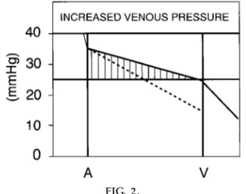
As the ability of the heart to pump blood falls, end-diastolic ventricular pressure will increase be-cause of the reduced ejection. This end-diastolic ventricular pressure is transmitted in a retrograde (backward) manner to the atria (increased atrial pres-sure) and further back to the venous and capillary beds. With the increase in capillary pressure, a net filtration pressure develops along the entire capillary and increases movement of fluid into the interstitial space (Fig. 2) (5). If the right heart is failing, then pressure at the venous end of peripheral capillaries will increase, resulting in peripheral edema (swelling in the feet). The retrograde transmission of pressure from the right ventricle is also responsible for the obvious jugular venous pressure pulse in patients with heart failure. If the left ventricle is failing, the in-creased filtration occurs in the lungs, resulting in interstitial edema and the classic rales during ausculta-tion. In addition, because the diffusion of oxygen is inversely proportional to the square of the diffusion distance, pulmonary interstitial edema can have marked effects on the movement of oxygen from the alveoli to the pulmonary capillary blood. This may result in reduced oxygen being carried by the blood, often
FIG. 1.
Changes in car diac output that occur during ex er cise in nor mal subjects and in patients with moderate heart failur e (HF). Although patients with heart failur e can ex er cise, the max imum ex er cise level (i.e., max i-mum car diac output) is r educed. This is the basis of ex er cise str ess testing in patients. Please note that the plateau is flat in compensated heart failur e; a descend-ing limb only appears in a decompensated heart.
FIG. 2.
exhibited as blue coloring under the fingernails. All of these are examples of backward failure, that is, dysfunction being transmitted in a direction opposite to the flow of blood.
Another way edema can be generated in the lung and periphery is as the result of a marked increase in blood volume. Renal failure, the inability of the kidney to excrete a normal urine volume, is often associated with heart failure and can be attributed among other reasons to reduced renal perfusion. This results in an accumulation of fluid (80% of which is stored in the venous circulation), an increase in capillary venous pressure, and net movement of fluid into the intersti-tial space. The reduced perfusion pressure (forward failure) also affects the liver, resulting in accumulation of potentially toxic metabolites, which circulate to ‘‘poison’’ the heart and peripheral organs, further contributing to heart failure. Treatment is through the use of diuretics, which lead to a reduction in venous pressures and reabsorption of the accumulated intersti-tial fluid.
The final misconception related to the clinical signs of heart failure is that the reduced muscle mass, ca-chexia, in itself is caused by the heart failure and may be a compensatory mechanism to supply substrate for oxidation in a failing heart. Rather, the cachexia is caused by cytokines, particularly tumor necrosis fac-tor-a (TNF-a), originally called cachexin (1). TNF-a itself or a combination of cytokines is responsible for the protein wasting in the periphery and is evidence of activation of an inflammatory process
MISCONCEPTIONS RELATED TO THE COMPENSATORY PHYSIOLOGICAL MECHANISMS THAT OCCUR IN HEART FAILURE
Although the cause of heart failure may be either myocardial or cardiac in origin, once a decrease in function occurs a number of physiological mecha-nisms are called into play to compensate for the defect. If the defect is small or the progression of the disease slow, these compensatory mechanisms may be sufficient to restore function to normal, especially in the absence of a stress such as exercise. It should be pointed out that the compensatory mechanisms are not sufficient to restore a normal level of maximum exercise performance. This reinforces the utility of
exercise testing in the early diagnosis of heart failure. Furthermore, these compensatory mechanisms in the long run are insufficient to maintain cardiac function. Ultimately, along with the evolution of the disease process itself, they contribute to the progression of heart failure. These compensatory processes include 1) increased secretion of catecholamines from sympa-thetic nerve endings in the heart, the periphery, and the adrenal glands; 2) recruitment of the Frank-Starling mechanism; and3) myocardial hypertrophy.
The best way to present these concepts is to remind students that they occur in a time-dependent fashion; i.e., secretion of norepinephrine caused by baroreflex unloading (a rapid process), then recruitment of the Frank-Starling mechanism as intravascular volume in-creases (an intermediate process), and finally hypertro-phy that involves protein synthesis (the slowest pro-cess). There are a number of misconceptions related to each of these three processes.
The misconceptions associated with the role of in-creased sympathetic tone during the development of heart failure are many. First, the tachycardia of heart failure is almost entirely caused by vagal withdrawal (2), as pointed out earlier. Second, there are marked alterations inb-adrenergic receptor affinity, number, and turnover, all of which serve to desensitize the b-adrenergic response. This implies that the sympa-thetic nervous system is unable to regulate inotropic state in the failing heart. In support of this concept is the recent therapeutic use of b-adrenergic receptor blocking agents in the treatment of heart failure. The rationale is to block a portion of the receptors, thereby allowing the remaining receptors to upregu-late so that, when stimuupregu-lated, they will transduce a positive inotropic effect. In addition to receptor desen-sitization, cardiac tissue cathecholamines fall to unde-tectable levels after the development of heart failure (7 ). This should be taken as evidence of depletion of norepinephrine from nerve terminals, however, rather than the disappearance of sympathetic nerve endings.
contractility (8). It should be remembered in this context that circulating norepinephrine comes primar-ily from peripheral adrenergic nerve endings and that the increase during the development of heart failure may reflect increased peripheral sympathetic nerve activity and neurally mediated increases in peripheral resistance. Be that as it may, it should be stressed that the best predictor of mortality in patients is the rise in circulating plasma catecholamines.
With regard to the Frank-Starling mechanisms, stu-dents often become confused on two points: 1) the presense (or absence) of a descending limb on the cardiac function curve and2) the distinction between increased preload as a compensatory mechanism and cardiac dilation. The increase in cardiac function that results from an increase in preload is caused by stretching of existing sarcomeres to cause a more optimal overlap and closer apposition of actin and myosin filaments and increased Ca21 sensitivity [see
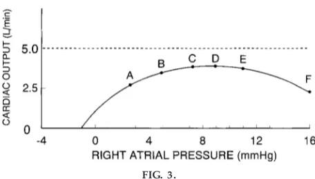
accompanying article in this issue by Solaro (6)]. It should be pointed out that, even in Guyton’s diagrams of mild heart failure, there is no descending limb on a cardiac output curve (Fig. 1). The plateau of the cardiac output curve is depressed but remains parallel to the x-axis. Only after the development of severe heart failure did Guyton find a true descending limb (Fig. 3). In fact, Guyton suggests that the presence of a descending limb is indicative of a late-stage failing heart. The old idea, now discredited, that cardiac muscle normally shows a descending limb on a Starling curve arose from studies of isolated hearts that may, in fact, have been failing.
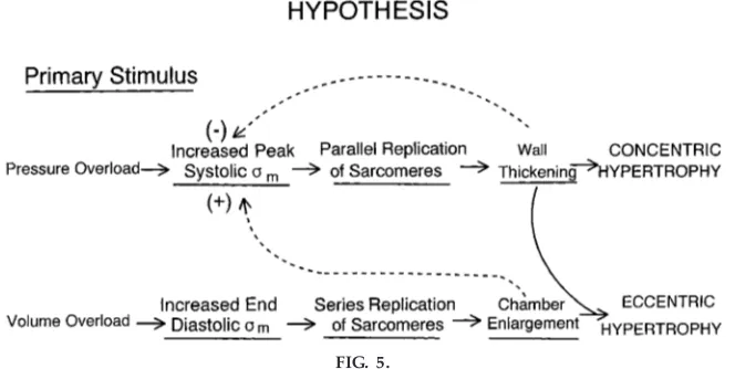
A good deal of confusion arises over the difference between preload and cardiac dilation. Cardiac dilation is an example of cardiac remodeling and is best appreciated as a shift in the passive length-tension diagram (ventricular compliance curve) as shown in Fig. 4. A movement from point Ato point B is an increase in preload and would serve to increase systolic function. On the other hand, a shift from point Ato point Cis clearly caused by some process that has made the end-diastolic volume much larger and may not be associated with increased systolic function. Initially, if the cardiac dilation is great, the LaPlace formula (T5Pr, where T is wall tension, P is pressure, andris radius) predicts that the wall tension needed to overcome the large ventricular volume may be so great as to result in even greater dysfunction (Fig. 5). Almost all forms of heart failure ultimately lead to a dilated heart, increased diastolic wall stress, and reduced function. The deleterious effects of too much cardiac dilation are the rationale behind the Battista procedure. During that surgery, a large por-tion of the left ventricle is removed, resulting in a reduced ventricular diameter, reduced wall stress, increased function, and at least some short-term relief from the heart failure. Unfortunately, it is still a matter of some debate as to the long-term outcome of this procedure.
FIG. 3.
Appearance of a descending limb on a Guyton curve. A descending limb is indicative of decompensated heart failur e. Up until point D ther e is a plateau on the car diac output curve. [Reprinted fr om Guyton (4).]
FIG. 4.
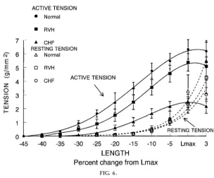
The major misconception concerning cardiac hypertro-phy is that it is ultimately good. Cardiac hypertrohypertro-phy is the slowest of the compensatory physiological mechanisms recruited during the development of heart failure because it involves a number of pro-cesses, including synthesis of new proteins and remod-eling of the ventricles. Hypertrophy was originally defined as an increase in weight of the ventricle in humans on autopsy and was associated with heart failure. Hypertrophy has also been associated with an increase in cardiac weight and larger skeletal muscle mass after exercise training. In fact, the hypertrophy of heart failure still results in a reduced contractile state at any level of preload when normalized for mass (Fig. 6). Even with substantial increases in the amount of muscle, the hypertrophied sick heart cannot create a normal level of cardiac output during exercise. Thus each unit of muscle is still in a negative inotropic state. In contrast, skeletal and cardiac muscle after chronic exercise conditioning are capable of greater levels of performance. This has led to the hypothesis that ‘‘hypertrophy is not hypertrophy is not hypertrophy’’ and also to the important question as to what differ-ences in transcriptional regulation direct the synthesis of new protein during the hypertrophy of exercise compared with the hypertrophy in a failing heart.
Hypertrophy can be either eccentric or concentric, and it has already been discussed that a large dilated
heart (eccentric hypertrophy) may not be able to overcome the increased wall stress caused by en-larged diastolic diameter. The limitation on concentric hypertrophy as a compensatory mechanism, in which myocytes increase in diameter and not length, is that concentric hypertrophy of individual myocytes in-creases the diffusion distance for oxygen from the nearest capillary. The maximum predicted diffusion distance in the heart is ,22–25 µm. When cardiac
cells hypertrophy beyond this diameter, the center of the myocyte becomes hypoxic and the contractile function of that myocyte decreases. Thus, even though the increase in cardiac mass during hypertrophy may compensate to some degree for the reduced contrac-tile state of each myocyte, ultimately the restructuring of the heart limits both eccentric and concentric hypertrophy as adaptive mechanisms, and these may actually contribute to cardiac dysfunction (Fig. 5).
MISCONCEPTIONS CONCERNING THE
INTEGRATED NEUROHUMORAL CONTROL OF THE CIRCULATION DURING THE
DEVELOPMENT OF HEART FAILURE
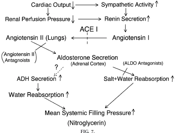
Perhaps the most useful diagram to explain the integrated neurohumoral control of the circulation during the development of heart failure is the one devised by Guyton (4) (Fig. 7) and modified by us.
FIG. 5.
Scheme illustrates concept that alterations in structur e of the heart serve to nor malize str ess (sm). Eccentric hypertr ophy may initially incr ease function
Students use this diagram to explain to themselves how blood volume is increased as heart failure devel-ops. A common misconception that students make at this point, however, is that the increase in blood volume that occurs during heart failure is bad; i.e., it exacerbates the reduction in cardiac output. How-ever, if blood volume is not allowed to expand initially, and perhaps as far as the intermediate stage of heart failure, cardiac pump function will be reduced because of an inability to recruit the Frank-Starling mechanism. Moreover, the driving force for eccentric hypertrophy, an increase in end-diastolic wall stress (3), will be lost. This may result in premature mortal-ity. Students may falsely reason that an increase in blood volume moves the heart onto the descending limb of the Frank-Starling curve, resulting in a further reduction in cardiac function. For that to occur, the mechanical function of the heart must have already deteriorated to the point of severe failure (Fig. 3). In the early stages of failure, however, function is de-scribed by a normal cardiac output relationship with a plateau (albeit reduced) rather than a descending limb (Fig. 1). Thus the major consequence of increased
blood volume early in the disease is not a reduced cardiac output but rather an increased venous pres-sure (Fig. 2), resulting in pulmonary and systemic edema (backward failure). These are important consid-erations for the medical student because they provide the rational basis for treating heart failure. Treatment is aimed at correcting defects in inotropic state and volume regulation.
The updated version of the original diagram devised by Guyton (Fig. 7) includes drugs that the first-year medical student has undoubtedly heard about, includ-ing angiotensin-convertinclud-ing enzyme inhibitors, aldoste-rone blocking agents, and angiotensin II receptor blocking agents, along with the original drugs dis-cussed by Guyton, which included digitalis and diuret-ics. All of these agents contribute to the pharmacologi-cal management of the increased blood volume that occurs as a normal compensatory mechanism (and is good) during the development of heart failure. Further-more, the critical and time-dependent use of these agents to prevent real volume overload (which is bad) in a decompensated heart may prolong survival. One
FIG. 6.
Effects of hypertr ophy and heart failur e on inotr opic state. Note that when nor malized for muscle mass, the hypertr ophied papillary muscle is still in a negative inotr opic state. RVH, right ventricular hypertr ophy; CHF, congestive heart failur e; Lmax, max imum length. [Reprinted fr om
has to be cautious to prevent the misconception that heart failure is curable. It is not, and heart failure will still be an epidemic cause of death in the 21st century.
In summary, in this review we have tried to address some misconceptions often encountered in discus-sions with first-year medical students. Examples are given using some recent clinical protocols such as the Battista operation and some newer types of drugs used in the treatment of heart failure. Finally, the concept of decompensated heart failure and the concept of a dynamic definition of heart failure have been reiterated to support the notion that decompen-sated heart failure is the end result of the disease process.
Address for reprint requests and other correspondence: T. H. Hintze, Dept. of Physiology, New York Medical College, Valhalla, NY 10595 (E-mail: [email protected]).
Refer ences
1. Beutler, B., and A. Cerami. The biology of cachexin/TNF, a primary mediator of the host response.Annu. Rev. Im m unol.7: 625–655, 1989.
2. Braunwald, E. Hea rt Disea se: A Textbook of Ca rdiova scula r Medicine. Philadelphia, PA: Saunders, 1980.
3. Gr ossman, W., D. Jones, and L. P. McLaurin.Wall stress and patterns of hypertrophy in the human left ventricle. J. Clin. Invest. 56: 56–64, 1975.
4. Guyton, A. C.Textbook of Medica l Physiology. Philadelphia, PA: Saunders, 1986.
5. Selkurt, E. E.Physiology. Boston, MA: Little, Brown, 1971. 6. Solar o, R. J.Integration of myofilament response to Ca21with
cardiac pump regulation and pump dynamics.Am . J. Physiol.
277 (Adv. Physiol. Educ.22): S155–S163, 1999.
7. Spann, J. F., Jr., R. A. Buccino, E. H. Sonnenblick, and E. Braunwald.Contractile state of cardiac muscle obtained from cats with experimentally produced ventricular hypertrophy and heart failure.Circ. Res. 21: 341–349, 1967.
8. Young, M. A., T. H. Hintze, and S. F. Vatner. Correlation between cardiac performance and plasma catecholamines in the conscious dog. Am . J. Physiol. 249 (Hea rt Circ. Physiol. 18): H49–H56, 1985.
FIG. 7.