MEASUREMENT OF HUMAN NASAL POTENTIAL DIFFERENCE
TO TEACH THE THEORY OF TRANSEPITHELIAL
FLUID TRANSPORT
Ulrich Kersting,1 Albrecht Schwab,2 and Alexandra Hebestreit3
1Institute of Clinica l Biochem istry a nd Pa thobiochem istry, Medica l University Clinic, 2Depa rtm ent of Physiology, a nd 3University Children’s Hospita l, University of Wu¨rzburg, 97078 Wu¨rzburg, Germ a ny
W
e describe a novel student course in membrane physiology in which students record their own nasal potential difference, i.e., the transepithelial potential difference of the respiratory mucosa in the nose. The nasal potential difference monitors directly, and in vivo, changes in the apical cell membrane potential of the respiratory mucosa induced by activators and inhibitors of ion channel activities. Basic principles of transepithelial fluid transport are taught by applying an appropriate perfusion protocol to the respiratory epithelium to either depolarize or hyperpolarize the membrane potential of the luminal cell side, thereby increasing or decreasing the nasal potential difference. This course was given at the Department of Physiology at the University of Wu¨ rzburg in 1997, and responses of the students as reported on questionnaires were mainly positive.AM. J. PHYSIOL. 275 (ADV. PHYSIOL. EDUC. 20): S72–S77, 1998.
Key words:student course; membrane physiology; chloride channel; cystic fibrosis
Transport of fluid is one of the main functions of many tissues, e.g., kidney, intestine, exocrine glands, and lung. Despite its clinical importance, students often have difficulties understanding the basic principles of transepithelial fluid transport. We therefore devel-oped a new course of membrane physiology in which students record the transepithelial electric potential difference of the respiratory mucosa in their own noses, i.e., the nasal potential difference. It is generally understood that changes in the nasal potential differ-ence can be caused by activation or inhibition of Na1
and Cl2channels in the apical cell membrane of the
respiratory epithelium and correlated to transepithe-lial ion transport (2–4, 6, 7). Fluid secretion depends on Cl2efflux at the apical cell membrane; Na1follows
Cl2electrostatically, and water follows salt transport
osmotically (5, 7, 9). Students are first introduced to the membrane concepts of ion gradients, diffusion potentials, and membrane conductances by
measur-ing the cell membrane potential of livmeasur-ing Xenopus la evisoocytes (8). Subsequently, the basic principles of transepithelial fluid transport are taught by record-ing the nasal potential difference. The clinical rel-evance of transepithelial fluid transport is exemplified by cystic fibrosis (CF), which is characterized by blockade of cAMP-activated apical Cl2channels (1, 7).
Measurements of nasal potential differences can be easily performed by nonelectrophysiologists and do not require extensive electrophysiological training.
METHODS
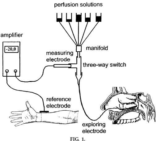
Ex perimental setup. The exploring electrode con-sists of a flexible umbilical vessel catheter with an outer diameter of ,1.2 mm (Sherwood Medical,
Schwalbach, Germany), which is continuously per-fused with isotonic NaCl solution. The umbilical vessel catheter is connected to the measuring
trode (KCl outflow electrode; World Precision Instru-ments, Sarasota, FL) by a conventional three-way switch (Connecta TH Luer-Lock; Ohmeda, Helsing-borg, Sweden). The tip of the umbilical vessel catheter is placed onto the respiratory mucosa between the inferior turbinate and the floor of the nose. The reference electrode is a silver/silver chloride elec-trode that is covered by gel (3M RedDot; Medica, Borken, Germany) and taped to the forearm. The KCl outflow electrode and the reference electrode are connected to the input of a battery-operated high-impedance voltmeter (World Precision Instruments). Figure 1 shows schematically the experimental setup.
Syringes (50 ml) serve as reservoirs for perfusion solutions. Every solution is filter sterilized before use. Infusion tubes connect the reservoirs via a manifold (Warner Instrument, Hamden, CT) and a three-way switch to the exploring electrode. The manifold exhibits a very small dead space of,100 µl, allowing a
rapid change of perfusion solutions. Flow of perfusion solutions is driven by gravity, and flow rate in each tube is adjusted by a valve to 0.5 ml/min. The control perfusion solution is an isotonic NaCl solution (308 mosmol/l). A low-Cl2solution is prepared by
substitut-ing NaCl with equimolar gluconate, givsubstitut-ing a final Cl2
concentration of 15 mmol/l. Amiloride (100 µmol/l) and isoproterenol (10 µmol/l) are dissolved in perfu-sion solutions shortly before class begins. Perfuperfu-sion solutions are stored frozen as 50-ml aliquots.
Ex perimental pr ocedur e. The setup for measure-ments of nasal potential difference is renewed daily during the course with sterile disposable infusion syringes, infusion tubes, valves, and three-way switches. A fresh sterile umbilical vessel catheter is used for each measurement. The measuring KCl outflow electrode is prepared shortly before the recording and connected to the three-way switch.
During the experiment, the student sits comfortably in a dental chair. The student’s head is tilted slightly back to facilitate the placement of the exploring electrode below the inferior turbinate. To create a suitable site of zero potential for the reference elec-trode, the epidermal surface of the skin of the forearm is treated with a special polishing gel and cleaned with 70% ethanol. Before measurements are taken, the exploring electrode, which is already perfused with NaCl solution, is placed in close contact to the reference electrode and adjustments are made to compensate for the recorded offset values. The explor-FIG. 1.
ing electrode is then passed along the floor of the nose until a stable nasal potential difference in the range of
210 to220 mV is obtained. Nasal potential difference reaches a maximum when the exploring electrode is placed between the inferior turbinate and the floor of the nose,,0.5 cm distal from the anterior tip of the
inferior turbinate. At this position the recording of the nasal potential difference is maximal and very stable. Maximal nasal potential difference can be lowered reproducibly by moving the tip of the exploring electrode to slightly sites more proximal or distal from the inferior turbinate. The correct placement of the exploring electrode may also be controlled by viewing through an otoscope. Perfusion solutions drop from the floor of the nose into the throat without discom-fort, according to the test persons. Each perfusion solution is applied with a constant flow rate of 0.5 ml/min for a time period of,5 min. Tightly
control-ling the perfusion rate assures that the amounts of amiloride and isoproterenol applied during the course stay well below pharmacologically relevant doses.
Because amiloride is contraindicated during preg-nancy and lactation period, pregnant or nursing stu-dents were excluded as test persons. Nasal potential differences were recorded in groups of about five students. One student from each group was expected to volunteer as a test person, and at least one student was expected to place the exploring catheter. All students were involved in preparing the experimental setup and data analysis. The students placed the exploring catheters after the lab instructors in each group demonstrated how to do so. This was, in principle, not difficult, but correct positioning of the tip of the exploring catheter below the inferior turbinate required some experience and was closely supervised by the lab instructors.
RESULTS
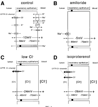
Figure 2 shows changes in transepithelial potential differences, apical cell membrane potentials, and ion transport of the Cl2-secreting respiratory epithelium
in the nose that were induced by the respective experimental maneuvers. The perfusion protocol was optimized to measure apical Na1and cAMP
depend-ing Cl2-channel activities in Cl2-secreting airway
epi-thelium (6). The numerical values of transepithelial potential differences in Fig. 2 correspond to data shown in Fig. 3.
The inferior turbinate of the nose was perfused with isotonic NaCl solution until a stable nasal potential value, e.g.,212 mV, was recorded.
After a stable baseline value was obtained, amiloride (100 µmol/l; dissolved in isotonic NaCl solution) was applied. Amiloride blocks Na1channels in the apical
cell membrane of the respiratory epithelium, thereby inhibiting cellular Na1 uptake. Consequently, the
membrane potential of the luminal cell side hyperpo-larized, which in turn led to a decrease in the nasal potential difference to25 mV (Fig. 2B).
The perfusion was then continued with a low-Cl2
solution (15 mmol/l Cl2), with amiloride still present.
The increased Cl2gradient across the luminal cell side
increased the efflux of Cl2 through cAMP-activated
Cl2channels. Increased Cl2efflux at the luminal cell
side depolarized the apical cell membrane potential, which in turn led to a rise in the nasal potential difference to224 mV (Fig. 2C).
To further stimulate Cl2efflux across the apical cell
membrane, isoproterenol (10 µmol/l) was added to the low-Cl2 solution, with amiloride still present.
Isoproterenol opens cAMP-activated CF transmem-brane conductance regulator (CFTR)-Cl2channels in
the apical cell membrane. The stimulated luminal Cl2
efflux further reduced the apical cell membrane poten-tial and led to an increase in the nasal potenpoten-tial difference to226 mV (Fig. 2D).
Finally, control Ringer solution was applied to check whether the basal nasal potential difference was kept constant during the experiment (212 mV).
During the course, which was held in winter, it became apparent that nasal mucosal infections de-crease the nasal potential difference. Therefore, Fig. 3 represents only data from students without any sign of mucosal inflammation or viral infection of the upper respiratory tract. Basal nasal potential difference was
212.4 6 1.3 mV (n 5 16). Inhibition of apical
Na1-channel activity by amiloride decreased the nasal
potential difference to24.6561.1 mV. Perfusion of the luminal side of the respiratory mucosa with a low-Cl2 solution increased the nasal potential
differ-ence to 224.12 6 0.95 mV. Isoproterenol further increased the nasal potential difference to 225.5 6
1.6 mV. Nasal potential difference decreased to basal
values, 212 6 6.8 mV, after perfusion with isotonic NaCl for,10 min.
At the end of the course, the students completed a questionnaire, anonymously rating the quality of the course on a scale from 1 (very good) to 5 (insufficient) (Fig. 4).
FIG. 2.
Schematic drawing of nasal potential differ ences, apical cell membrane potentials, and ion transporters at r espiratory epithelium. InB-D, only those ion transporters that ar e affected by the r espective ex perimental pr otocols ar e shown. Water movement is not shown.A: per fusion with isotonic NaCl (contr ol). CFTR, cystic fibr osis transmembrane conductance r egulator.B: application of amiloride (100
mmol/ l). C: per fusion with a low-Cl2 solution (15 mmol/ l Cl2). D: addition of isopr oter enol (10mmol/ l) to low-Cl2solution. Nasal potential differ ence is
DISCUSSION
Recording the nasal potential difference is a suitable model for teaching membrane physiology for several reasons. First, it is an easy and reliable technique that can be established in almost every laboratory at low cost. Second, it shows all the advantages and risks of a ‘‘live’’ experiment without being an animal experi-ment. Finally, the students gain hands-on experience regarding the effects of inhibitors and activators of transepithelial ion transport. Placement of the umbili-cal vessel catheter below the nasal inferior turbinate does not pose any danger to the subject but requires concentration and the ability of the experimenter to take responsibility. These demands greatly increased the motivation of the students in our course on membrane physiology.
After the practical part of the course was completed, the changes in the nasal potential differences were discussed in detail. The observed changes in the nasal
potential differences were explained by changes in the apical cell membrane potentials, which were induced by alterations of luminal ion transport by amiloride, low-Cl2solution, and isoproterenol. On the
basis of their personal measurements of the nasal potential difference, the students could realize the FIG. 3.
Changes of nasal potential differ ences after applica-tion of amiloride, low-Cl2solution, and isopr oter enol
in students. Stable values of nasal potential differ ences wer e r ecor ded 5 min after per fusion solutions wer e switched.
FIG. 4.
Students anonymously ranked course quality on a scale fr om 1 (very good) to 5 (insufficient).
importance of the transepithelial potential difference as a driving force for transepithelial fluid transport through the paracellular space. So far, the students were showing a lively interest not only during the measurements but also during this theoretical part of the course. As the survey shows, the students rate our new course of membrane physiology very positively.
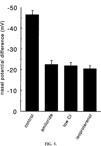
The clinical significance of disturbed transepithelial transport was stressed by discussing the pathophysiol-ogy of CF, the most common inherited lethal disease in Europeans and Caucasian North Americans. For comparison, Fig. 5 shows mean data of nasal potential differences from CF patients. The hyperpolarization in low-Cl2and isoproterenol-containing solution is
miss-ing because of the blockade of luminal Cl2 efflux.
Inhibition of fluid secretion in the lung of CF patients induced dehydration of the mucous, thereby causing lung damage. During our course in membrane physiol-ogy, the main symptoms of CF were discussed on the pathophysiological basis of disturbed transepithelial ion transport.
We thank Prof. S. Silbernagl for technical support and for assistance in the preparation of this manuscript.
Address for reprint requests: U. Kersting, Inst. of Clinical Biochemis-try and PathobiochemisBiochemis-try, Medical Univ. Clinic, Versbacher Str. 5, 97078 Wu¨ rzburg, Germany.
Refer ences
1. Dodge, J. A., D. J. Br ock, and J. H. Widdicombe. Cystic Fibrosis Current Topics. Chichester, UK: Wiley, 1994. 2. Hofmann, T., O. Bo¨hmer, G. Hu¨ ls, H. G. Terbrack, P.
Bittner, V. Klingmu¨ ller, E. Heer d, and H. Lindemann.
Conventional and modified nasal potential-difference measure-ment in cystic fibrosis. Am . J. Respir. Crit. Ca re Med. 155: 1908–1913, 1997.
3. Knowles, M. R., J. L. Carson, A. M. Collier, J. T. Gatzy, and R. C. Boucher.Measurements of nasal transepithelial electric potential differences in normal human subjects in vivo. Am . Rev. Respir. Dis. 124: 484–490, 1981.
4. Knowles, M. R., A. M. Paradiso, and R. C. Boucher.In vivo nasal potential difference: techniques and protocols for assess-ing efficacy of gene transfer in cystic fibrosis.Hum . Gene Ther. 6: 445–455, 1995.
5. Liedtke, C. M. Regulation of chloride transport in epithelia.
Annu. Rev. Physiol. 51: 143–160, 1989.
6. Middleton, P. G., D. M. Geddes, and E. W. F. W. Alton.
Protocols for in vivo measurement of the ion transport defects in cystic fibrosis nasal epithelium.Eur. Respir. J.7: 2050–2056, 1994.
7. Quinton, P. M. Cystic fibr osis: a disease in electr olyte transport.FASEB J.4: 2709–2717, 1990.
8. Schwab, A., U. Kersting, H. Oberleithner, and S. Silber-nagl. Xenopus la evis oocyte: using living cells to teach the theory of cell membrane potential. Am . J. Physiol.268 (Adv. Physiol. Educ.13): S26–S31, 1995.
9. Spring, K. R. Mechanism of fluid transport by epithelia. In:
Ha ndbook of Physiology. The Ga strointestina l System . Intesti-na l Absorption a nd Secretion. Bethesda, MD: Am. Physiol. Soc., 1991, sect. 6, vol. IV, chapt. 5, p. 195–207.
FIG. 5.