DOI: 10.2478/10004-1254-61-2010-1937 Short Communication
TOXICOLOGICAL METHODS FOR TRACING DRUG
ABUSE: CHROMATOGRAPHIC, SPECTROSCOPIC
AND BIOLOGICAL CHARACTERISATION OF
ECSTASY DERIVATIVES
Hafi d BELHADJ-TAHAR
1,2, Pierre PAYOUX
2, Mathieu TAFANI
2, Yvon COULAIS
2,
Serge CALET
3, and Azzedine BOUSSEKSOU
4Centre Anti-Poisons, CHU Purpan1, EA3033, Faculté de Médecine Université Paul Sabatier2,
Laboratoire Holis Technologies3, CNRS, Laboratoire de Chimie de Coordination4, Toulouse, France Received in January 2009
Accepted in September 2009
Analysis often reveals variability in the composition of ecstasy pills from pure 3,4-methylenedioxymethamphetamine (MDMA) to mixtures of MDMA derivatives, amphetamine, and other unidentifi ed substances. For a comprehensive toxicological analysis one needs to know all steps to MDMA synthesis which may originate impurities. The aim of this study was to synthesise and determine the chemical-physical and in vitro biological properties of a series of MDMA derivatives.
3,4-methylendioxyphenyl-2-nitropropene (MDNP) was obtained by condensation of piperonal with an excess of nitroethane in the presence of ammonium acetate. MDNP was then reduced to
methylenedioxyamphetamine (MDA) by LiAlH3. All compounds were analysed using HPLC and
spectroscopic technique [Raman, nuclear magnetic resonance (NMR), or infrared (IR)] at all the steps of synthesis. In addition, we assessed the biological potentials of these compounds by measuring in vitro their (i) blood cell/whole blood partition coeffi cient, (ii) binding to plasmatic proteins (Fbp), and (iii) membrane adsorption. Chemical structure was determined with antibody fl uorescence polarisation immunoassay (FPIA). This study showed the presence of solid impurities, particularly of a neurotoxic compound of Al3+ in the fi nal products. FPIA identifi ed the aminoethane group close to the substituted benzene ring,
but did not detect the two major precursors of MDMA: MDNP and piperonal. Raman spectroscopy is an attractive alternative technique to characterise ecstasy pills and it can identify stereoisomeric forms such as cis-MDNP and trans-MDNP, which exhibit signals at 1650 cm-1 and 1300 cm-1, respectively.
KEY WORDS:IR, MDA, MDMA, MDNP, NMR, Raman, toxicology
Ecstasy is a synthetic drug used recreationally worldwide, especially by young people, which makes it a major public health concern (1). A recent French study with 1666 ecstasy pills carried out by a non-governmental organisation Rave Mission revealed that the pill composition varied from pure 3,4-methylenedioxymethamphetamine (MDMA) to a mixture of ecstasy derivatives, amphetamine, and 20 % of other substances unidentifi ed by Rapid
Product Control (RCP) (2). There are several possible ways to synthesise ecstasy, all of them starting with piperonal to obtain either a ketone (3,4-methylenedioxyphylpropanone) or an amine chemical form (3,4-methylenedioxyamphetamine) (2). Various impurities and by-products were identifi ed in powdered tablets: 3,4-methylenedioxyamphetamine (MDA), MDA or MDMA dimers, substituted pyridines, lead, aluminium, and nitroethane (3,4).
Some of these compounds are toxic and could be involved in documented cases of fulminant toxic hepatitis and neurotoxic effects (4).
This suggests that a comprehensive toxicological analysis of an ecstasy pill requires expert knowledge of all synthetic steps and identifi cation of impurities. Raman spectroscopy is a powerful technique to characterise ecstasy pills. In addition, it is an in situ, non-destructive technique that identifi es chemical bonds through intramolecular vibration spectrum (5-7). The technique has proven successful in several published studies of illegal drugs (8,9). In addition, Raman spectroscopy was used to probe photoexcitated phase transition with second irradiation using nanosecond pulsed laser leading for example to colour changes (10). These properties could be exploited to identify organic metals in a sample.
The aim of our study was to fi rst synthesise a series of MDMA derivatives, then to characterise them using HPLC and spectroscopic techniques [Raman, nuclear magnetic resonance (NMR), or infrared (IR)], and fi nally to establish their biological effects on blood cells, proteins, anisotropic polar membrane, and antibodies.
MATERIALS AND METHODS
Chemicals
Piperonal, lithium aluminium hydride (LiAlH3, 1 mol L-1 in tetrahydrofuran), sodium borohydride (NaBH4), ammonium acetate [(NH4(CH3COO)], trifl uoroacetic anhydride [(CF3CO)2O], nitroethane (CH3CH2NO2), and other solvents (all of pro analysis grade) were purchased from Acros Organics (Geel, Belgium). All reagents were used without further purifi cation.
Synthesis
The synthesis started with piperonal (11), which was dissolved with excess of nitroethane in the presence of ammonium acetate, leading to 3,4-methylendioxyphenyl-2-nitropropene (MDNP). MDA was obtained by reducing MDNP with LiAlH3. Typically, MDNP was prepared by refl uxing a mixture of 25 mmol of piperonal (3.75 g), 133 mmol of nitroethane, 12.5 mmol of ammonium acetate, and 60 mL of toluene. The mixture was heated overnight with continuous removal of water into a Dean-Starck
tube. The solvent was then evaporated in vacuum and the residue re-crystallised from solvent mixture CH3OH:H2O (1:10).
Dropwise we carefully added 1.15 mmol of LiAlH3 to 100 mL of the stirred solution of 0.05 mol L-1 MDNP in tetrahydrofuran. The resulting mixture was heated for fi ve hours. LiAlH3 was decomposed by H2O and the solid was removed by fi ltration. MDA was dissolved in 20 mL of dichloromethane and washed with 500 mL of 1 mol L-1 sodium hydroxide solution.
To obtain 3,4-methylendioxyphenyl-2-nitropropane, we added NaBH4 (0.02 mol) to a solution of MDNP (0.01 mol) in methanol (25 mL) by stirring at 5 °C to 10 °C. After allowing the reaction to continue for another hour at room temperature, the mixture was concentrated. Excess of NaBH4 was decomposed with acetic acid and extracted with chloroform.
CHARACTERISATION
HPLC
The precursor and fi nal products were determined by HPLC equipped with a UV detector. The mobile phase consisted of methanol:H2O:trifl uoroacetic acid (60:40:0.01). Eluation was in isocrat mode at the fl ow rate of 1 mL min-1. HPLC separation was performed on a reverse-phase C18 column (LiChrospher 60, RP-select B ,125 mm x 4 mm, with 5 µm sized particles).
NMR spectroscopy
1H and 13C NMR spectra were acquired at 250.13 MHz on a Bruker WM250 spectrometer. Chemical shifts are given in ppm vs. tetramethylsilane (TMS,1H and 13C) using (CD
3)2SO as solvent (d1H=2.62 ppm and d13C=39.6 ppm).
Raman spectroscopy
Spectra were acquired using a Dilor XY1800 triplemate spectrograph coupled to a Princeton Instruments CCD detector. The 647.1 nm line of a Kr+ laser (Coherent Radiation Innova) was used as the excitation source with laser power of 50 mW. Raman signals were collected at room temperature, at 180 °C to the incident laser beam in the 170 cm-1 to 2400 cm-1 frequency range. Rayleigh scattering was removed using a holographic notch fi lter.
Infrared spectroscopy
IR spectra were recorded on KBr discs with a Perkin-Elmer 983 spectrometer. For all the studied compounds we scanned four accumulations between 400 cm-1 and 4000 cm-1 with 2 cm-1 resolution.
Biological characterisation
The biological potentials of compounds were assessed from in vitro measurements of their fraction bound to plasma proteins coeffi cient (Fbp), determination of blood cell/whole blood partition coeffi cient (Fcb), membrane adsorption coeffi cient (Fad), and interaction with specific anti-MDMA antibody. These biological methods were described elsewhere (12).
Determination of Fbp
200 µL of plasma spiked with the studied compound (0.1 nmol L-1) was incubated in a water bath at 37 °C for 15 min and placed in an ultrafi ltration cell. After centrifugation in an SM24 centrifuge (Dupont Instruments; Wilmington, DE, USA) at 1978 g for 20 min, the ultrafi ltrate was analysed using HPLC. The unbound to protein fraction (Fup) was determined by calculating the ratio of the concentration in ultrafi ltrate and the initial compound concentration in plasma. The fraction bound to plasma proteins was deducted from the following equation: Fbp=100 % -Fup.
Determination of Fcb
20 µL of physiological solution of each compound at concentration of 1 mmol L-1 was added to 2 mL fresh blood taken on EDTA tripotassium salt (K3EDTA). After incubation for 15 min at 37 °C, the blood sample was centrifuged (at 2000 g for 15 min) and 200 µL plasma were taken to determine the concentration in the ultrafi ltrate (see determining Fbp above). Calculations were performed using the following equation:
Fcb= [Cbt-(Cuf/Fup)]/[Cbt h]
The free fraction, which is neither bound to blood cells nor to proteins, was determined using the following equation:
Ffr= (1-Fcb)(1-Fbp),
where Fcb is the blood cell/whole blood partition coefficient; Cbt the concentration in total blood (0.01 mmol L-1); C
uf the concentration in ultrafi ltrate; Fup the unbound-to-protein fraction coeffi cient; h is
haematocrit; Fbp binding to plasma proteins coeffi cient, and Ffr the free fraction coeffi cient.
Determination of Fad
200 µL of physiological solution of each compound (1 µmol L-1) were centrifuged through permeable anisotropic membrane (Amicon) at 1978 g for 20 min (SM24 centrifuge, Dupont Instruments; Wilmington, DE, USA) and the ultrafi ltrate was analysed using HPLC. The coeffi cient of membrane adsorption was estimated using the following equation:
Fad= [Cis-Csu]/Cis,
where Fad is the membrane adsorption coeffi cient; Cis the concentration in initial physiological solution; and Csu the concentration in the ultrafi ltrate.
Interaction with specifi c anti-MDMA antibody
Physiological solution of each compound (0.01 mmol L-1) was analysed using an automated FPIA system (Abbott AxSYM®, USA).
RESULTS AND DISCUSSION
In this study, we have described the synthesis route starting from piperonal and ending with 3,4-methylenedioxyamphetamine (MDA) (11). We have also reported the synthesis of 3,4 methylendioxyphenyl-2-nitropropane, which is not considered precursor of MDMA. We used 3,4 methylendioxyphenyl-2-nitropropane to study its interaction with specifi c anti-MDMA antibody.
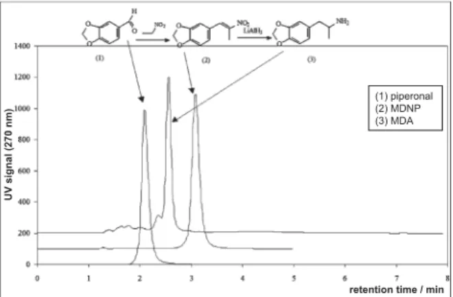
MDNP and MDA were obtained with high chemical purity. The synthesis reactions were followed by HPLC determination of retention times of the obtained products. Piperonal, MDA and MDNP exhibited high lipophilicity (Figure 1). The retention
Figure 1 HPLC patterns of piperonal and synthesised MDNP
and MDA
retention time / min
UV signal (270 nm)
(1) piperonal (2) MDNP (3) MDA
times of piperonal, MDA, and MDNP were 2 min, 2.5 min, and 3.1 min, respectively, and correlated to their lipophilicity (13).
IR and NMR spectroscopies showed a clear profi le of piperonal, MDNP, and MDA. NMR detected the following signals: 1H NMR: d=1.22 (d, J=6.2 Hz, 3 H, CH 3), 2.71 (dd, J=13.4 and 8.8 Hz, 1H, CH 2), 3.07 (dd, J=13.4 and 5.2 Hz, 1H, CH 2), 3.43 (m, 1H, CH), 6.10 (s, 2 H, CH2), 6.80 (dd, J=7.8 and 1.3 Hz, 1H, C 4H), 6.97 (d, J=1.3 Hz, 1H, C 6H), 6.97 (d, J=7.8 Hz, 1H, C 3H). 13C NMR: 17.6 (CH 3), 39.7 (CH2), 48.2 (CH), 101.0 (CH2 dioxole), 108.4 (C3H), 109.6 (C6H), 122.5 (C4H), 130.6 (C5), 146.1 (C1), 147.5 (C2).
IR (Figure 2) revealed signals related to benzene ring (absorption at 1603 cm-1, 1505 cm-1, 1680 cm-1, and 1489 cm-1), to ether oxide bridge at 1489 cm-1, and to MDMA derivatives.
For piperonal, the band located at 1677 cm-1 revealed the presence of aldehyde, for MDNP the band at 1510 cm-1 indicated a nitro compound in position alpha of an unsaturated bond, and MDA was characterised by bands at 3358 cm-1 and 1634 cm-1 pointing to amines, and by the disappearance of the nitro signals.
Figure 2 shows the vibrational modes of piperonal, MDNP, and MDA plotted in the range of 250 cm-1 to 1750 cm-1. While piperonal and MDNP showed high quality Raman spectra (Figure 3), MDA showed a spectrum with a rather large background, possibly due to a fl uorescence in the system at either 632 nm or 541 nm (Figure 4). Milhazes et al. (9) obtained MDA spectrum at 541 nm.
Table 1 lists the biological characteristics of the ecstasy derivatives and impurities in the fi nal product. Of solid impurities found, a neurotoxic compound of Al3+ is of particular interest (4). Save for F
bp, all other biological parameters seem to be proportional to their retention time determined by HPLC and to their lipophilicity. Retention times for piperonal was 2 min, for MDA and 2.5 min, and for MDNP 3.1 min and corresponded to Fad and Fbp.
Table 1 shows that the percent of free compounds in plasma is less than 40 % of total blood concentration: Ffr=23 % for piperonal, Ffr=27 % for MDA, and Ffr=38 % for MDNP.
FPIA antibody recognised the aminoethane group close to the substituted benzene ring, but did not detect the two major precursors of MDMA, that is, MDNP and piperonal (Figure 5). This limitation must be taken into consideration, as detection of all ecstasy
Figure 2 Infrared spectra of (a) piperonal, (b) MDNP, and
(c) MDA
derivatives is required to optimise performance of detection methods.
CONCLUSION
This study has detected solid impurities and by-products in the synthesised products: MDA, unreacted piperonal, MDNP, and aluminium. While HPLC, Raman, and IR spectroscopy identifi ed ecstasy derivatives, FPIA did not detect MDNP and piperonal.
a
b
Table 1 Biological characteristics of ecstasy derivatives and impurities in the fi nal product
Fbp / % Fcb / % Ffr / % Fad / % Derivatives and impurities FPIA
Piperonal 63 38 23 47 Negative MDNP 88 68 38 90 Piperonal(s),Toluene(l), NH 4Ac (s),EtNO 2 (l) Negative MDA 71 30 27 63 MDNP (s) , THF (l), Al3+ (s) Positive (l) liquid; (s) solid
MDNP: 3,4-methylendioxyphenyl-2-nitropropene; MDA: 3,4-methylenedioxyamphetamine; FPIA: fl uorescence polarization immunoassay; Fbp: blood cell/whole blood partition coeffi cient; Ffr: free fraction coeffi cient; Fcb: binding to plasmatic proteins coeffi cient; Fad: membrane adsorption coeffi cient; THF: tetrahydrofuran; NH4Ac: ammonium acetate; EtNO2: nitroethane
Figure 3 Raman spectra of piperonal and MDNP
Figure 4 Raman spectrum of MDA
Figure 5 Molecules tested in vitro with fl uorescence polarization
immunoassay (FPIA). (1) piperonal; (2) MDNP; (3) 3,4-methylendioxyphenyl-2-nitropropane; and (4) MDA.
Raman spectroscopy has turned out to be a particularly interesting alternative technique for characterising ecstasy pills, as it can determine stereoisomeric forms of compound such as cis-MDNP and trans-MDNP at 1650 cm-1 and 1300 cm-1, respectively.
Our findings call for further study of Raman spectroscopy to better characterise other drug derivatives.
Acknowledgements
This work was supported by Association Française de Promotion de la Recherche Médicale (AFPReMed). The authors thank Ms Aurore Palmaro for help in editing the text.
REFERENCES
1. Leung KS, Cottler LB. Ecstasy and other club drugs: a review of recent epidemiologic studies. Curr Opin Psychiatry 2008;21:234-41.
2. Belhadj-Tahar H, Molnar Y, Payoux P, Coulais Y, Costes JP, Robert L, Esquerre JP, Bousseksou A. Contribution of the Raman spectroscopy in the characterization of ecstasy derivatives. In: 40th International Meeting of The
International Association of Forensic Toxicologists (TIAFT), Paris, France, August 26-30th
. Paris: TIAFT; 2002. Wave number / cm-1 Wave number / cm-1 Wave number / cm-1 excitation: 643 nm, 50 mW excitation: 643 nm, 50 mW excitation: 643 nm, 50 mW
3. Cheng JY, Chan MF, Chan TW, Hung MY. Impurity profi ling of ecstasy tablets seized in Hong Kong by gas chromatography-mass spectrometry. Forensic Sci Int 2006;16:87-94.
4. Belhadj-Tahar H, Houin G, Frances B, Molnar G, Zwick A, Bousseksou A, Costes JP, Esquerre JP, Coulais Y. Relationship between chemical structure of ecstasy and positive response to fl uorescence polarisation immunoassay: Interest of 1H, 13C RMN, IR, and Raman spectrocopic
methods [Relation entre structure chimique et positivité du dépistage de l’ecstasy par immunochimie FPIA: intérêt des spectroscopies 1H, 13C RMN, IR et Raman, in French].
Annales de Toxicologie Analytique 2003;17:179-80. 5. Smith WE. Practical understanding and use of surface
enhanced Raman scattering/surface enhanced resonance Raman scattering in chemical and biological analysis. Chem Soc Rev 2008;37:955-64.
6. Bell SE, Spence SJ. Disposable, stable media for reproducible surface-enhanced Raman spectroscopy. Analyst 2001;126:1-3.
7. Bousseksou A, McGarvey JJ, Varret F, Real JA, Tuchagues JP, Dennis AC, Boillot ML. Raman spectroscopy of the high- and low-spin states of the spin crossover complex Fe(phen)2(NCS)2: an initial approach to estimation of vibrational contributions to the associated entropy change. Chem Phys Lett 2000;318:409-16.
8. Bell SE, Fido LA, Sirimuthu NM, Speers SJ, Peters KL, Cosbey SH. Screening tablets for DOB using
surface-enhanced Raman spectroscopy. J Forensic Sci 2007;52:1063-7.
9. Milhazes N, Martins P, Uriarte E, Garrido J, Calheiros R, Marques MP, Borges F. Electrochemical and spectroscopic characterisation of amphetamine-like drugs: application to the screening of 3,4-methylenedioxymethamphetamine (MDMA) and its synthetic precursors. Anal Chim Acta 2007;596:231-41.
10. Bonhommeau S, Molnár G, Galet A, Zwick A, Real JA, McGarvey JJ, Bousseksou A. One shot laser pulse induced reversible spin transition in the spin-crossover complex [Fe(C4H4N2){Pt(CN)4}] at room temperature. Angew Chem Int Ed Engl 2005;44:4069-73.
11. Nichols DE, Hoffman AJ, Oberlender RA, Jacob P, Shulgin AT. Derivatives of 1-(1,3-benzodioxol-5-yl)-2-butanamine: representatives of a novel therapeutic class. J Med Chem 1986;29:2009-15.
12. B e l h a d j - T a h a r H , B o u m a h d i R , D a r b i e u M H . Conceptionalization of diagnostic agents: From empirical
in vivo screening to rational in vitro predictive parameters.
Altern Lab Anim 2000;28:303-14.
13. Mrkvicková Z, Kovaríková P, Balíková S, Klimes J. Determination of lipophilicity of novel potential antituberculotic agents using HPLC on monolithic stationary phase and theoretical calculations. J Pharm Biomed Anal 2008;48:310-4.
Sažetak
TOKSIKOLOŠKE METODE OTKRIVANJA OPOJNIH DROGA U TRAGOVIMA:
KROMATOGRAFSKA, SPEKTROSKOPSKA I BIOLOŠKA KARAKTERIZACIJA DERIVATA ECSTASYJA
Analize često otkriju neujednačenost sastava tableta ecstasyja od čistoga 3,4-metilendioksimetamfetamina (MDMA) do mješavina njegovih derivata, amfetamina i drugih neutvrđenih tvari. Stoga je za kvalitetnu toksikološku analizu potreban uvid u sve korake sinteze MDMA, s obzirom na to da se ondje vjerojatno kriju izvori nečistoće (prekursori, katalizatori). Cilj ovog ispitivanja bio je sintetizirati derivate MDMA te napraviti njihovu kemijsko-fi zikalnu i biološku in vitro karakterizaciju.
3,4-metilendioksifenil-2-nitropropen (MDNP) dobiven je kondenzacijom piperonala u suvišku nitroetana uz dodatak amonijeva acetata. Njegovom redukcijom s pomoću LiAlH3 dobiven je 3,4-metilendioksiamfetamin (MDA). Svi spojevi iz pojedinih koraka sinteze karakterizirani su s pomoću tekućinske kromatografi je visoke djelotvornosti (HPLC) i spektroskopskih tehnika [Ramanove spektroskopije, nuklearne magnetske rezonancije (NMR-a) te infracrvene spektroskopije (IR-a)]. Usto je ocijenjen i njihov biološki učinak
in vitro mjerenjem (i) koefi cijenta raspodjele krvna stanica/puna krv, (ii) vezanja za bjelančevine u plazmi (Fbp) te (iii) adsorpcije na membranu. Kemijska je struktura utvrđena s pomoću fl uorescentnoga polarizacijskog imunokemijskog testa (FPIA). Analiza je u konačnim proizvodima utvrdila prisutnost krutih nečistoća, napose spojeva neurotoksičnog aluminija (Al3+). FPIA je prepoznao aminoetansku skupinu blizu
supstituiranoga benzenskog prstena, ali ne i dva glavna prekursora za MDMA: MDNP i piperonal. Posebno je zanimljiva Ramanova spektroskopija budući da (i) pruža privlačnu alternativu za karakterizaciju sastava tableta ecstasyja te (ii) može otkriti stereoizomerne cis/trans-oblike spoja poput cis-MDNP-a odnosno
trans-MDNP-a, čiji se signal vidi na 1650 cm-1 odnosno 1300 cm-1.
KLJUČNE RIJEČI: IR, MDA, MDMA, MDNP, NMR, Ramanova spektroskopija, toksikologija
CORRESPONDING AUTHOR: Dr Hafi d Belhadj-Tahar
Centre Antipoison et de Toxicovigilance CHU-Purpan, 31059 Toulouse, France E-mail: belhadj-tahar@afpremed.org