Sequential Isolation of Saturated, Aromatic, Resinic and Asphaltic Fractions
Degrading Bacteria from Oil Contaminated Soil in South Sumatera
Munawar
1,2*), Pingkan Aditiawati
2, and Dea Indriani Astuti
21. Biology Department, Faculty of Mathematic and Natural Science, Sriwijaya University, Indralaya 30662, Indonesia 2. Microbiology Study Program, School of Life Sciences and Technology, Bandung Institute of Technology,
Bandung 40116, Indonesia
*)
E-mail: [email protected]
Abstract
Sequential isolation has been conducted to obtain isolates of saturated, aromatic, resin, and asphaltene fractions degrading bacteria from oil contaminated sites. Five soil samples were collected from South Sumatera. These were analyzed using soil extract medium enriched with oil recovery or Remaining-Oil recovery Degradated (ROD) as sole carbon and energy sources according to the isolation stage. ROD at the end of every isolation stage analyzed oil fractions by use of the SARA analysis method. Six isolates of bacteria have been selected, one isolate was fraction saturates degrading bacteria that are Mycobacterium sp. T1H2D4-7 at degradation rate 0.0199 mgs/h with density 8.4x10⁶ cfu/g from stage I. The isolate T2H1D2-4, identified as Pseudomonas sp. was fraction aromatics degrading bacteria at accelerate 0.0141 mgs/h with density 5.1x10⁶ cfu/g are obtained at stage II. Two isolates namely Micrococcus sp. T3H2D4-2 and Pseudomonas sp. T1H1D5-5 were fraction resins degrading bacteria by accelerate 0.0088 mgs/h at density 5.6x10⁶ cfu/g and 0.0089 mgs/h at density 5.7x10⁶ cfu/g are obtained at stage III. Isolation of stage IV has been obtained two isolates Pseudomonas sp. T4H1D3-1and Pseudomonas sp. T4H3D5-4 were fraction asphaltenes degrading bacteria by accelerate 0.0057 mgs/h at density 5.6x10⁶ cfu/g and accelerate 0.0058 mgs/h at density 5.7x10⁶ cfu/g.
Abstrak
Isolasi Bertahap Bakteri Pendegradasi Fraksi Jenuh, Aromatik, Resin, dan Aspal dari Tanah Terkontaminasi Minyak di Sumatra Selatan. Penelitian isolasi bertahap telah dilakukan untuk mendapatkan bakteri pendegradasi fraksi jenuh, aromatik, resin dan aspal. Isolasi dilakukan terhadap lima sampel tanah terkontaminasi minyak dari Sumatera Selatan. Medium isolasi menggunakan Soil extract diperkaya oil recovery atau oil recovery sisa degradasi (OSD) sebagai satu-satunya sumber karbon dan energi sesuai tahapan isolasi. OSD setiap akhir tahap isolasi difraksinasi menggunakan SARA Analysis untuk mengetahui fraksi jenuh, aromatik, resin dan aspal. Hasil penelitian mendapatkan enam isolat bakteri terpilih berdasarkan kecepatan degradasi tertinggi pada setiap tahap, satu isolat bakteri pendegradasi fraksi jenuh yaitu Mycobacterium sp. T1H2D4-7 dengan laju degradasi 0,0199 mg/jam dan kepadatan 8,4x10⁶ cfu/gram dari tahap I. Isolat T2H1D2-4 teridentifikasi sebagai Pseudomonas sp. merupakan bakteri pendegradasi fraksi aromatik dengan laju degradasi 0,0141 mg/jam dan kepadatan 5,1x10⁶ cfu/gram diperoleh pada tahap II. Dua isolat yaitu Micrococcussp. T3H2D4-2 dan Pseudomonassp. T1H1D5-5 merupakan bakteri pendegradasi fraksi resin yang masing-masing mempunyai laju degradasi 0,0088 mg/jam dengan kepadatan 5,6x10⁶ cfu/gram, dan 0,0089 mg/jam dengan kepadatan 5,7x10⁶ cfu/gram diperoleh dari tahap III. Isolasi tahap IV diperoleh dua isolat yaitu Pseudomonas sp. T4H1D3-1 dan Pseudomonas sp. T4H3D5-4 yang merupakan bakteri pendegradasi fraksi aspal, masing-masing mempunyai kecepatan degradasi 0,0057 mg/jam dengan kerapatan 5,6x10⁶ cfu/gram, dan 0,0058 mg/jam dengan kerapatan 5,7x10⁶ cfu/gram.
Keywords: aromatics, asphaltenes, hydrocarbon‐degrading bacteria, saturates, resins, sequential isolation
1.
Introduction
Environmental pollution with petroleum and petroleum products has been recognized as one of the most serious
contamination in soil was caused by, among others, the occurrence of oil spills or spills during transport, pipeline leaks, tank-cleaning of crude oil storage tanks, and also because of accidents like oil well blow-outs. Petroleum contaminated soil, can affect the biota either above or below ground level. These impacts can lead to death (lethal effects), physical damage to biota ( sub-lethal effect), and habitat degradation due to contamination of a-biotic factors on the soil. In this regard, the decree from the Minister of Environment No. 128 of 2003 [4] required the processing of petroleum-contaminated soil in an attempt towards environmental restoration, through bioremediation. The use of bioremediation to change the characteristic and composition of the oil contaminants to become harmless is by reducing the mobility, mass, and concentration of petroleum contaminants in the soil.
Generally the fractions of petroleum hydrocarbons are saturates, aromatics, resins, and asphaltenes fractions [5-6]. The content of the four fractions in oil-contaminated soil is determined by the length of time oil is exposed to the soil. Fractions of petroleum hydrocarbons that are volatile (saturates and aromatics fractions by atomic C<8) evaporate in the environment. Thus the fraction of hydrocarbon-contaminated soil contained on the relative has a greater molecular weight and the level of biodegradability is more difficult [7]. The process of biodegradation of petroleum hydrocarbons is influenced by several factors, the composition and concentration of hydrocarbon compounds, petrophilic bacteria ( hydrocarbon-degrading bacteria), environmental factors during the process of biodegradation, and necessary nutrients [8]. To perform bioremediation of oil-contaminated soil, the mixed bacterial cultures in the form of a consortium have potential application in the bioremediation of the polluted site by removing all the fractions of hydrocarbon pollutants from the environment [9].
Some research on the isolation of hydrocarbon-degrading bacteria have been done (Sutiknowati, 2011; Sivaraman et al., 2011; Mandri and Lin, 2007; Mittal and Singh, 2009), but they only obtained bacteria that can degrade specific hydrocarbons fractions, but cannot degrade all the fractions. Fraction aromatic (naphthalene, phenantrene, trichlorodibenzofuran, and benzo[a]pyrene) degrading bacteria isolated from “beach simulator tank” was added with nutrients N and P [10]. Pseudomonas mendocina was isolated from bilge oil contaminated water at Mormugao harbor. This strain effectively degraded saturated fractions (tetradecane, hexadecane and octadecane) leaving a residual concentration of about 73%, 54% and 40% respectively in 120 hours [11]. Three bacterial isolates have been obtained from soil contaminated with diesel and engine oils, they were identified as Flavobacterium sp., Acinetobacterium calcoaceticum and Pseudomonas
aeruginosa. All isolates were capable of degrading saturates fraction (n-paraffin) up to 80% in a 2 week period [12]. Pseudomonas strain PS-I has been isolated from soils contaminated with crude oil spill, this isolate could degrade saturates (alkanes) (70.69%) and aromatics (45.37%) fractions [13]. Therefore, sequential isolation needs to isolate the bacterial strains that can degrade all fractions of hydrocarbons petroleum (saturates, aromatics, resins, and asphaltenes fractions).
The area of South Sumaterais one area associated with the oil industry, both the public and private sectors. One side effect of oil industry activity is contamination of soil by oil. Sequential isolation is the isolation method based on a succession of bacterial communities that process biodegradation of hydrocarbon petroleum contaminants. Hopefully, through sequential isolation, we can obtain petrophilic bacteria capable of using the carbon from all fractions of petroleum components. Subsequently this may serve as the basis for the development of biological agents in the bioremediation of oil-contaminated soil.
2. Methods
Samples of oil-contaminated soil was collected from five locations: Abab (D1), Benakat (D2), Limau (D3), Raja (D4), and Talangjimar (D5).Contaminated soils at each sampling site and map of sampling sites are seen in Figure 1. Five locations in sequence have been contaminated by petroleum for five, four, three, two, and one year respectively. Sampling was conducted using multiple sampling methods, by determining the five sites based on stratified sampling, while the determination of the three sub-locations of the depth of the surface (0 cm), middle (15 cm), and bottom (30 cm) at each location with a stratified sampling. From each sampling point 0.5 to 2.0 kg of contaminated soil was taken using a soil sampling tool (auger), samples from the same depth were pooled and labeled, then inserted into the sample container aseptically [8,14].
Figure 1. Map of Sampling Sites of Petroleum-Contaminated Soil (left), Oil-Contaminated Soils at Each Sampling Site (Right)
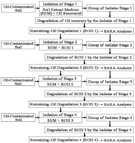
(26-31 °C) for 3 x 24 hours, and then colonies having different morphological features were purified by the quadrant streaking method. The Remaining-Oil recovery Degradated (ROD) was sterilized and reused in stage II as the sole carbon source. Isolation of stage II was performed in the same way as isolating stage I but using the ROD stage I as the sole carbon source, and so on until reaching the isolation of stage IV (Figure 2). Oil Recovery and the ROD of each stage of isolation analyzed saturates, aromatics, resins, and asphaltenes fractions using the SARA Analysis method (Figure 3).
Bacterial isolates obtained at each stage of isolation were verified to determine the best ability of the isolates in degrading the oil fractions. Each bacterial isolate was inoculated 2.5% (v/v) cell density of 2.5 x10⁸ cfu/mL into a 100 mL glass bottle containing 12.5 mL of Mineral Medium (MM) with crude oil, a composition according to the Atlas [15] grams per Liter: K2HPO4 4.5, (NH4)2SO4 1.0, MgSO4.7H2O 0.2, NaCl 0.1, CaCl2 1.0, FeCl3 0.02 and enriched with 250 mg oil recovery as the sole carbon and energy source, and one glass bottle was not inoculated to function as the control. Cultures were incubated at room temperature (26-31 °C) at 100 rpm in an orbital shaker for six days, this experiment used completely randomized designs with 3 times replications for each isolate. Furthermore, cell numbers were counted using the spread method, and analysis of the fraction of saturates, aromatics, resins, and asphaltenes using the SARA Analysis method as shown in Figure 3.
Fraction analysis was performed after the cultures were taken prior to the analysis of cell numbers. 20 mL n-pentane was then added to two hundred and fifty to five hundred milligrams of oil recovery and ROD 1 up to ROD 4 in the culture bottles, homogenized and separated from the medium by a separator funnel. The asphaltenes fractions was obtained by filtering the fraction of n-pentane using a filter paper of known weight. Furthermore, the remaining filtrate collected
Figure 2. Typical Scheme for Isolation of Hydrocarbon-Degrading Bacteria by Sequential Isolation Method
from the separation of the asphaltenes is commonly known as maltenes. It contains the remaining three fractions, saturates, aromatics and resins. These three fractions are separated using open- chromatographic column with silica gel (60-100 mesh). Saturates fraction on percolation in n-pentane eluant, are not absorbed on activated silica under the conditions specified. The saturates fraction of the oil was eluted from column with 20 mL of n-pentane at 5 mL/min. The solvent was removed by evaporation to recover the saturates fraction. Aromatics fraction are absorbed by activated silica in the presence of n-pentane, and desorbed by toluene after removal of the saturates under the conditions specified. The aromatics fraction of the oil is eluted from the chromatographic column using 20 mL toluene at 5 mL/min. The resin fraction of the oil is eluted from the chromatographic column using 20 mL 90:10 toluen : methanol solution at 5 mL/min. All fractions were determined by the gravimetry method and then reported as milligrams [16-17]. The degradation rate of the fraction is determined using the following equation:
(1)
KF0 where is the initial concentration of fraction, KFt is
the concentration fraction after t time, Δt is the difference. The data of the rate of degradation fraction of saturates, aromatics, resins, and asphaltenes was analyzed by F test to determine the isolates with the highest activity to degrade each fraction tested by the New Duncan's Mutiple Range Test (DNMRT) at significant level (α) <5%.
Identification and characterization of selected isolates were performed by morphological colonies on agar stab, agar slant, agar plate using temperature growth, Gram and endospores staining, motility, and biochemical [1,18-21]. Results obtained were used for further identification on the basis of Bergey's Manual of Determinative Bacteriology [22] and Bergey's Manual of Systematic Bacteriology [23].
3. Results and Discussion
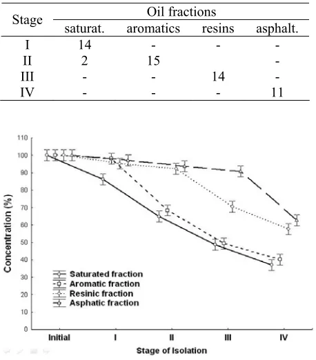
The results of the sequential isolation from five locations were grouped according to the stages of isolation. We obtained 14 isolates of bacteria at stage I, all of these isolates are degrading the fraction of saturates. In stage II, we obtained two saturates degrading isolates and 15 aromatics degrading isolates. Isolation of stage III obtained 14 resins degrading isolates, and more isolation results are presented in Table 1. Thus there is indication of the existence of a trend pattern between the stages of isolation with the degraded fraction. In stage I, all isolates tend to degrade saturates; isolates from stage II tend to degrade the
aromatics fraction. Isolates obtained in stage III tend to degrade the resins fractions, and isolates from stage IV tend to degrade the asphaltenes fraction. The trend occurs because each group of isolates differ in starting work to degrade each fraction, the group of saturates degrading bacteria starts first, then is followed by aromatics degrading bacteria, resins degrading bacteria, and asphaltenes degrading bacteria, respectively. It is also supported by concentrations of saturates, aromatics, resins, and asphalt fractions at each stage isolation (Figure 4). Figure 4 shows that instage I ROD-1 the saturated fraction decreases dramatically, while the other factions experience no real decline. Respectively it shows that at stage II ROD-2, stage III ROD-3, and stage IV ROD-4 the aromatics, resins, and asphaltenes fractions decrease dramatically. This condition explains that the fraction of saturates starts to degrade rapidly at stage I so bacterial isolates obtained at this stage are saturates fraction degrading isolates. Likewise, aromatics, resins, and asphaltenes fractions degraded starting at stage II, III, and IV respectively, so that at stage II the isolates of bacteria degrading are aromatics fractions, at stage III and IV bacterial isolates obtained is for resins degrading bacteria and asphaltenes degrading bacteria respectively. These results are in accordance with the theory put forward by Taki et al. [7] that the order of ease of biodegradation of petroleum hydrocarbon fractions from the easiest to the most difficult is the fraction of saturates, aromatics resins, and asphaltenes.
Table 1. Number of Isolates base on Isolation Stage
Oil fractions Stage
saturat. aromatics resins asphalt. I 14 - - - II 2 15 -
III - - 14 -
IV - - - 11
Figure 4. Average Concentration of Saturates, Aromatics, Resins, and Asphalthenes Fractions in the Initial Condition and at Each Stage of Isolation
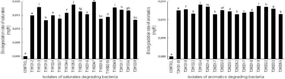
Result of verification test indicated that there were 16 saturates degrading isolates, the rate of each saturates degrading isolates are listed in Figure 5. The highest rate was the isolate T1H2D4-7 from Raja, this isolate has the rate of degradation of the aromatics fraction of 0.0199 mgs/h on the number of cells 8.4x10⁶ cfu/g.
As 15 isolates were obtained from aromatic fractions, all isolates from stage II and the rate of each aromaticsdegrading isolates are listed in Figure 5. The isolateT2H1D2-4 was the only one isolate that has the highest degradation rate of aromatic fraction, from Benakat. This isolate has the rate of degradation of the
aromatic fraction of 0.0141 mgs/h on the number of cells 5.1x10⁶ cfu/g.
Figure 6 shows the resins fraction degrading isolates from stage III, from 13 isolates of resin degrading isolates there are two with the highest rate of degradation of isolate T3H2D4-2 and T1H1D5-5 from Raja and Talangjimar respectively. The degradation rate of each is 0.0088 mgs/h at cell number 5.6 x10⁶ cfu/g and 0.0089 mgs/h on the number of cells 5.7x10⁶ cfu/g respectively.
Figure 5. Degradation Rate of Saturates Degrading Isolates (Left), Degradation Rate of Aromatic Degrading Isolates (Right),
the Stem Given the Same Lower Case Notation Indicates Non-Significant Difference at the α <0.05
Figure 6. Degradation Rate of Resin Degrading Isolates (Left), Degradation Rate of Asphalthene Degrading Isolates (Right),
the Stem Given the Same Lower Case Notation Indicates Non-Significant Difference at the α <0.05
Figure 7. Cell Morphologies of Selected Isolates, (a) Mycobacterium sp. (T1H2D4-7), (b) Pseudomonas sp. (T1H1D5-5),
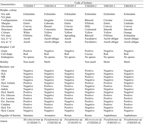
Table 2. The Results of the Characterization and Identification of Six Selected Bacterial Isolates
Code of Isolates Characteristics
T1H2D4-7 T2H1D2-4 T1H1D5-5 T3H2D4-2 T4H1D3-1 T4H3D5-4
Morpho. colony
- NA stab Echinulate Echinulate Echinulate Echinulate Echinulate Echinulate
- NA plate
Configurations Circular Irregular Circular Rhizoid Circular Circular
Margins Entire Laborate Entire Filiform Entire Undulate
Elevations Convex Raised Convex Hilly Convex Convex
Structures Smooth Concentric Smooth Contoured Smooth Smooth
Colours White Yellow Yellow Yellow Yellow Orange
- NA slant Filiform Effuse Spreading Rhizoid Filiform Echinulate
- NA 37 °C Aerob Aerob obligat Aerob Facultative Aerob obligat Aerob obligat
- NA 41 °C Aerob Aerob obligat Aerob Facultative Aerob obligat Aerob obligat
Morphol. Cell
- Gram Positive Negative Negative Positive Negative Negative
- Cell shape Rod Rod Rod Coccus Rod Rod
- Endospores No spores No spores No spores No spores No spores No spores
Motility Non motil Motil Non motil Motil Motil
Biochem. test
- Sitrat Negative Negative Negative Positive Negative Negative
- H2S Negative Negative Negative Negative Negative Negative
- MR Negative Negative Negative Positive Negative Negative
- VP Negative Negative Negative Negative Negative Negative
- Hyd. Gelatin Positive Negative Negative Positive Positive Negative
- Urease Negative Positive Positive Negative Positive Positive
- Indole Negative Negative Negative Negative Negative Negative
- Hyd. Starch Positive Negative Negative Negative Negative Negative
- Fer. Glucose Positive Negative Positive Positive Positive Positive
- Fer. Lactose Positive Positive Positive Positive Positive Positive
- Fer. Sucrose Positive Negative Negative Positive Negative Positive
- Catalase Positive Positive Positive Negative Positive Positive
- Hyd. Fat Positive Positive Positive Negative Negative Positive
- Hyd. Casein Negative Negative Negative Negative Negative Negative
Degrader of fraction Saturates Aromatics Resins Resins Asphaltenes Asphaltenes
Genera Mycobacterium sp
(T1H2D4-7)
Pseudomonas sp (T2H1D2-4)
Pseudomonas sp (T1H1D5-5)
Micrococcus sp (T3H2D4-2)
Pseudomonas sp (T4H1D3-1)
Pseudomonas sp (T4H3D5-4)
The group of asphaltenes fractions degrading isolates are 11, all isolates are from stage IV (Figure 6). Isolates that have the highest rate are isolates T4H1D3-1 namely0.0057 mgs/h at number of cells 5.6x10⁶ cfu/g, this isolate is from Limau and T4H3D5-4 namely 0.0058 mgs/h at number of cells 5.7x10⁶ cfu/g, this isolate is from Talangjimar.
Characterization and identification of bacterial isolates were performed on six selections based on the highest rate in fractions degradation. Six bacterial isolates included one isolate of saturates degrading bacteria, one isolate of aromatics degrading bacteria, two isolates of resins degrading bacteria, and two isolate of asphaltenes degrading bacteria. The cell morphology of six isolates can be seen at Figure 7, and then more results of the characterization and identification are listed in Table 2.
4. Conclusion
number of cells 5.7x10⁶ cfu/mL respectively. Two isolates namely Pseudomonas sp. (T4H1D3-1) from Limau and Pseudomonas sp. (T4H3D5-4) from Talangjimar are asphaltenes degrading with a rate of 0.0057 mgs/h on the number of cells 5.6x10⁶ cfu/mL and 0.0058 mgs/h on the number of cells 5.7x10⁶ cfu/mL respectively.
Acknowledgements
We would like to thank Prof. Dr. Djoko T. Iskandar who has provided input during the research and writing of this article, Pusat Penelitian Lingkungan Hidup Universitas Sriwijaya which has helped with some of the costs of research, the Head of Chemical Laboratory Services, Faculty of Mathematic and Natural Science, Universitas Sriwijaya who has given permission and assistance during the analysis of oil fractions, the Head of Laboratory of Microbiology Sekolah Ilmu dan Teknologi Hayati, Institut Teknologi Bandung who gave permission and assistance in performing characterization and identification of bacterial isolates.
References
[1] T.K.C. Udeani, A.A. Obroh, C.N. Okwusa, P.U. Achukwu, N. Azubike, Afr. J. Biotechnol. 8/12 (2009) 6301.
[2] A.J. Effendi, J. Infrastruct. Built Environ. 2/2 (2006) 41.
[3] A. Ueno, Y. Ito, Y. Isao, H. Okuyama, World J. Microbiol. Biotechnol. 23/12 (2007) 1739.
[4] Keputusan Mentri Negara Lingkungan Hidup, Tentang Tata Cara dan Persyaratan Teknis Pengolahan Limbah Minyak Bumi dan Tanah Terkontaminasi oleh Minyak Bumi Secara Biologis, No. 128, 2003.
[5] J.D.V. Hamme, Ph.D Thesis, Waterloo University, Canada, 2000.
[6] S.T.J. Pollard, S.E. Hrudey, M. Rawluk, B.J. Fuhr, J. Environ. Monit. 6 (2004) 713.
[7] H. Taki, Y. Takahata, S. Harayama, Research paper, Kanazzawa University Repository for Academic Resources, 2003, unpublished.
[8] B.G. Chokshi, Master Thesis, Faculty of California Polytechnic, State University, San Luis Obispo, 2003.
[9] A. Moneke, C. Nwangwu, Afr. J. Microbiol. Res. 5/12 (2011) 1457.
[10] L.I. Sutiknowati, J. Ilmu dan Teknologi Kelautan Tropis, 3/1 (2011) 91.
[11] C. Sivaraman, A. Ganguly, M. Nikolausz, S. Mutnuri, Int. J. Environ. Sci. Tech. 8/3 (2011) 461. [12] T. Mandri, J. Lin, African J. Biotechnol. 6/1
(2007) 23.
[13] A. Mittal, P. Singh, Indian J. Exp. Biol. 47 (2009) 760.
[14] E.A. Greene, J.G. Kay, K. Jaber, L.G. Stehmeier, G. Voordouw, Appl. Environ. Microbiol. 66/12 (2000) 5282.
[15] R.M. Atlas, Handbook of Media for Environmental Microbiology, 2nd ed., Taylor & Francis, Singapore, 2005, p.664.
[16] I.H. Auflem, Ph.D. Thesis, Department of Chemical Engineering, Norwegian University of Science and Technology, The Trondheim, 2002. [17] D. Vazquez, G.A. Mansoori, J. Petrol. Sci. & Eng.
26 (2000) 49.
[18] G. Pineda-Flores, G. Boll-Argüello, C. Lira-Galeana, A.M. Mesta-Howard, Biodegradation 15 (2004) 145.
[19] J.A. Khan, S.H.A. Rizvi, Adv. Appl. Sci. Res. 2/3 (2011) 455.
[20] A. Sebiomo, S.A. Bankole, A.O. Awosanya, Afr. J. Microbiol. Res. 4/21 (2004) 2257.
[21] S. Badrunnisa, M. Shantaram, V.R. Pai, Int. J. Appl. Biol. Pharm. Technol. 2/3 (2011) 444. [22] J.G. Holt, N.R. Krieg, P.H.A. Sneath, J.T. Staley,
S.T. Williams, Bergey’s Manual of Determinative Bacteriology, 9th. ed., A Wolters Kluwer Company, Tokyo, 2000, p.787.