Guidelines for
HIV Diagnosis
and Monitoring of
Antiretroviral Therapy
Guidelines for
HIV Diagnosis
and Monitoring of
Antiretroviral Therapy
e
9
R
vis
ed V
ers
ion
200
SEA-HLM-382
Laboratory support is critical in all the areas of HIV diagnosis and
management. Diagnosis of HIV infection cannot be established by
any means other than blood tests by the laboratory. CD4 lymphocyte
count is a prerequisite for the initiation of antiretroviral therapy and
for monitoring treatment outcome. Both immunological and
microbiological monitoring of antiretroviral therapy is therefore
exclusively dependent on an efficient laboratory service. While
laboratory support to HIV/AIDS programmes is very important, the
infrastructure, expertise and networking require strengthening in
most countries of our Region. These Guidelines aim to assist Member
Countries in scaling up ART and responding to the rapidly evolving
HIV/AIDS epidemic.
Guidelines for
HIV Diagnosis and Monitoring
of Antiretroviral Therapy
© World Health Organization 2009
All rights reserved.
Requests for publications, or for permission to reproduce or translate WHO publications – whether for sale or for noncommercial distribution – can be obtained from Publishing and Sales, World Health Organization, Regional Office for South-East Asia, Indraprastha Estate, Mahatma Gandhi Marg, New Delhi 110 002, India (fax: +91 11 23370197; e-mail: publications@searo. who.int).
The designations employed and the presentation of the material in this publication do not imply the expression of any opinion whatsoever on the part of the World Health Organization concerning the legal status of any country, territory, city or area or of its authorities, or concerning the delimitation of its frontiers or boundaries. Dotted lines on maps represent approximate border lines for which there may not yet be full agreement.
The mention of specific companies or of certain manufacturers’ products does not imply that they are endorsed or recommended by the World Health Organization in preference to others of a similar nature that are not mentioned. Errors and omissions excepted, the names of proprietary products are distinguished by initial capital letters.
All reasonable precautions have been taken by the World Health Organization to verify the information contained in this publication. However, the published material is being distributed without warranty of any kind, either expressed or implied. The responsibility for the interpretation and use of the material lies with the reader. In no event shall the World Health Organization be liable for damages arising from its use.
This publication does not necessarily represent the decisions or policies of the World Health Organization.
Contents
Acronyms and abbreviations ...v
Preface to First Edition ... vii
1. HIV and laboratories: An overview ... 1
2. Diagnosis of HIV infection ... 7
3. Immunological monitoring of antiretroviral therapy: CD4 T lymphocytes counts ... 17
4. Virological monitoring of ART ... 27
5. Laboratory monitoring of side-effects of ART ... 35
6. Tuberculosis in HIV/AIDS ... 39
7. New technologies in HIV diagnosis and ART monitoring ... 47
8. Laboratory infrastructure and networking ... 53
9. Quality system in the laboratory ... 57
10. Bio-safety practices, accidental exposures and post-exposure prophylaxis ... 61
11. Collection, storage, packaging and transport of biological specimens ... 71
Suggested further reading ...81
Annexes 1. Suggested sources of availability of laboratory equipment and supplies ... 85
3. Procedures carrying potential risks of HIV, HBV and
other bloodborne agents ... 89
4. Summary of CD4 T lymphocytes enumeration technologies:
flow cytometry ... 91
5. Summary of CD4 T lymphocytes enumeration technologies:
dedicated and manual assays ... 93
6. Summary of main characteristics of viral load technologies
based on Nucleic Acid Testing (NAT) ... 95
7. Summary of main characteristics of viral load technologies
not based on nucleic acid (non-NAT) ... 97
8. Good laboratory practices ... 99
9. Algorithm for determination of infection status in
HIV-exposed children <18 months of age ... 101
Acronyms and abbreviations
AIDS acquired immunodeficiency syndrome ALT alanine aminotransferase
ART antiretroviral therapy ARV antiretroviral
CD4 CD4 T lymphocyte CMV cytomegalovirus CNS central nervous system DOT directly observed therapy
ELISA enzyme-linked immunosorbent assay EQAS external quality assessment scheme FBC full blood count
GCLP good clinical laboratory practice
Hb haemoglobin
HIV human immunodeficiency virus IDU injecting drug users
IFT immunofluorescence test LA latex agglutination
MTCT mother-to-child transmission (of HIV) NGO nongovernmental organization
NNRTI non-nucleoside reverse transcriptase inhibitor
NsRTI Neucleoside Analogue Reverse Transcriptase Inhibitor NtRTI Nuncleotide Analogue Reverse Transcriptase Inhibitor OIs opportunistic infections
QA quality assurance QC quality control RT reverse transcriptase
RTV-OU ritonavir-boosted protease inhibitor SOP standard operating procedures TB tuberculosis
TLC total lymphocyte count UN United Nations
UNAIDS Joint United Nations Programme on HIV/AIDS VCT voluntary counselling and testing
WBC white blood cells
Preface to First Edition
HIV/AIDS is part of one of the greatest health crises ever faced by humanity. This pandemic has already killed 20 million people. Today, 40 million people are living with HIV. Each year, three million die of HIV/AIDS. However, most of these deaths could be prevented if they had access to antiretroviral therapy (ART).
In September 2003, WHO declared that failure to provide antiretroviral therapy to patients in developing countries will lead to a global public health emergency. Accordingly, WHO in collaboration with the Joint United Nations Programme on HIV/AIDS (UNAIDS) and partners set a target of providing three million people in developing countries with antiretroviral treatment by the end of 2005 (called the “3 by 5” Initiative). While this is an interim target, the long-term goal is of universal access to ART for all those who need it.
The primary objective of antiretroviral therapy is to prolong the survival, as well as improve the quality of life, of people living with HIV/AIDS. By bringing down the HIV viral load to sustained undetectable level, it is expected that ART will contribute also to HIV prevention.
Laboratory support is critical in all the areas of HIV diagnosis and management. Diagnosis of HIV infection cannot be established by any means other than blood tests by the laboratory. CD4 lymphocyte count is a prerequisite for the initiation of antiretroviral therapy and for monitoring treatment outcome. Both immunological and microbiological monitoring of antiretroviral therapy is, therefore, exclusively dependent on an efficient laboratory service.
While laboratory support to AIDS programmes is very important, the infrastructure, expertise and networking require strengthening in most countries of our Region.
In order to assist Member countries in building laboratory capacity, WHO has developed the Regional Guidelines on HIV Diagnosis and Monitoring of Antiretroviral Therapy. WHO is committed to provide all possible technical support to the countries, including for the purposes of strengthening laboratory support. I sincerely hope that these Guidelines will be helpful to Member Countries in scaling up ART and responding to the rapidly evolving HIV/AIDS epidemic.
1
HIV and laboratories: An overview
The human immunodeficiency virus (HIV) has changed the social, moral, economic and health fabric of the world in a short span. Today HIV/AIDS is the greatest health crisis faced by the global community. As per the 2008 UNAIDS Report this pandemic has till date killed nearly 25 million people. More than 30 million people are living with HIV, and an additional 7400 are added to this pool every day. It is expected that, if not treated, three million people will die of HIV/AIDS every year. It is estimated that of the millions of people living with HIV/AIDS (PLWHA) in developing countries, 6.7 million people require antiretroviral therapy (ART). Most of these are in 34 high-burden countries of Africa and Asia.
Figure 1: A global view of HIV infection
HIV/AIDS in the South-East Asia Region
It is estimated that more than 4.2 million people are living with HIV in the countries of the South-East Asia Region. India ranks as the country with the second largest number (2.4 million) of such people in the world and is next only to South Africa. Four countries (India, Thailand, Myanmar and Indonesia) are considered high-burden countries. Currently, only 100 000 people with HIV in the countries of the Region are receiving ART, which is 31% of the number of people who need it.
The understanding of the transmission of HIV has given rise to various interventions which can prevent the occurrence of new cases. Reduction of viral load by efficient antiretroviral therapy is also a powerful tool in the overall interventions against HIV. To accelerate global efforts in augmenting antiretroviral therapy WHO is advocating and supporting the long-term goal of universal access to all those who need it. The basic principles are expanding access to ART and ensuring quality and adherence.
Role of the laboratory
Areas in which laboratories will play a critical role in implementing HIV programmes at the country level include detection of anti-HIV antibody as well as monitoring of ART.
Detection of HIV
The presence of HIV 1/2 infections in individuals can be ascertained only through the use of laboratory tests on body fluids such as blood, plasma, etc. The detection of HIV-2 has implications for ART. WHO and UNAIDS have established an algorithm for these of various tests for screening, surveillance and diagnostic purposes. These are being widely followed and their successful utilization has shown their utility.
Monitoring of ART
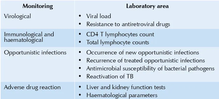
cost of treatment. Hence, laboratories are bound to play a critical role in the successful implementation of any ART programme. Various areas which need to be monitored are shown in Table 1.1: ART aims at reducing the viral load and augmenting the immune potential of the patient.
Table 1.1: Monitoring of disease progression and ART
Monitoring Laboratory area
Virological • Viral load
Resistance to antiretroviral drugs
•
Immunological and
haematological • • CD4 T lymphocytes countTotal lymphocyte counts
Opportunistic infections • Occurrence of new opportunistic infections Recurrence of treated opportunistic infections
•
Antimicrobial susceptibility of bacterial pathogens
•
Reactivation of TB
•
Adverse drug reaction • Liver and kidney function tests Haematological parameters
•
HIV-1viral load measurement has been found to be useful in monitoring treatment. It requires the establishment of a baseline plasma viral load before starting ART. The viral load in the case of successful ART becomes undetectable in four to six months of therapy. It can be measured using a variety of commercial kits. A number of home-brew assays for viral load are in use. However, these need rigorous standardization and continued quality assurance which often is not possible in a developing country setting. The assessment of viral load is a very expensive, complex and sophisticated procedure and hence it may be considered only when feasible.
Functions of laboratories
Table 1.2: Recommended tiered laboratory capabilities for diagnosis and treatment of HIV disease in resource-limited settings
Laboratory tests: Diagnosis and
monitoring Peripheral Intermediate Central
HIV antibody testing (Rapid and/or ELISA)
Yes Yes Yes
Haemoglobin Desirable Yes Yes
Total lymphocyte count (TLC) Desirable Yes Yes
Pregnancy testing Desirable Yes Yes
Basic microscopy for TB and malaria (sputum smear for TB and blood film for malaria diagnosis)
Desirable Yes Yes
Full blood count (FBC) No Yes Yes
CD4 T lymphocyte count No Yes (with flow cytometry
Liver and renal functions tests No Yes Yes
Diagnostic (including India Ink for cryptococcal meningitis), syphilis and other sexually transmitted infections.
No Yes Yes
Diagnostic tests for other major treatable HIV coinfections and AIDS-related opportunistic diseases (hepatitis B virus, hepatitis C virus serology, bacterial cultures and diagnostic procedures for cryptococcosis, toxoplasmosis and other major OIs.
Laboratory tests: Diagnosis and
monitoring Peripheral Intermediate Central
Full chemistry (including but not restricted to liver enzymes, renal function, glucose, lipids, amylase, lipase and serum electrolytes)
No No Yes
HIV viral load measurement and HIV drug resistance testing
No No Desirable
Participation in external quality assessment scheme
Yes Yes Yes
Support by WHO
HIV prevention and control programmes are being supported by WHO with globally coordinated activities. A strategic framework has been developed with the following five pillars:
Global leadership, strong partnership and advocacy.
•
Urgent, sustained country support.
•
Simplified, standardized tools for delivering ART.
•
Effective, reliable supply of medicines and diagnostics.
•
Rapid identification and reapplication of new knowledge and
•
successes.
2
Diagnosis of HIV infection
Objectives of HIV diagnosis
(1) Blood and blood products safety: This is achieved by mandatory testing of all donated blood units and blood products.
(2) Screening of donors of sperms, organs and tissues.
(3) Diagnosis of HIV infection in clinically suspected cases.
(4) Voluntary counselling and testing (VCT).
(5) Epidemiological surveillance using unlinked anonymous HIV testing. Here the result of the test cannot be linked with the identity of the person.
(6) Research and surveys.
For all individuals who are tested for HIV, the pre-test and post-test counselling should be an integral part of the HIV testing services.
Diagnosis of HIV infection can be carried out by detecting any of the following:
Antibodies to HIV
•
P24 HIV antigen
•
HIV nucleic acid (RNA/DNA)
•
The most commonly used method for the diagnosis of HIV infection is detection of anti-HIV antibodies in serum/plasma. It is economical, rapid and can be performed easily in most laboratories. HIV antibody assays are now commercially available in various formats. It is necessary to differentiate between HIV-1 and HIV-2 infections as the treatment varies for the two types. HIV-2 is intrinsically resistant to NNRTI drugs. Although some of the rapid tests can indicate the presence of anti-HIV-2 antibodies in a sample, the frequent occurrence of cross-reactions makes virus speciation on serological grounds problematic. Furthermore, some studies have reported high sensitivity (91% to 100%) and specificity (81% to 100%) for HIV-2 antibody testing by Western blot (immunoblot). On the contrary, over-estimation of the incidence of dual infections caused by HIV-1 and HIV-2 based on serological testing has been reported in India. Since the role of western blot is not conclusive in the diagnosis of HIV-2 infections, use of molecular techniques may be considered.
Figure 2.1: Laboratorymarkers during HIV-1 disease progression
0 1 3 6 1 2 3 4 5 6 7 8 9 10 11
Months Years
Time post infection
Acute Asymptomatic phase AIDS
Phase
Relative
value
0
2
4
6
8
10
HIV-antibodies HIV-antibodies
Seroconversion
CD4+ T cell count
HIV CTL
HIV CTL
Plasma viral load Plasma viral load
Detection of anti-HIV antibodies
ELISA is the most widely used technique for the detection of antibodies to HIV. HIV antibody tests have been classified as first- to fourth-generation tests based on the principle used in the assays as well as the type of antigens used.
Table 2.1: Generation of ELISAs.
Generation Antigens/antibodies Comment/characteristic
First Antigens from HIV lysates Lack of sensitivity and specificity Second Recombinant proteins and/or
synthetic peptides
Improved sensitivity and production of combined HIV-1/ HIV-2 assays
Third Use labelled antigen as conjugate
Very high sensitivity and able to detect IgM antibody; reduced the window period considerably Fourth Detection of both HIV antigen
(p24) and antibody
Further reducing the window period
Enzyme Linked Immunosorbent Assay (ELISA): ELISA requires a washer and reader and is suitable for use in laboratories where the sample load is high. Using antigens employed in the third-generation ELISA systems, several rapid tests have been developed and are widely used. The commonly employed rapid anti-HIV tests are based on the principle of immunofilteration (flow through), immunochromatography (lateral flow), dot immunoassay, or particle agglutination (e.g. gelatinorlatex). Rapid tests are visual tests that do not require the ELISA reader. These tests are available in smaller test packs and each device has procedural control system. Therefore, these are suitable for a laboratory that tests smaller sample numbers as well as for stand-alone sample. They are technically simple to perform and most of them have sensitivity and specificity comparable to ELISA. Moreover, most rapid test-kits can be stored at ambient temperature (20 °C to 25 °C) and the diagnostic performance is comparable to that of traditional ELISAs.
HIV infection during window period can be detected by demonstrating the presence of virus and virus components. PCR and detection of p24 antigen may be helpful.
Diagnosis of HIV infection in children and infants
Diagnosis of HIV infection in babies born to HIV-infected mothers cannot be established by conventional antibody tests. The presence of anti-HIV antibody in the newborn may not necessarily indicate primary infection. It may be due to the presence of passively transmitted anti-HIV antibodies from the mother to the uninfected child. These maternal antibodies may persist in the infant for as long as 18 months. Hence, virological assays such as HIV DNA-PCR or HIV RNA assays represent the gold standard for diagnosing of HIV infection in children younger than 18 months. Some DNA and RNA PCR assays support the use of dried blood spots (DBS) samples, which have considerable advantages in settings where sample transportation and storage are problematic.A definitive diagnosis of HIV-1 infection can only be made on the basis of two positive HIV-1 DNA or RNA assay results at six weeks after birth (sensitivity >98%). For infants born to HIV-1–infected mothers, it has been recommended that diagnostic testing with HIV-1 DNA or RNA assays be performed within the first 14 days of life, at one to two months of age, and again at three to six months of age. If any of these test results are positive, repeat testing is recommended to confirm the diagnosis of HIV-1 infection. However, if an infant is breastfeeding he or she remains at risk of acquiring HIV infection throughout the breastfeeding period and, therefore, negative virological test results can be assumed to reliably indicate HIV infection status only after six weeks after complete cessation of breastfeeding (Annex 9).
Alternatively, if the first PCR is negative in a non-breastfed baby, confirm with a second PCR test at six months. Definitive exclusion of HIV-1 infection is based on two negative virologicaltest results, one obtained at one month of age and one obtained at four months of age, or two negative HIV-1 antibody test results from separate specimens obtained at six months of age. For exclusion of infection, the child should have no other laboratory or clinical evidence of HIV-1 infection and should not be breastfed.
However, given the expense and complexity of nucleic acid testing, the World Health Organization strongly encourages the development of technologically simpler, less expensive assays that can be used to diagnose HIV-1 infection in early infancy. The ultra-sensitive p24 (Up24 assay) assay is gaining support as a tool for detection of HIV-1 infection in infants following mother-to-child transmission. The ultrasensitive p24 antigen assay is an ELISA and is, therefore, suited to health facilities where serological testing is routinely performed. The performance of the Up24 assay for HIV diagnosis in infants and young children has been evaluated in a number of studies in countries with different HIV subtypes yielding sensitivities and specificities ranging from 96% to 99% when compared to HIV DNA PCR testing.
By the age of 12 months most uninfected HIV-exposed children lose maternal antibodies and testing HIV antibody-positive at this age can be considered indicative of HIV infection. This should be confirmed by repeat antibody testing after the age of 18 months. If an HIV-exposed child between 9 and 18 months of age had never been breastfed or had stopped breastfeeding for at least six weeks and had a negative HIV antibody test result, then the child should be considered uninfected with no further testing required unless symptoms develop.
Specimen collection
Optimal time of specimen collection
Blood specimens can be collected at any convenient time
Correct specimen type and method of collection
Whole blood or anticoagulated blood
DBS may also be collected. However, it is
advisable to collect multiple spots for repeat assay or quality control
Adequate quantity Approximately 3−5 ml
Specimen transport and storage
Time between specimen collection and processing
Whole blood specimens should be transported
•
at ambient temperature (20 °C to 25 °C) and processed as soon as possible or within 24 hours.
If serum/plasma has been separated, it can be
•
stored in a refrigerator (2 °C to 8 °C) for a week or can be transported at 2 °C to 8 °C
For longer storage, store in a freezer at –20 °C
Criteria for rejection of sample
The following are the criteria for the sample rejection:
Hemolyzed samples
•
Samples showing turbidity
•
Specimens not stored and transported properly
•
Sample that does not carry appropriate label
•
Samples that have leaked
•
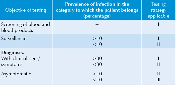
Table 2.2: UNAIDS and WHO recommendations for HIV testing
strategies according to test objective and prevalence of infection in the sample population
Objective of testing
Prevalence of infection in the category to which the patient belongs
(percentage)
Testing strategy applicable
Screening of blood and blood products
it should be tested by a confirmatory assay (e.g. Western Blot). However, if the confirmatory test fails to resolve the serodiagnosis, follow-up testing should be undertaken at four weeks, three months, six months and 12 months. After 12 months, such indeterminate results should be considered negative. However, the molecular assays (HIV-1 and HIV-2 NAT) can be used to resolve specimens giving repeated (>2 times) indeterminate results.
Reporting procedure
Report Negative – if initial/screening test shows non-reactive result.
Positive – if the sample shows reactive results concordantly by the three tests as per the algorithm.
HIV-1 positive/HIV-2 positive or dual reactive as per the algorithm should be mentioned.
In case of dual reactivity, confirmation by molecular assays may be considered.
Indeterminate – if the sample shows discordant results by the three tests. The follow-up testing is recommended as mentioned above.
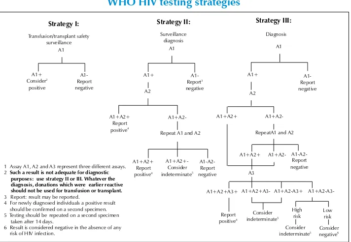
Figure 2.2: Schematic representation of the UNAIDS and WHO HIV testing strategies
Strategy I: Strategy II:
Strategy III:
1Assay A1, A2 and A3 represent three different assays. 2Such a result is not adequate for diagnostic
purposes:use strategy II or III. Whatever the diagnosis, donations which wereearlier reactive should not be used for transfusion or transplant. 3 Report: result may be reported.
4 For newly diagnosed individuals a positive result should be confirmed on a second specimen. 5Testing should berepeated on a second specimen
taken after 14 days.
6 Result is considered negative in the absence of any risk of HIV infection.
A1
Repeat A1 and A2
Selection of test kits
A wide variety of test kits are now available for HIV diagnosis. The selection of appropriate test kits/assays/reagents is critical to ensure quality in laboratory services. Every country or laboratory must, therefore, define a policy for selection, evaluation and procurement of the most appropriate kit. Selection of assays/reagents is a complex process that needs to be planned carefully. The overall performance of an assay/reagent depends upon a number of local factors. Quite often the manufacturer’s quoted sensitivity and specificity figures may not reflect the actual working figures. Therefore, selection of an assay/reagent needs to consider the testing needs of a centre and the resources available to meet those needs. Procurement systems may have a significant impact on the selection of kits. Stock control is vital, especially where continuity of supply cannot be guaranteed. Ongoing monitoring systems are essential to identify problems either with the assay/reagent or the laboratory.
The steps that are involved in the selection of a test kit/reagent for a testing laboratory are:
Sensitivity and specificity of the test: Sensitivity is the ability of (1)
an assay to identify all infected individuals (true positives). The specificity of an assay is the ability of an assay to identify uninfected individuals (true negatives). Kits/assays should have a sensitivity >99% for Rapid test/ELISA) and specificity >98%.
Ease of testing, competency of the staff performing the test and (2)
infrastructure available.
Type of sample: serum/plasma/whole blood/dried blood spot/other (3)
body fluid
Type of test controls provided: Reliable kit systems that provide (4)
internal controls are preferred. Similarly, for rapid tests, kits that provide a sample addition check are generally preferred.
Number of tests per kit. (5)
Shelf life of kit as per the kit recommendation: Kits with a longer (6)
expiry date are preferred over those that have more recent expiry dates.
Storage and handling requirements during transport and their (7)
Resource availability: Finally, the choice of a test kit/reagent would (8)
depend a great deal on the availability of financial resources, existing systems in place in the laboratory and the time scale in which results are expected to be delivered.
Time required to complete the test: If HIV results are required to (9)
be obtained within a short time and only a few samples need to be tested in a laboratory, rapid and simple HIV kits may be preferred over other assays. On the contrary, if a large number of samples are to be tested, assays such as ELISA may be preferred.
Evaluation and final selection of kit
Define specific requirements for assays/reagents for the country
Prepare a protocol for laboratory assessment
Collect all available relevant data pertaining to kits/reagents
Assess on paper each assay/reagent against specific requirements and list most suitable assays
Perform laboratory assessment of most suitable candidate kits Evaluate results
Select assay/reagent
Evaluation and procurement
Monitoring subsequent performance
Monitoring the performance of a reagent/kit is a continuous process which begins from the time of procurement until all the kits are used or reach their expiry dates. Each country should draw up a plan for periodic monitoring of reagents/kits at various testing levels in the country, much akin to those that are already existing for vaccines in many countries. It may be advisable to have periodic “post-marketing surveillance” of the kits carried out in the central laboratory which gets these kits for evaluation from various peripheral testing laboratories.
Technical support from WHO
Realizing the inadequacies in the mechanisms at the country level to procure reagents/kits, WHO has established a mechanism called AIDS Medicines and Diagnostic Services (AMDS) with the following features:
AMDS is created to expand access to quality, effective anti-retroviral
•
therapy (ART) by facilitating increased supply of drugs and diagnostics in developing countries.
AMDS will provide to manufacturers information and forecasting
•
about global demand/market.
AMDS will provide to buyers sources, process and patents on drugs
•
and diagnostics and assist them in obtaining the best prices for individual or pooled demand.
AMDS will provide technical support to countries in improving their
•
procurement mechanisms.
AMDS will assist countries, NGOs and other non-profit
•
organizations.
3
Immunological monitoring of
antiretroviral therapy:
CD4 T lymphocytes counts
What are CD4 T lymphocytes?
Cellular components of blood comprise red blood cells and white blood cells. Two populations of leucocytes constitute the latter — the granulocytes and non-granulocytes, including the lymphocytes. Surface receptors of the lymphocytes provide identity to sub-populations of lymphocytes which differentiate into unique clusters. This property gives the subtypes of lymphocytes a nomenclature of clusters of differentiation followed by the number of the unique subtype (CD1, CD2, CD3, CD4…). CD stands for cluster of differentiation; CD numbers are now used to identify cell surface antigens that can be distinguished by monoclonal antibodies. CD4 T lymphocytes (CD4+ T-cells), commonly known as T helper cells, play a vital role in maintaining the integrity of the human immune system.
Importance of CD4 T lymphocytes
A primary target of HIV is CD4 T lymphocytes which are preferentially depleted during the course of the disease. It is well recognized now that accurate and reliable enumeration of CD4 T lymphocytes is very crucial for monitoring the rate of progression to AIDS, both for initiating prophylaxis for opportunistic infections as well as monitoring the impact of antiretroviral therapy (ART).
specimens need to be tested. To obtain an absolute CD4 T lymphocytes count, two concepts (Annex 4 and 5) are utilized:
Dual-platform (DP) approach
The DP approach uses two instruments to generate absolute CD4 T lymphocytes: an FCM for generating a percentage CD4 T lymphocytes among lymphocytes and a haematological analyser to enumerate the absolute lymphocyte counts. An absolute CD4 T-cell count is derived by multiplying % CD4 T lymphocytes by the absolute lymphocyte count. Examples of these DP FCM instruments include BD FACSVantage, FACSCalibur/FASCScan/FACSort or Beckman-Coulter Epics XL/XL-MCL.
Single-platform (SP) approach
This technique enables absolute CD4 T lymphocytes counts to be derived directly without the need for a haematological analyzer, i.e., the use of volumetric counting (Partec CyFlow), microfluorometry (Guava) and, most commonly, the addition of reference fluorescent beads of a known density (eg., BD-Truecount and Coulter Flowcount) to the sample.
Other alternative methods
Flow cytometry is a widely used method for estimation of CD4 T lymphocytes. Flow cytometer and reagents are expensive and hence are a cause for concern in developing countries. For those countries and settings where the infrastructure is not available or difficult to set up for such FCM technologies, a number of alternative assays have been developed and most of them are commercially available. These assays have been evaluated against the gold standard method and reported in literature.
Total lymphocyte count
There are limitations in the use of TLC as a substitute for CD4 T lymphocytes counts. For example:
Lack of correlation with CD4 counts in asymptomatic patients
•
Variation due to intercurrent infections or drugs.
•
Microscope-based CD4 counting systems
These alternative assays are, however, fairly labour intensive and thus less appropriate for a large number of samples. Moreover, qualified personnel are required for making accurate measurements.
Microbead system (Dynal Biotech, Oslo, Norway) uses two types
•
of beads. The first bead removes monocytes from the sample and the second (CD4) estimates CD4 T lymphocytes that get stained with acridine orange to make the cell nuclei visible for counting under a fluorescent microscope. The initial cost of equipment for a fluorescence microscope is low (US$ 6000) with a running cost of US$ 3–5 per test. A modified DynaBeads system with an alternate stain for the cells can be used with a light microscope.
Evaluations of Dynal bead assay was highly comparable with the standard FCM and found to be accurate and reproducible. However, this is a manual and labour-intensive assay. To scale these up to match expanding access to ART may prove a challenge. The system could be cheaper than other alternatives and will be useful in small settings if it is backed up with flow cytometry for quality assurance.
Affordable flow cytometry
Combined use of CD4 and CD45 conjugated antibodies using Panleucogating (PLG) methodology (eg. Beckman Coulter FlowCARE CD4 Reagent) has been found to be workable and cost-effective.
Modified single platform volumetric flow cytometry
Cyflow from Partec
out based on an SP method with single antibody reagent and ten-minute incubation. The system showed good correlation with the CD4 T lymphocytes counts obtained by conventional flow cytometry. However, an experienced technician is required for accurate measurement. The CyFlow capital equipment costs approximately US$ 20 000 and the cost per test is US$2.
Guava technology
A SP system that uses CD3 antibodies to measure T lymphocytes and CD4 antibodies to estimate absolute T-cells expressing CD4. The system showed good correlation with conventional flow cytometry and is easy to operate. It uses smaller blood volume. It requires very minimal infrastructure facilities and it is easy to train technologists to perform the test. The cost per test is around US$ 2. However, the equipment costs about US$ 26 000.
The recent evaluation studies with these two modified flowcytometry systems have shown a good correlation with the standard FCM.
Selection of alternative methodology for CD4 T
lymphocytes count
The following specifications should be considered for the selection of the better technology for CD4 T lymphocytes count:
The equipment should be simple to operate, easy to maintain and
•
require minimal training.
Methodology needs minimal infrastructure laboratory facilities.
•
Methodology should have the internal QC procedures.
•
Test kits should be cost-effective and available anytime.
•
Easy access to the technical specialist/service engineer support.
•
Supplying company should be in a position to supply the critical parts
•
of the equipment with short turnaround time.
Utility of CD4 T lymphocytes count in monitoring
According to the WHO recommendation, HIV-infected adults should start ARV therapy when infection has been confirmed and one of the following conditions is present (Table 3.1):
Table 3.1: WHO recommendations for initiating ARV therapy in
HIV-infected adults and adolescents according to CD4 T lymphocytes counts and total lymphocyte counts*
WHO Stage CD4 T lymphocytes counts Total lymphocyte counts
IV Irrespective Irrespective
III <350 cells/uL Irrespective
II <200 cells/uL <1200 cells/uL
I <200 cells/uL Not applicable
* It should be noted that in HIV-related symptoms (Stage II, III), a total lymphocyte count of 1200 cells/ul can be substituted for the CD4 T lymphocytes count when the latter is not available. However, asymptomatic HIV-infected patients (Stage I) need not be treated because the total lymphocyte count correlates poorly with the CD4 T lymphocytes count in asymptomatic patients.
Sample guidelines
Blood samples are collected by venipuncture into EDTA (preferably K2) containing tubes, mixed well and processed within the time-frame (the sample stability varies depending upon the equipment and the type of kit). For example, the samples should be processed within 48 hours (stored between 20 °C–25 °C) after the collection for the FACS Count. Blood samples should preferably be collected within a fixed time in the day (morning or evening) so as to avoid diurnal variations. Blood sample that is not suitable should be rejected. For example, blood sample that cannot be performed within the time-frame, or if blood sample is hemolyzed or frozen or clotted or is without proper labelling. Data on storage temperature for Partec instrument needs to be incorporated.
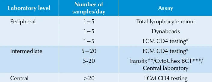
Suggestions for testing at different levels
Table 3.2: Guidelines on appropriate assay for different clinical laboratory levels
Laboratory level Number of
samples/day Assay
Peripheral 1−5 Total lymphocyte count
1−5 Dynabeads
1−5 FCM CD4 testing*
Intermediate 5−20 FCM CD4 testing*
5-20 Transfix**/CytoChex BCT***/ Central laboratory
Central >20 FCM CD4 testing
* Modified or affordable flowcytometry methodologies could be considered after the required validation.
** Transfix is a whole blood stabilizing reagent from United Kingdom Quality Assessment Service (UKNEQAS). When Transfix is added to fresh blood, the sample retains its integrity for almost one week. The use of Transfix is therefore suitable to increase the catchment area of central laboratories. (***CytoChex BCT is also a blood stabilizing reagent used to delay blood sample degradation. It is a product manufactured by Streck, Inc. USA).
Gold standard for CD4 T lymphocytes count:
Flow cytometry
Principle
Samples
Cells to be measured must be suspended in a liquid. So, ideally, this is simple for whole blood. Anticoagulant blood is stained with appropriate monoclonal antibody that binds to the specific antigens, i.e. CD4 that are to be measured. Normally, the monoclonal antibody is directly conjugated with fluorochrome.
Advantages and disadvantages
Table 3.3: Advantages and disadvantages of using flow cytometer
for CD4 T lymphocytes testing
Advantages Disadvantages
Reference method Expensive instrument
High reliability
High accuracy, precision and reproducibility
Expensive maintenance
Handling large number of samples Need well-trained technician
Cost
At present, the cost of CD4 testing varies from country to country and ranges between US$ 12−US$ 30. However, the recent development of a simplified
panleucogating method together with the use of generic monoclonal antibody reagents drastically cut the cost of CD4 test by more than one-fourth.
Quality system
Reporting result
All data are reported in terms of percentages and absolute counts of CD4 T lymphocytes, or %CD4 T. It should be noted that the absolute counts are used in adult HIV-infected patients, whereas only the percentages are used in paediatric HIV-infected patients. In the report, the mean and reference of both percentage and absolute CD4 T lymphocytes (normal ranges) should also be given. Each clinical laboratory should validate normal ranges.
Total lymphocyte counts
Principle
Total lymphocyte counts are easily obtained by automated haematological analyser on aspirated blood samples appropriately diluted with a solution (e.g., acid or detergent) that lyses the red blood cells but preserves leukocyte integrity. A typical automated haematological analyzer performs white blood cell counts by either impedance or light-scattering technology or both. Cells in suspension are made to flow through a small orifice across which an electric current is flowing as in impedance technology. As a cell enters the orifice, the flow current is reduced. Electronic circuits detect the decrease in current and thus the presence of the cell. In a light-scattering haematological analyzer, cells flow through a light beam rather than through an orifice. Different cell types intercepted by light will show different patterns based on the size and shape of the cells. In this way, the device can count the number of cells per second of flowing cells through the orifice or light beam, and since the volume flow rate can be measured one can thus determine the number of white blood cells per ul and or total lymphocyte count per ul of the blood sample.
Samples
Advantages and disadvantages
Table 3.4: Advantages and disadvantages of using
total lymphocyte count for ARV therapy
Advantages Disadvantages
Inexpensive unit cost High variation (CV up to 15%)
Simple and commonly available test No EQAS
Less complicated instruments compared to FCM
Fault results due to nucleated red blood cells, platelet aggregation and nonlysed red blood cells.
Cost
The cost of total lymphocyte counts is cheap compared to CD4 testing. It varies from country to country but is around US$ 1−US$ 2.
Quality system
Currently no external quality assessment scheme on white blood cell counting is available. Only internal quality controls supplied by each manufacturer are recommended. It is important that accurate daily internal quality control and proficiency tests, if applicable, are employed to ensure reliability and value of the total lymphocyte count data.
Reporting result
In haematology cell counter the total lymphocyte count is calculated from direct measurement of lymphocyte count, and expressed as cells x 103/ul. Normal
value for total lymphocyte count is 2.6 x 10³/ul (range 1.0-4.9 x 10³/ul). Normal
ranges should be validated by each clinical laboratory.
4
Virological monitoring of ART
Viral load testing
HIV-1viral load measurement is useful for monitoring treatment. A baseline plasma viral load is established before starting ART. Periodic monitoring is essential. It is predicted that with successful therapy a fall of 1.5 to 2 log in plasma viral load occurs within 4-6 weeks. With successful ART, it should become undetectable in four to six months of therapy.
Viral load is measured using a variety of commercial kits based on nucleic acid testing (NAT) and non-NAT (Annex 6 and 7). The Roche Amplicor HIV-1 monitor test, version 1.5, is used widely. The assay uses gag specific primers for the highly conserved region. The lower limit of detection with the standard assay kit is 400 RNA copies/ml and the upper limit is 750 000 RNA copies/ml. The Amplicor ultra-sensitive kit detects down to 50 copies/ml of plasma.
A real time PCR is being tested in some laboratories. The cheaper cost of the test and the advantage of avoiding batch testing are some of the plus points. The test uses primers and probe sets specific for the LTR region of the HIV genome, which is conserved across subtypes. The primers need to be selected carefully as some subtypes may be estimated in relatively lower copy numbers with some of the primers. The lower limit of detection is in the range 50 IU/ ml. The virological assays are useful tools in monitoring for the emergence of resistance in HIV against antiretroviral drugs.
are based on HIV 1 subtype B—and their appropriateness for use in countries where subtype C is not prevalent needs to be considered before choosing an assay.
HIV drug resistance assays
The increase in the use of ART is expected to lead to the emergence of drug-resistant mutants of HIV-1. This has been the experience from other parts of the world. The inherent mutability of the reverse transcriptase (RT) gene of HIV allows for drug resistance to emerge under selection pressure. The protease gene also undergoes mutations, which manifest as a failure to respond to protease inhibitors.
A survey of drug resistance of indigenous HIV strains is essential to ascertain the usefulness of the antiretrovirals, especially in public health programmes. Drug resistance should be suspected if the plasma viral load does not show a greater than 1 log fall within eight weeks of therapy.
WHO has prioritized the prevention, surveillance and monitoring of HIV drug resistance and has developed an approach to prevent the emergence and transmission of HIV drug-resistant virus. HIV drug resistance surveys are targeting untreated individuals, particularly patients who had recently become HIV-infected. Public health action would be essential if such a survey indicated HIV drug resistance to be >15%.
The examination of plasma RNA for resistant HIV strains indicates circulating mutants, if any, including emergent polymorphisms. The proviral DNA testing reveals mutants that have emerged in individuals about a year back and have slowly replaced the native wild type derived provirus.
Two types of antiretroviral drug resistance assays that exist today are: Phenotypic assays and Genotypic assays. These assays can be carried out only in laboratories with recommended biosafety measures (minimum biosafety level 2) in place.
Phenotypic assays
the presence of varying concentrations of the drug is measured. Results are expressed as concentrations of drug required to inhibit 50% of growth (IC50) in comparison to a wild type control strain.
Genotypic assays
The genotypic assays detect changes in the sequence of the relevant HIV1 gene. This measures resistance by detecting mutations in the HIV1 genome that leads to one or more specific amino acid substitutions in the HIV1 reverse transcriptase or protease enzymes. These specific changes cause drug resistance. Viral RNA from plasma or proviral DNA can be used for testing. The amplified products are then sequenced. The sequences are analysed using special software such as Stanford University Online software (http://hivdb.stanford.edu/), Los Alamos National Laboratories HIV Sequence Database (hiv-web.lanl.gov), http://www. hivresistanceweb.com/request/pda.shtml, etc.. Single genome amplification (SGA) may provide more accurate genotyping characteristics than conventional genotyping assay, where population-based sequencing is done. Importantly, the EQAS programme samples should be compatible to the locally circulating strains.
Table 4.1: Advantages and disadvantages of genotypic assays
over phenotypic assays
Category Genotypic assay Phenotypic assay
Advantages • Less expensive than phenotypic assays Short turnaround time
•
(<1 week)
May detect presence of
•
resistance mutations before they have resulted in phenotypic resistance
Interpretation similar to
•
susceptibility testing of bacteria
Assesses the total effect
•
of mutational changes Good reproducibility
•
The threshold to define
•
susceptibility is arbitrary
Disadvantages • Interpretation requires knowledge of mutational changes
May show discrepancy
•
with phenotypic assay
Very expensive to set up
•
and run
Turnaround time is more
•
(3 weeks) than that of genotypic assays. Slower to show
•
A third approach to resistance testing is the ”virtual“ phenotype. This assay is really a genotype resistance that is interpreted with the aid of a large database of samples with paired genotypic and phenotypic data. Viruses with genotypes that are similar to the patient’s virus are identified by searching the database, and the average IC50 of these matching viruses is calculated. This information is then used to estimate the likely phenotype of the patient’s virus. The major advantage of this approach is that it reduces complex genotypic data to simple phenotypic categories based on a rational, data-driven analysis of similar genotypes. The major disadvantage of this approach is that the confidence placed in the result depends on the number of matches, and on picking the right codons to incorporate into the database search. Correlation between actual and virtual phenotype will be weaker for newer drugs or in cases where there are fewer matches due to unusual genotypes. However, it needs more validation studies.
Monitoring of opportunistic infections in
HIV disease
Morbidity and mortality in HIV disease is due to the occurrence of life-threatening opportunistic infections (OIs) during the natural course of the disease. These are the direct consequence of a decline in CD4 count. A wide variety of opportunistic infections (Table 5.1) are encountered in patients with AIDS which are caused by various microorganisms. Very often these represent reactivation of organisms that have been dormant in the host for several years. The incidence of these diseases increases as the patient’s CD4 count declines. The pattern/repertoire of opportunistic infections may vary in different geographical areas. The knowledge of important OIs specific for particular areas/ countries is useful for correct diagnosis and management of OIs.
Table 5.1:Common opportunistic infections in HIV/AIDS
Disease Pathogen Infection type
Bacterial
Tuberculosis and non-tuberculous infections
Mycobacterium tuberculosis
Mycobacterium avium
complex
Pulmonary or meningeal or other extra-pulmonary or systemic
Salmonellosis Salmonella sp. Typhoid fever or diarrhoea
Disease Pathogen Infection type Fungal
Candidiasis Candida albicans Oral thrush or vulvovaginitis
Cryptococcosis Cryptococcus neoformans Meningoencephalitis, or pulmonary or systemic disease
Aspergillosis Aspergillus sp. Pulmonary disease or sinusitis
Histoplasmosis Histoplasma capsulatum Pulmonary disease or disseminated
Penicilliosis Penicilium marneffei Pneumonitis or disseminated disease
Parasitic
Pneumocystis jiroveci
pneumonia
Pneumocystis jiroveci Pneumonia
Toxoplasmosis Toxoplasma gondii Encephalitis
Cryptosporidiosis Cryptosporidium sp. (mainly C.hominis and C. parvum)
Diarrhoea
Isosporosis Isospora belli Diarrhoea
Viral
CMV infection Cytomegalovirus (CMV) Retinitis or encephalitis or esophagitis or colitis
Herpes Herpes simplex virus (HSV), types 1 & 2
Recurrent oral or genital ulceration or bronchitis or pneumonitis or esophagitis
Herpes zoster Human herpes virus type 3 Vesicular skin lesions
Warts and carcinoma in situ
Human papilloma virus (HPV) Genital lesions and cervical carcinoma
PML JC virus Encephalitis
Utility of microbiological monitoring
more than one opportunistic infection. Patients who had more than one opportunistic infection were 2.6 times more likely to die than those who did not have an opportunistic infection.
Diagnosis of opportunistic infections
Infrastructure facilities should be established based on the level of the set-up required (Table 5.2). Although intermediate laboratories have sufficient facilities, further identifications and confirmations may be carried out by the central laboratory. The flow of specimens should be worked out from the lower to the higher level, and the flow of technical and scientific information and QA/QC procedures should be from the higher to the lower level.
Table 5.2:Suggested laboratory diagnostic procedures to be
performed at different levels
Disease Diagnosis at different levels
Peripheral Intermediate Central
Tuberculosis and non-tuberculous infections
AFB smear AFB smear, culture AFB smear, culture and sensitivity, PCR
Salmonellosis − Culture and
sensitivity
Culture and sensitivity Bacterial pneumonia − Culture and
sensitivity
Culture and sensitivity Candidiasis Gram’s staining Gram’s staining Culture and
sensitivity Cryptococcosis Negative staining,
LA Test
Culture Culture
Aspergillosis 10% KOH mount Culture Culture
Histoplasmosis 10% KOH mount Culture Culture
Penicilliosis 10% KOH mount Culture Culture
PCP − − IFT
Toxoplasmosis LA test LA test IFT
Cryptosporidiosis − Wet mount,
Modified acid-fast staining
Modified acid-fast staining, IFT
Isosporosis − Wet mount,
Modified acid-fast
Disease Diagnosis at different levels
Peripheral Intermediate Central
CMV infection − − IFT, cell culture,
PCR
Herpes − − Cell culture, PCR,
IFT
PML − − PCR for JC virus
HPV VIA VIA/PAP smear PAP smear/biopsy
and PCR
IFT: immunoflourescent technique, LA: latex agglutination, VIA : visual inspection on application of acetic acid.
Role of the central laboratory in sharing data
5
Laboratory monitoring of
side-effects of ART
Toxicity is related to the inability to tolerate the side-effects of medications and to significant organ dysfunction that may result on account of it.All the available antiretroviral agents have potential toxicities (Table 5.1). Careful monitoring of patients by laboratory investigations play a major role in better clinical management. Toxicity should be monitored clinically based on patient reports and physical examination, supplemented by a limited number of laboratory tests depending on the symptoms that arise and the specific combination regimen that is used.
Table 5.1:Adverse drug reactions of individual
antiretroviral agents and laboratory tests
Agent Affects quality of life Serious, may require intervention Serious and life-threatening Laboratory tests
NsRTI (nucleoside reverse transcriptase inhibitor)
Didanosine
Liver function tests, amylase, lipase, lactate
Lamivudine (3TC)
Nausea, vomiting Pancreatitis in children, increased
Agent Affects quality of life Serious, may require intervention Serious and life-threatening Laboratory tests
Emtricitabine Nausea, vomiting, diarrhoea
Skin rashes, neuropathy, paresthesis
Lactic acidosis Lactate
NtRTI (nucleotide reverse transcriptase inhibitor)
Tenofovir (TDF)
Diarrhoea, headache, asthenia
Renal failure Lactic acidosis Renal failure tests
NNRTI (non-nucleoside reverse transcriptase inhibitor)
Delavirdine (DLV)
Nausea, vomiting Skin rash, hepatotoxicity psychosis, skin rash, hepatotoxicity, dyslipidaemia
Liver function tests, lipid profile
PI (protease inhibitor)
Amprenavir
Liver function tests, lipid profile
Indinavir (IDV)
Requires adequate daily fluid intake to prevent crystalluria;
Liver function tests, lipid profile, renal function tests,
Dyslipidaemia Liver function tests, lipid profile
Nelfinavir (NFV)
Diarrhoea Diarrhoea may lead to severe dehydration, dyslipidaemia
Liver function tests, lipid profile
Dyslipidaemia Liver function tests, lipid profile
Saquinavir (SQV)
Nausea, vomiting, abdominal pain
Agent Affects quality of life Serious, may require intervention Serious and life-threatening Laboratory tests
Liver function tests, lipid profile
Dyslipidaemia Liver function tests, lipid profile
Monitoring of adverse reactions
Monitoring toxicities of the drug can be done clinically based on patient reporting and physical examination. However, inclusion of limited laboratory investigations in the ARV monitoring will determine the severity of the toxicity and this will help physicians to change the dose and specific drug combination in the regimen. When the toxicity is related to an identifiable drug in the regimen, the offending drug can be replaced with another drug that does not have the same side-effect.
Monitoring of ART
The different levels of testing capabilities and types of specimen to be collected for monitoring of ART toxicity are described in Table 5.2.
Table 5.2: Suggested testing capabilities at different levels
Laboratory tests Level 1: Peripheral Level 2: Intermediate Level 3: Central
Haemoglobin Haemoglobinometer1 Haematology analyser2 Haematology
analyser Total and differential
cell count
Microscopic - manual Haematology analyser Haematology analyser Complete blood
count
Microscopic - manual Haematology analyser Haematology analyser Liver and renal
function markers
− Medium throughput autoanalyser3
− Medium throughput autoanalyser and ion selective electrode4
Patients with hepatitis B (HBV) and C (HCV) viruses should be
•
monitored more closely for liver toxicity. HBV and HCV screening and testing may be undertaken in the particular community (eg. injecting drug users or IDUs) where prevalence is high.
The critical values/panic values (abnormal values) of the laboratory
•
results should be reported only after consultations with the treating physician.
Role of central laboratories
6
Tuberculosis in HIV/AIDS
The WHO South-East Asia Region bears over a third of the global tuberculosis (TB) burden and ranks second after sub-Saharan Africa in the estimated number of people living with HIV/AIDS. Each year nearly three million cases of TB and 500 000 TB deaths are estimated to occur in the Region. Of the estimated six million adults living with HIV in the Region, about 60%-70% are likely to be infected with Mycobacterium tuberculosis (M. tuberculosis). The extent to which HIV will contribute to the TB epidemic depends on the degree of overlap between the population groups infected with TB and those with HIV.
Pulmonary TB accounts for 60%-80% and extrapulmonary TB for 20%-40% of the total tuberculosis manifestations in HIV patients. Before the AIDS pandemic, non-tuberculosis mycobacteria (NTM) rarely caused serious illness, even in compromised individuals. The prolonged immuno-suppression of the cell-medicated immune system caused by HIV provides the opportunity for these relatively avirulent organisms to cause disease.
Impact of TB on HIV
Drug-resistant TB
When a strain of M. tuberculosis becomes resistant to two or more “first-line” antibiotic drugs (i.e. isoniazid and rifampicin), it is called multidrug-resistant TB or MDR-TB. This can take up to two years to be treated, with drugs that are a hundredfold more expensive than those used in first-line treatment. When the bacteria becomes resistant to three or more “second-line” antibiotics as well (i.e. MDR-TB plus resistant to any of the fluoroquinolones and any one of the injectable second-line anti-TB drugs such as amikacin, kanamycin or capreomycin), it is classified as extremely drug-resistant TB or XDR-TB. Drug resistance usually arises when TB patients do not or cannot take their medicine as prescribed and drug-resistant mutations of the bacteria are allowed to replicate. People can also acquire MDR- and XDR-TB from others.
According to the Fourth WHO Report on Drug Resistance in Tuberculosis (2008), MDR TB has been shown to be almost twice as common in TB patients living with HIV compared to TB patients without HIV. MDR-TB and XDR-TB are highly lethal in HIV-positive people, whose weakened immune systems render them unlikely to fight off the infection; studies show case fatality rates going over 90%. HIV-infected TB cases are more likely to be smear-negative, and delayed diagnosis of drug resistance has contributed to high death rates in people living with HIV.
Newer methods for rapid diagnosis of infection and drug susceptibility testing are now available to ease this problem. Acquired rifamycin resistance has been associated with HIV infection and anti-TB drug malabsorption has been documented in patient cohorts in settings of high HIV prevalence. Drug-resistant TB is, therefore, a major threat to the effectiveness of both TB treatment and anti-retroviral treatment programmes.
Laboratory diagnosis of TB
Types of specimen
Sputum is the specimen of choice for diagnosis of pulmonary TB. Two specimens of sputum (one on-the-spot and one after an overnight collection) are to be examined over a period of two days. Specimens should be collected in sterile universal containers, and should have a fixed label for noting patient’s details on the side. All forms of TB other than pulmonary TB are paucibacillary in nature. Depending upon the form of disease manifestation, several specimens such as sputum and/or gastric lavage, bronchoalveolar lavage(BAL), lymph nodes and other biopsy specimens, pus, ascitic fluid, and pleural and cerebrospinal fluid should be examined. If delay is anticipated, biopsy specimens may be collected in a suitable transport medium for sending them to the laboratory (to retain the viability of the organisms).
Techniques for detection of infection and drug
susceptibility testing
The following are some of the commonly used laboratory tests for the diagnosis of TB and drug susceptibility testing (DST).
1.
Sputum smear microscopy
Microscopy of sputum is of great value in the detection of open or infectious cases of TB, and is still the front-line tool for diagnosis of active TB. The establishment of a good sputum microscopy service is of prime importance in developing countries for the detection and treatment of open cases. Since the test can help determine if a person is infectious, it can also be used to monitor an active TB patient’s response to treatment.
Smears are stained by the Ziehl-Neelsen (ZN) method, or by one of its various modifications. Grading of the positive smears gives a broad indication of the severity of the disease and the response to therapy. Although this is the simplest and cheapest laboratory test for the diagnosis of active TB, it has a few major shortcomings.
Limitations
The sensitivity of the AFB smear test is known to be poor, varying
•
As the test is based on sputum, it has particular difficulty in detecting
•
extrapulmonary TB which occurs frequently in HIV-infected individuals.
The test will also identify certain types of bacteria that are not
•
M. tuberculosis (e.g. NTM, Nocardia, etc.) and so cannot always distinguish between TB and other infections.
Differentiation between M. tuberculosis and NTM
The identification of strains isolated as belonging to M. tuberculosis complex or NTM can be ascertained by performing a few simple tests, i.e. susceptibility to p-nitrobenzoic acid (PNB), niacin production test, catalase activity requirement and the morphological appearance. Important differences between M. tuberculosis and NTM are shown in Table 6.1.
Table 6.1: Differentiation between M. tuberculosis and NTM
Characteristic M. tuberculosis NTM
Growth rate Slow grower Slow/rapid grower
Temperature 37 °C 25–45 °C
Colony morphology Dry, rough Dry
Colony on solid media Eugonic Dysgonic
Colour of colony Buff Yellow, orange or creamy
Emulsification Difficult Easy
Cord formation + –
Niacin test + −
Nitrate reduction test + +/−
Growth on p-nitrobenzoic acid (500 ug/ml)
− +
2.
Sputum Culture
by modified Petroff’s method, where sputum is decontaminated with 4% NaOH and then inoculated on to Lowenstein-Jensen slopes. Alternatively, sputum specimens can be decontaminated and transported in 1% CPC (useful where specimens may take several days to reach the laboratory). The incubated cultures are examined once a week for eight consecutive weeks or until they become positive or contaminated. However, the usefulness of this technique is limited by several factors.
Limitations
Conventional culture methods are very slow (Lowenstein-Jensen
•
cultures take 20-56 days for diagnosis and 4-6 weeks after initial culture for DST; liquid cultures in 7H11 medium take 17-21 days for diagnosis and 3-6 weeks for DST).
The sensitivity of culture is limited by the need to have at least 1000
•
bacilli/ml present in the sample to be cultured. HIV positive patients and children have difficulty in producing sputum, and sputum culture will not detect extrapulmonary forms of TB frequently found in HIV-positive patients.
Culture-based tests are difficult to implement in the field. They require
•
dedicated facilities and staff, with specific requirements for training, quality assurance, biosafety and equipment.
3.
Culture-based rapid methods for detection of M. tuberculosis
Some more rapid culture methods have been developed (e.g. BACTEC-MGIT) based on liquid culture media that are commercially available, though most of them are difficult to implement in the field due to the complexity of the technique or the required equipment. There are also some emerging simplified culture techniques (e.g. MODS) that can reduce time to diagnosis or DST that seem more appropriate for use in resource-limited settings.
(a) Rapid liquid TB culture medium: BACTEC MGIT 960 Automated (Becton Dickinson, US)
![Figure 1: A global view of HIV infection 33 million people [30–36 million] living with HIV, 2007](https://thumb-ap.123doks.com/thumbv2/123dok/2586563.1660309/11.499.73.429.434.618/figure-global-view-infection-million-people-million-living.webp)