Clinical and Cerebrospinal Fluid Abnormalities as Diagnostic Tools of
Tuberculous Meningitis
Fiona Lestari,1 Sofiati Dian,2 Ida Parwati3
1Faculty of Medicine Universitas Padjadjaran, 2Department of Neurology Faculty of Medicine,
Universitas Padjadjaran/Dr. Hasan Sadikin General Hospital,Bandung, 3Department of Clinical Pathology Faculty of Medicine Universitas Padjadjaran/Dr. Hasan Sadikin General
Hospital,Bandung
Abstract
Background: Tuberculous meningitis (TBM) is the most severe form of extrapulmonary tuberculous (TB) disease and remains difficult to diagnose. The aim of the study was to determine the diagnostic value of clinical and laboratory findings of cerebrospinal fluid (CSF) examinations for diagnosing TBM using bacterial culture result as the gold standard.
Methods: A prospective cross sectional study was carried out to 121 medical records of hospitalized TBM
patients in neurological ward at Dr. Hasan Sadikin General Hospital Bandung, from 1 January 2009–31 May 2013. The inclusion criteria were medical records consisted of clinical manisfestations and laboratory findings. The clinical manisfestations were headache and nuchal rigidity, whereas the laboratory findings were CSF chemical analysis (protein, glucose, and cells) and CSF microbiological culture. Validity such as sensitivity, specificity, positive predictive value (PPV), negative predictive value (NPV) for clinical and laboratory findings were calculated, using bacterial culture result as the gold standard.
Results: The most clinical findings of TBM was nuchal rigidity and it had the highest sensitivity value, but
the lowest spesificity value. Decreased of CSF glucose had the highest sensitivity value compared to other laboratory findings, but the value was low.
Conclusions: The clinical manisfestations and the laboratory findings are not sensitive and specific enough
for diagnosing TBM. [AMJ.2016;3(1):132–6]
Keywords: Cerebrospinal fluid, clinical manisfestations, diagnostic tools, laboratory findings, tuberculous
meningitis
Correspondence: Fiona Lestari, Faculty of Medicine, Universitas Padjadjaran, Jalan Raya Bandung-Sumedang Km.21,
Jatinangor, Sumedang, Indonesia, Phone: +62 81931207624 Email: [email protected]
Introduction
Tuberculous (TB) is one of the major health
problems in the world, especially in developing
countries.1,2 Manifestations of TB can be
pulmonary and or extrapulmonary, which 20.4% cases are extra-pulmonary TB.3,4 Based
on data from Centers for Disease Control and
Prevention (CDC) in 2011, it was indicated that 5.7% extrapulmonary TB involved the
Central Nervous System (CNS).4,5 The most severe manifestation of CNS TB is Tuberculous
Meningitis (TBM) which causes high mortality
in children and adult.5-8 The mortality rate of TBM in Bandung, the capital city of West Java,
Indonesia, is 50% in the first week of admission to the hospital and increases to 67% after
one month treatment in the hospital.9 Early
diagnosis and accurate treatment are promptly needed in order to improve the outcomes.8,10,11
Standardized diagnostic criteria for TBM
have not been established, because the
clinical manifestations of TBM are not specific,
especially in the early stages of disease.12 Patients usually come to the hospital after having headache, fever, nuchal rigidity, irritability, vomiting or even after having many
neurologic symptoms and signs within a few
days.9,12 Many patients come with history of typical systemic symptoms of TB infection,
such as cough, lethargy, weight loss, and night sweating that might be suggestive of TB, but also non-specific.12 Lumbar puncture is the
in most patients with TBM shows clear
appearance, increased protein, decreased glucose concentration (a CSF glucose to plasma
ratio or absolute value) and pleocytosis with
lymphocyte predominance.12
The aim of the study was to analyze the sensitivity and specificity of TBM clinical manifestations and cerebrospinal fluid
abnormalities compared to bacterial culture result.
Methods
A restrospective cross sectional study was
carried out to medical records of TBM patients
in neurological ward at Dr. Hasan Sadikin
General Hospital Bandung, top referral hospital for West Java Province, Indonesia
from 1 January 2009 to 31 May 2013.
The inclusion criteria in this study
were medical records of hospitalized TBM
patients, consisted of clinical manisfestations
and laboratory findings. The clinical manisfestations were headache and nuchal rigidity, whereas the laboratory findings were
CSF chemical analysis (protein, glucose, and
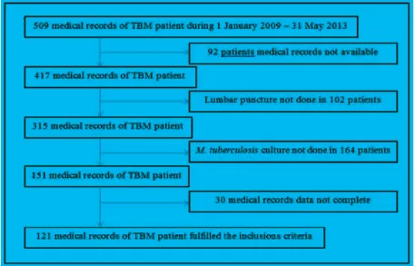
Figure 1 The Inclusion Criteria among 509 TBM Patients
cells), CSF microbiological culture. From 509
available medical records, only 121 medical
records which met the inclusion criteria (Figure
1). The operational variables in this study
were defined as nuchal rigidity defined by a resistance to flexion of the neck due to muscle
spasm of the extensor muscles; increased CSF
protein defined by positive in concentration >100 mg/dL; decreased CSF glucose defined
by positive in CSF to plasma glucose ratio
of <50%; CSF pleocytosis with lymphocytic predominance defined by positive in CSF cells count 10 500 /µL and lymphocyte >50%; CSF abnormalities defined by positive for all three CSF findings in increased CSF protein,
decreased CSF glucose and CSF pleocytosis
with lymphocytic predominance.
Sensitivity, specificity, positive predictive value (PPV) and negative predictive value (NPV) were calculated for each variable using
bacterial culture result as the gold standard.
All of the clinical data were entered and
calculated using computer. Prior to this study,
ethical approval was obtained from the Health Research Ethics Committee of Dr. Hasan
Universitas Padjadjaran, Bandung, Indonesia
(No.LB.02.01/C02/9324/VIII/2013).
Results
This study discovered that from 121 TBM cases, most of the patients had nuchal rigidity and
headache. From the laboratory findings, the
highest percentage of laboratory abnormalitiy
was the decrease of the glucose level in CSF (Table 1). Moreover, only 28.93% had positive
bacterial culture.
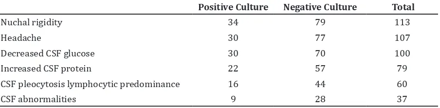
Among 6 variables identified and measured,
the symptom of nuchal rigidity had the highest positive culture result, compared to other variables.
This study discovered that nuchal rigidity
was the highest sensitivity among 6 variables, but the lowest specificity value. This study revealed that CSF abnormalities was the best variable which incorrectly identified
the negative cultural result. All variables in
this study showed low percentages for PPV, on the other hand the NPV showed higher
percentages (Table 3).
Discussion
In this study, nuchal rigidity had the highest sensitivity and CSF abnormalities had the
highest specificity among 6 analyzed variables. Among 121 patients, 93.39% patients had positive sign of nuchal rigidity and 88.43%
patients complained of headache. In previous
TBM study, it was reported on 77.2% patients with nuchal rigidity and 67% patients with
headache.8 There was an increase number of nuchal rigidity sign and headache in TBM patients. According to Rock et al.7, adult TBM
patients normally come with classic signs
of meningitis such as fever, headache, and meningismus/nuchal rigidity.7
This study revealed that sensitivity for
all of the variables was quite high but less specificity. Diagnosing TBM persistence is still difficult.13 Sensitivity of nuchal rigidity
in this study was 97% positive when cultural result was positive either. Nevertheless, specificity for nuchal rigidity was very low as 8%. It described that nuchal rigidity was not specific to identify the positive culture of TBM. Ideally, the greater sensitivity and specificity
Table 1 Clinical Manisfestations and Laboratory Findings
Total n (%)
Clinical Manifestation
Nuchal rigidity 113 (93.39)
Headache 107 (88.43)
Laboratory Findings
Decreased of CSF* glucose 100 (82.64)
Increased of CSF protein 79 (65.29)
CSF pleocytosis lymphocytic predominance 60 (49.59)
CSF abnormalities 37 (30.58)
Positive bacteria in CSF culture 35 (28.93)
Note: *CSF= cerebrospinal fluid
Table 2 Clinical Manifestations and Laboratory Findings according to Bacterial Culture
Positive Culture Negative Culture Total
Nuchal rigidity 34 79 113
Headache 30 77 107
Decreased CSF glucose 30 70 100
Increased CSF protein 22 57 79
CSF pleocytosis lymphocytic predominance 16 44 60
of test will make a better diagnostic tool from
identifying a disease. A prospective cross
sectional study which comparing signs of meningeal inflammation (nuchal rigidity, head
jolt accentuation of headache, Kernig’s sign
and Brudzinski’s sign) to the reference CSF white cell count >5 cells as the gold standard
concluded that physical signs of meningeal
inflammation do not accurately discriminate between patients with meningitis from those without it accurately regarding the poor
accuracy.11 Moreover, headache and nuchal rigidity in meningitis caused by M. tuberculosis and in meningitis caused by other etiological factors cannot be differentiated.
Laboratory findings of CSF examinations
revealed that CSF glucose have the highest
sensitivity value but the lowest specificity
value. A prospective cross-sectional study in the Philipines5, using culture or acid fast staining or basal meningeal enhancement
on computerized axial tomography (CT) head contrast as gold standard for analyzing the laboratory findings showed similar
results.5 This study revealed that 35 patients have positive result for M. tuberculosis.
The culture results explained that 28.93% patients have definite diagnosis of TBM, and the remaining patients are categorized as
probable or possible diagnosis TBM based
on the scoring systems. However, the absence of mycobacterial findings in culture result
does not exclude the patients from diagnosis of TBM.12 One study in Philipine5 reported
that among 68 patients who were diagnosed with TBM, 5.9% culture positive or acid fast staining were found. Another study by Chaidir
et al.8 discovered that 36% TBM patients have positive culture. Study in Shanghai13 reported
that 12% of 25 patients TBM have positive culture. The CDC informed that culture was
used as the gold standard for laboratory
confirmation of TB disease.14 Culture result
positive established that patients have positive TBM infections. Ideally, sensitivity of a good
culture media is 100% which it will grow the etiologic factor in whole TBM infections cases.
Nonetheless, culture is an imperfect gold standard. Literature studies informed that M.
tuberculosis culture has low sensitivity. It is limited because of the low concentration of
bacilli in CSF, characteristic of mycobacterial
itself with the need of high enrichment media,
or because the patients have already taken the anti tuberculosis drugs before the lumbar puncture done.5,13,15,16 The chances of positive diagnosis can be increased by doing more lumbar punctures.12
There are some limitations of this study. First, acid fast staining and Polymerase Chain
Reaction (PCR) method were not used to
identify bacteria in CSF. Polymerase Chain
Reaction was not routinely done because it is expensive. Second, some medical records were not written completely enough and available to be analyzed in this study. Third, there is
no separation calculation for the Human
Immunodeficiency Virus (HIV)-co infection patient, which it possibly affects the clinical presentation and laboratory findings in TBM
patients.
The ideal diagnostic tests which are validated, rapid, sensitive, and specific
are absolutely needed so that appropriate and accurate therapy can be started early, toxicities of unnecessary treatment can be avoided, morbidity and mortality prevalences
can be lowered. In conclusion, from clinical findings and laboratory examinations, we found that the sensitivity was quite high but lack for specificity. Combination of all CSF examinations abnormalities showed higher specificity, but less sensitivity. Culture result has low sensitivity.
Table 3 Sensitivity, Specificity, PPV and NPV of Clinical Manifestations and Laboratory Findings.
Sensitivity (%) Specificity (%) PPV* (%) NPV** (%)
Nuchal rigidity 97 8 30 88
Headache 86 10 28 64
Decreased CSF glucose 86 19 30 76
Increased CSF protein 63 34 28 69
CSF pleocytosis lymphocytic
predominance 46 49 27 69
CSF abnormalities 26 67 24 69
References
1. WHO. Global tuberculosis report 2012.
Geneva: World Health Organization, 2012.
2. Tai MLS. Tuberculous meningitis: diagnostic and radiological features, pathogenesis and biomarkers. Neuroscience & Medicine.
2013;4(2):101–7.
3. Pedoman Nasional Pengendalian Tuberkulosis. In: Indonesia KKR, editor. 2nd ed. Jakarta: Kementrian Kesehatan Republik Indonesia Direktorat Jenderal Pengendalian Penyakit dan Penyehatan
Lingkungan; 2011. p. 1–99.
4. CDC. Reported tuberculosis in United
States 2011. Atlanta: Central for Disease
Control and Prevention; 2012. p. 1–154.
5. Pasco PM. Diagnostic features of tuberculous meningitis: a cross-sectional
study. BMC Res Notes. 2012;5:49.
6. Galimi R. Extrapulmonary tuberculosis:
tuberculosis meningitis new developments. Eur Rev Med Pharmacol Sci. 2011;15(4):365–86.
7. Rock RB, Olin M, Baker CA, Molitor
TW, Peterson PK. Central nervous system tuberculosis: pathogenesis and clinical aspects. Clin Microbiol Rev.
2008;21(2):243–61.
8. Chaidir L, Ganiem AR, Van der Zanden A, Muhsinin S, Kusumaningrum T,
Kusumadewi I, et al. Comparison of real
time IS6110-PCR, microscopy, and culture for diagnosis of tuberculous meningitis in a cohort of adult patients in Indonesia.
PloS One. 2012;7(12):e52001.
9. Basuki A, Dian S, editors. Neurology in daily
practise. 2nd ed. Bandung: Bagian/UPF Ilmu Penyakit Saraf Fakultas Kedokteran UNPAD/ RS. Hasan Sadikin; 2011.
10. Christie LJ, Loeffler AM, Honarmand S, Flood JM, Baxter R, Jacobson S, et al. Diagnostic challenges of central nervous
system tuberculosis. Emerg Infect Dis. 2008;14(9):1473–5.
11. Waghdhare S, Kalantri A, Joshi R, Kalantri S. Accuracy of physical signs for detecting meningitis: a hospital-based diagnostic accuracy study. Clin Neurol Neurosurg.
2010;112(9):752–7.
12. Marais S, Thwaites G, Schoeman JF, Torok
ME, Misra UK, Prasad K, et al. Tuberculous meningitis: a uniform case definition for
use in clinical research. Lancet Infect Dis.
2010;10(11):803–12.
13. Quan C, Lu C-Z, Qiao J, Xiao B-G, Li X. Comparative evaluation of early diagnosis of tuberculous meningitis by different
assays. J Clin Microbiol. 2006;44(9):3160–
6.
14. CDC. Diagnosis of tuberculosis. Atlanta: Central for Disease Control and Prevention;
2005. p. 75–107.
15. Velenzuela PB, Mendoza MT, Ang C,
Guzman JD. Validation of the Thwaites’
diagnostic rule in the diagnosis of tuberculous meningitis in adults at the Philippine General Hospital. Philippine J
Microbiol Infect Dis. 2008;37(1):11–9.
16. Thwaites GE, Tran TH. Tuberculous