Vol 42, No 4, July 2017
Pain Relief with Wet Cupping Therapy in Rats
is Mediated by Heat Shock Protein 70 and
ß-Endorphin
Imam Subadi1, MD, PhD; Boya Nugraha2, MS, PhD; Hening Laswati1, MD, PhD; Harjanto Josomuljono3, MD, PhD
1Department of Physical Medicine and Rehabilitation, Faculty of Medicine, Airlangga University, Surabaya, Indonesia;
2Department of Rehabilitation Medicine, Hannover Medical School, Hannover, Germany;
3Departmentof Physiology, Faculty of Medicine, Airlangga University, Surabaya, Indonesia
Correspondence:
Imam Subadi, MD, PhD; Faculty of Medicine Airlangga University, Jl. Prof. Dr. Moestopo 47 Surabaya-Indonesia
Tel: +62 812 3250655
Fax: +62 31 5038838
Email: [email protected] Received: 06 February 2016
Revised: 25 July 2016 Accepted: 21 August 2016
Abstract
Background: Wet cupping therapy is a complementary therapy in pain management. The mechanism of this therapy, however, needs further elucidation. Cells injured by wet cupping therapy seem to stimulate the expression of heat shock protein 70 (HSP70). Its benefit in pain reduction could be mediated by the expression of ß-endorphin. This study aimed at determining the correlation between HSP70 and ß-endorphin after wet cupping therapy.
Methods: Sixteen male Wistar rats were divided into control (CG; n=8) and treatment (TG; n=8) groups. The rats in both groups were injected with complete Freund’s adjuvant (CFA) at the footpad. In the TG, wet cupping therapy was done at the left and right paralumbar regions 48 hours after the CFA injection. Twenty-four hours after therapy, the hot plate test was done to assess pain threshold. Thereafter, immunohistochemistry from the skin subjected to wet cupping therapy was conducted for HSP70 and ß-endorphin.
Results: The expression of HSP70 was significantly higher in the keratinocytes of the TG (20.25±3.53; P<0.001) than in the keratinocytes of the CG (10.50±2.44; P<0.001). The expression of ß-endorphin was significantly higher in the keratinocytes of the TG (22.37±3.52; P<0.001) than in the keratinocytes of the CG (5.12±1.72; P<0.001). The results also revealed a
high correlation between HSP70 and ß-endorphin (β=0.864;
P<0.001). Pain threshold after wet cupping therapy was
significantly higher in the TG (22.81±6.34 s; P=0.003) than in the CG (11.78±3.56 s).
Conclusions: The benefit of wet cupping therapy in terms of pain reduction in rats could be mediated by the expression of HSP70 and ß-endorphin.
Please cite this article as: Subadi I, Nugraha B, Laswati H, Josomuljono H. Pain Relief with Wet Cupping Therapy in Rats Is Mediated by Heat Shock Protein 70 and ß-Endorphin. Iran J Med Sci. 2017;42(4):384-391.
Keywords ● Pain ● Rat ● HSP70 heat-shock proteins
● ß-endorphin ● Complementary therapies
Introduction
Pain prevalence ranges from 8% to over 60% worldwide and as such constitutes a major clinical, social, and economic problem.1,2 Chronic pain reduces health-related quality of life3
by creating disturbances in sleep, sexual activity, social activity, and work.4,5
Original Article
What’s Known
• Wet-cupping therapy is a
complementary and alternative therapy. Many studies have shown that cupping therapy can relieve pains such as headache, low back pain, brachialgia paraesthetica nocturna, carpal tunnel syndrome, and cervicalgia. The mechanism is still unclear.
What’s New
• In an animal model of pain,
wet-cupping therapy reduced pain. This study showed that wet-cupping therapy expressed heat shock protein 70 and
β-endorphin. We conclude that the
The most common treatment modalities for patients with chronic pain are pharmacological and non-pharmacological therapies. Pharmacological therapies draw upon nonsteroidal anti-inflammatory drugs (NSAIDs), steroids,6 opioids,7 and herbalmedicines.8 Until
now, NSAIDs have been used as common drugs for the treatment of chronic pain.6 However,
prolonged treatment with NSAIDs will lead to complications such as dyspepsia and peptic ulcer.9,10 Non-pharmacological therapies include
aerobic exercise, cognitive behavioral therapy,11
balneotherapy,12 and cryotherapy.13
Complementary and alternative medicines, including cupping therapy, have been utilized for many a year in different regions and countries such as China, Arab world, Central Europe, and some parts of Africa.14 There are
different types of cupping therapy such as needle cupping, moving cupping, medicinal (herbal) cupping, and bleeding cupping (wet cupping).15 Wet cupping therapy is the most
common cupping therapy16 and has been
demonstrated to be beneficial in headache,17
brachialgia paraesthetica nocturna,18 carpal
tunnel syndrome,19 and low back pain.20,21 As
the election of a suitable treatment for various types of chronic pain is becoming increasingly based on the mechanism of the treatment,22 a
thorough understanding of the mechanism of wet cupping therapy in reducing pain is of vital importance.
Heat shock protein 70 (HSP70) is a chaperone protein that is expressed in response to stress. Wet cupping therapy can induce stress due to cell injury (Asea et al. [2007]). HSP70 binds to protein substrates and stabilizes them to avoid denaturation or aggregation until the condition improves.23 Stress can trigger the expression
of heat shock proteins,24,25 including HSP70,
as well as corticotropin-releasing hormone (CRH).26 CRH binds to corticotropin-releasing
hormone receptor-1 (CRHR1) and stimulates the transcription of the proopiomelanocortin (POMC) gene and the expression of adrenocorticotropic
hormone as well as β-endorphin.27 ß-endorphin
is the main endogenous opioid and is coded by the POMC gene.28 ß-endorphin has been known
to have analgesic effects. Its mechanism occurs in both peripheral and central nervous systems by binding to opioid receptors, particularly of the mu subtype.29
We hypothesized that wet cupping therapy could confer pain relief and that its analgesic effects could be mediated by the expression of HSP70 and ß-endorphin due to cell injury. An increase in HSP70 would positively correlate with a rise in ß-endorphin. Accordingly, we sought to
determine the correlation between HSP70 and ß-endorphin after wet cupping therapy.
Patients and Methods
The present study was a posttest-only control group design. The study protocol was approved by the Ethics Committee of the Faculty of Veterinary Medicine, Universitas Airlangga, Surabaya, Indonesia (Ethics #189-KE). Sixteen 3-month-old male Wistar rats were randomized into 2 groups: Control (n=8) and treatment (n=8). In the control group, the rats were treated only with complete Freund’s adjuvant (CFA) (Sigma-Aldrich, Saint Louis, Missouri, USA). In the treatment group, the animals were treated with CFA and wet cupping therapy.
Animal Experiment
The rats were housed in cages (4–5 per cage) for 7 days under a 12:12 hour light/dark cycle (lights off at 19:30 hours) and at constant temperature (24 °C). Food (Pelet BR 511, Comfeed, Indonesia) and tap water were available ad libitum.
CFA (100 μL) was injected into the ventral
surface of the right hind paw centered in the footpad of the rats in both groups. In the treatment group, 48 hours after the CFA injection, the rats were treated with wet cupping therapy. The skin of the left and right paralumbar regions of the rats was punctured with a lancet, and then sterile cups (2 cm in diameter) were placed and negative pressure was applied (-200 mm Hg) for 5 minutes. Twenty-four hours after cupping therapy, all the rats were tested for pain threshold by using the hot plate test (Hot/ Cold Plate Cat #35100, Ugo Basile, Varese, Italy). Following the pain threshold test, the animals were sacrificed by cervical dislocation and their paralumbar skin was directly dissected for immunohistochemistry.
Pain Threshold Test
Pain threshold was assessed by using hot plates under light conditions. Pain threshold was counted from the time of placing the rat on the heated surface (51 °C) until a nocifensive response, which was demonstrated by licking the hind paw or attempting to jump out of the hot plate within a 30-second cutoff time. One measurement was taken using a stopwatch for each animal to obtain paw withdrawal latency.
Immunohistochemistry Assay
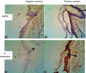
Figure 1: Immunohistochemistry, showing the distribution of heat shock protein 70 (HSP70) and ß-endorphin in rat skin (light microscope, magnification 400). A. Expression of HSP70 in the control group (dots arrow), showing a small number. B. Expression of HSP70 in the treatment group (arrow), showing an elevated number. C. Expression of ß-endorphin in the control group (dots arrow), showing a small number. D. Expression of ß-endorphin in the treatment group (arrow), showing an elevated number.
A
C
B
D
immunohistochemistry using antibody monoclonal anti-HSP70 (HSP70/HSC70 antibody [W27]: sc-24, Santa Cruz Biotechnology, Dallas, Texas, USA) and antibody monoclonal anti-ß-endorphin (anti-endorphin beta antibody, clone 3-E7. MAB5276. Chemicon, Merck Millipore Co., Darmstadt, Germany), respectively. The cells positive for HSP70 and ß-endorphin expressions were counted using a light microscope (OlympusCX21, New York, USA).
Statistics Analysis
The Mann–Whitney U-test was used to compare the data between the control and treatment groups for the expression of HSP70
and ß-endorphin as well as for the pain threshold test. The Spearman correlation test was applied to demonstrate the correlation between the expression of HSP70 and ß-endorphin. The significance of the results was set at P <0.05. Statistic package SPSS 17 was employed.
Results
Expression of Heat Shock Protein 70 and ß-Endorphin
The expression of HSP70 and ß-endorphin according to immunohistochemistry demonstrated positive cells (keratinocytes), which were brown in color, in the treatment
Figure 2: Number of the keratinocytes that expressed heat shock protein 70 (HSP70) (A) and the number of the keratinocytes that expressed ß-endorphin (B) in the control group and the treatment group of wet cupping therapy. **P<0.001, t test.
group (figure 1). The negative reaction of the expression of HSP70 (1A) and ß-endorphin (1C) did not show the dark brown color of keratinocytes. Meanwhile, the positive reaction of the keratinocytes produced a dark brown color both for HSP70 (figure 1B) and ß-endorphin (figure 1D).
Counting the number of the positive cells in the keratinocytes revealed a significantly higher number of cells that expressed HSP70 (P<0.001) and ß-endorphin (P<0.001) in the treatment group than in the control group (figures 2 A and B, respectively).
Correlation of Expression between Heat Shock Protein 70 and ß-Endorphin
A positive correlation was demonstrated between HSP70 and ß-endorphin in the
treatment group (β=0.864; P<0.001).
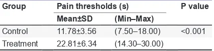
Pain Threshold Test
The pain threshold test was done 24 hours after wet cupping therapy in the treatment group, while it was carried out in the control group 72 hours after the CFA injection. In the treatment group, the rats had longer pain thresholds than the rats in the control group (P<0.001) (table 1).
Discussion
Pain, particularly chronic pain, is a debilitating disease. Patients with chronic pain often tend to exhibit other symptoms such as depression and anxiety,30,31 stiffness, sleep disturbances,
and other complaints related to quality of life.4,5
Needless to say, all these problems affect their daily life activities and working performances.5
What compounds the problem even further is that chronic pain imposes a high treatment cost.4
Taken together, chronic pain begets problems in many domains, from health to social life and economic matters.
Due to the complexity of their symptoms, patients with chronic pain are usually prescribed to have multimodal treatments, which consist of pharmacological and non-pharmacological modalities, including drugs, exercise, and psychological treatment.32 However, many
patients with chronic pain still seek other
alternative medicines.33 Some common
complementary or alternative medicines include acupuncture and chriopractice.33 Another
alternative is cupping therapy, which has been practiced for many years now.14 Wet cupping
therapy is the most common type of cupping therapy.16
The present study demonstrated that wet cupping therapy reduced pain by increasing pain threshold. To the best of our knowledge, this is the first study of its kind to demonstrate this effect of wet cupping therapy in an animal study. The difference in pain thresholds between our treatment and control groups was almost 50%, which means that the effect was considerable. This marked effect of wet cupping therapy among patients with chronic pain has also been demonstrated in a previous study.19
The result obtained in terms of pain reduction is in agreement with the benefit of wet cupping therapy in chronic regional pain syndrome,19
chronic low back pain,21 and headache.17
Furthermore, although there have been reports of hematoma as a regular minor adverse effect at the site of the application of glass cupping,19
we detected no serious adverse events.
The selection of an appropriate treatment modality for different types of chronic pain syndromes is becoming increasingly based on the mechanism of the modality.22 As the
mechanism of this benefit is still far from clear, an understanding of the mechanism of wet cupping therapy in conferring pain reduction is of great significance and needs further elucidation. According to previous studies, wet cupping therapy can lessen pain among patients with neck and shoulder pain by stimulating the peripheral nervous system,34 removing antioxidants, and
decreasing oxidative stress.15 Since nociceptive
activation contributes to chronic pain,35 it seems
that wet cupping therapy reduces pain by its antinociceptive effect. In the current study, wet cupping therapy increased the expression of ß-endorphin and HSP70 in the keratinocytes at the site of cupping.
The expressions of HSP70 and ß-endorphin in keratinocytes had a positive correlation. HSP70 is a chaperon protein that can be upregulated by stress.36 It seems that negative pressure
produced by wet cupping therapy leads to stress to the cells and stimulates the expression of HSP70.24,25 Additionally, in wet cupping therapy,
a lancet is used to puncture the skin before applying negative pressure. This treatment leads to the damage of the cell membrane of keratinocytes. The damaged tissues trigger an inflammation reaction and release inflammatory
Table 1: Pain thresholds of the control and treatment groups according to the hot plate test
Group Pain thresholds (s) P value
Mean±SD (Min–Max)
Control 11.78±3.56 (7.50–18.00) <0.001 Treatment 22.81±6.34 (14.30–30.00)
mediators such as bradykinin, prostaglandin, leukotriene, serotonin, histamine, and substance P.37 The expression of prostaglandin
E2 stimulates the expression of HSP70,38 and
the excess of HSP70 is secreted from the cells.25
Furthermore, extracellular HSP70 acts as a cytokine and triggers the expression of CRH.27
In mammals, the activation of the hypothalamic-pituitary-adrenal (HPA) axis is the main endocrine response to stress, which is signed by a rise in CRH. In corticotrophshypophyse cells, CRH binds to CRHR1 and stimulates the transcription of the POMC gene. POMC is the progenitor of adrenocorticotropic hormone and ß-endorphin.27 Nuclear factor kappaB (NFkB)
is a transcription factor associated with the expression of inflammatory cytokines such as interleukin (IL)-1, IL-6, and tumor necrosis
factor-alpha (TNF-α).39 These cytokines are
highly expressed in inflammation states.39 The
induction of NFkB in corticotrophshypophyse cells activates the POMC gene via CRH.40 NFkB
also binds to the POMC gene and stimulates the transcription of the POMC.41 POMC is
a precursor protein that produces different types of biologically active peptides, including ß-endorphin, via a series of enzymatic steps.42
ß-endorphins are synthesized and stored in the anterior pituitary gland and the immune system; they include T-lymphocytes, B-lymphocytes, monocytes, and macrophages.29,43 In the
peripheral nervous system, ß-endorphins bind to the opioid receptor (particularly of the mu subtype) at both pre- and post-synaptic nerve terminals and exert analgesic effects.29 In the
present study, ß-endorphins were determined in keratinocytes, where the wet cupping treatment was located. Interestingly, the expression of ß-endorphin and the mu-opioid receptor could also be detected on keratinocytes both at mRNA and protein levels.44 The interaction between the
nervous system keratinocytes and immune cells is mediated by the opiate receptor system.44
Nonetheless, this interaction, which leads to pain reduction after wet cupping therapy, needs to be studied further.
The effect of wet cupping therapy seems to be systemic as it was able to raise pain thresholds in the hot plate test. The systemic effect of pain reduction could involve the central nervous system. Therefore, it would be of interest to determine its mechanism in the central nervous system given that ß-endorphin and the mu-opioid receptor are also expressed in the central nervous system.45
The current study provides more evidence-based data on wet cupping therapy for pain reduction and its mechanism through the
expression of HSP70 and ß-endorphin. Nevertheless, this study has several limitations, first and foremost among which is that wet cupping therapy was applied in only a single treatment. (It was applied only 48 hours after the CFA injection.) Therefore, it would be of interest and importance to conduct prospective studies to determine the longer effects bearing in mind that it will also be more relevant for patients with chronic pain. Inflammatory pain was a model for the current study. Thus, it would also be interesting to determine the effect of wet cupping therapy in a neuropathic pain model. Wet cupping therapy is mostly applied to healthy subjects as a preventive strategy; it is, therefore, prudent that the benefit and mechanism of wet cupping therapy as a preventive strategy be determined in an animal model.
Conclusion
Taken together, the present study showed that wet cupping therapy conferred pain relief. We assume that this benefit was mediated by a rise in the expression of HSP70 and ß-endorphin.
Acknowledgement
Taken together, the present study showed that wet cupping therapy conferred pain relief. We assume that this benefit was mediated by a rise in the expression of HSP70 and ß-endorphin. Imam Subadi and Raghwendra Mishra contributed equally to this work.
Conflict of Interest: None declared.
References
1. Phillips CJ. Economic burden of chronic pain. Expert Rev Pharmacoecon Outcomes Res. 2006;6:591-601. doi: 10.1586/14737167.6.5.591. PubMed PMID: 20528505.
2. Imani F, Safari S. “Pain Relief is an Essential Human Right”, We Should be Concerned about It. Anesth Pain Med. 2011;1:55-7. doi: 10.5812/kowsar.22287523.2306. PubMed PMID: 25729655; PubMed Central PMCID: PMC4335733.
3. Katz N. The impact of pain management on quality of life. J Pain Symptom Manage. 2002;24:S38-47. doi: 10.1016/ S0885-3924(02)00411-6. PubMed PMID: 12204486.
treatment. Eur J Pain. 2006;10:287-333. doi: 10.1016/j.ejpain.2005.06.009. PubMed PMID: 16095934.
5. Smith BH, Elliott AM, Chambers WA, Smith WC, Hannaford PC, Penny K. The impact of chronic pain in the community. Fam Pract. 2001;18:292-9. doi: 10.1093/ fampra/18.3.292. PubMed PMID: 11356737. 6. Fishman S, Ballantyne J, Rathmell JP,
Bonica JJ. Bonica’s Management of Pain. 4th ed. Philadelphia: Lippincott, Williams &
Wilkins; 2010. 1661 p.
7. Inturrisi CE, Lipman AG. Opioid Analgesics. In: Fishman S, Ballantyne J, Rathmell JP, Bonica JJ, editors. Bonica’s Management of Pain. 4thed. Philadelphia: Lippincott,
Williams & Wilkins; 2010. p 1172.
8. Kaye AD, Anwar M, Baluch A. Alternative and herbal pharmaceuticals. In: Vadivelu N, Urman RD, Hines RL, editors. Essentials of Pain Management. London: Springer; 2011. p 151.
9. Pountos I, Georgouli T, Bird H, Giannoudis PV. Nonsteroidal anti-inflammatory drugs: prostaglandins, indications, and side effects. Int J Interferon Cytokine Mediat Res. 2011;3:19-27. doi: 10.2147/IJICMR.S10200. 10. Fujimori S, Gudis K, Sakamoto C. A Review
of Anti-Inflammatory Drug-Induced Gastrointestinal Injury: Focus on Prevention of Small Intestinal Injury. Pharmaceuticals (Basel). 2010;3:1187-201. doi: 10.3390/ ph3041187. PubMed PMID: 27713295; PubMed Central PMCID: PMC4034028. 11. van Koulil S, van Lankveld W, Kraaimaat FW,
van Helmond T, Vedder A, van Hoorn H, et al. Tailored cognitive-behavioural therapy and exercise training improves the physical fitness of patients with fibromyalgia. Ann Rheum Dis. 2011;70:2131-3. doi: 10.1136/ard.2010.148577. PubMed PMID: 21926189.
12. Nugraha B, Neues-Lahusen M,
Candir F, Gutenbrunner C. Effect of a series of H2S mineral water bathing on pain in patients with fibromalgia syndrome- A pilots study. Physikalische
Medizin, Rehabilitationsmedizin, Kurortmedizin. 2011;21:284-9. doi: 10.1055/s-0031-1291336.
13. Nugraha B, Gunther JT, Rawert H, Siegert R, Gutenbrunner C. Effects of whole body cryo-chamber therapy on pain in patients with chronic low back pain: a prospective double blind randomised controlled trial. Eur J Phys Rehabil Med. 2015;51:143-8. PubMed PMID: 25296740.
14. Chirali IZ. Traditional Chinese Medicine
Cupping Therapy. 3rd ed. London: Elsevier
Health Sciences UK; 2014. 372 p.
15. Tagil SM, Celik HT, Ciftci S, Kazanci FH, Arslan M, Erdamar N, et al. Wet-cupping removes oxidants and decreases oxidative stress. Complement Ther Med. 2014;22:1032-6. doi: 10.1016/j.ctim.2014.10.008. PubMed PMID: 25453524.
16. Cao H, Han M, Li X, Dong S, Shang Y, Wang Q, et al. Clinical research evidence of cupping therapy in China: a systematic literature review. BMC Complement Altern Med. 2010;10:70-9. doi: 10.1186/1472-6882-10-70. PubMed PMID: 21078197; PubMed Central PMCID: PMC3000376. 17. Ahmadi A, Schwebel DC, Rezaei M. The
efficacy of wet-cupping in the treatment of tension and migraine headache. Am J Chin Med. 2008;36:37-44. doi: 10.1142/ S0192415X08005564. PubMed PMID: 18306448.
18. Ludtke R, Albrecht U, Stange R, Uehleke B. Brachialgia paraesthetica nocturna can be relieved by “wet cupping”--results of a randomised pilot study. Complement Ther Med. 2006;14:247-53. doi: 10.1016/j. ctim.2006.07.004. PubMed PMID: 17105694.
19. Michalsen A, Bock S, Ludtke R, Rampp T, Baecker M, Bachmann J, et al. Effects of traditional cupping therapy in patients with carpal tunnel syndrome: a randomized controlled trial. J Pain. 2009;10:601-8. doi: 10.1016/j.jpain.2008.12.013. PubMed PMID: 19380259.
20. AlBedah A, Khalil M, Elolemy A, Hussein AA, AlQaed M, Al Mudaiheem A, et al. The Use of Wet Cupping for Persistent Nonspecific Low Back Pain: Randomized Controlled Clinical Trial. J Altern Complement Med. 2015;21:504-8. doi: 10.1089/ acm.2015.0065. PubMed PMID: 26069973; PubMed Central PMCID: PMC4522952. 21. Farhadi K, Schwebel DC, Saeb M,
Choubsaz M, Mohammadi R, Ahmadi A. The effectiveness of wet-cupping for nonspecific low back pain in Iran: a randomized controlled trial. Complement Ther Med. 2009;17:9-15. doi: 10.1016/j.ctim.2008.05.003. PubMed PMID: 19114223.
22. Imani F, Rahimzadeh P. Interventional pain management according to evidence-based medicine. Anesth Pain Med. 2012;1:235-6. doi: 10.5812/aapm.4514. PubMed PMID: 24904805; PubMed Central PMCID: PMC4018708.
Cell Mol Life Sci. 2005;62:670-84. doi: 10.1007/s00018-004-4464-6. PubMed PMID: 15770419; PubMed Central PMCID: PMC2773841.
24. Rylander MN, Diller KR, Wang S, Aggarwal SJ. Correlation of HSP70 expression and cell viability following thermal stimulation of bovine aortic endothelial cells. J Biomech Eng. 2005;127:751-7. doi: 10.1115/1.1993661. PubMed PMID: 16248304.
25. Asea AA, Milani V, Calderwood SK. Heat shock proteins in physiology and pathology: the Berlin meeting. Cell Stress Chaperones. 2007;12:205-8. doi: 10.1379/CSC-289.1. PubMed PMID: 17915552; PubMed Central PMCID: PMC1989779.
26. Grammatopoulos DK. Insights into mechanisms of corticotropin-releasing hormone receptor signal transduction. Br J Pharmacol. 2012;166:85-97. doi: 10.1111/j.1476-5381.2011.01631.x. PubMed PMID: 21883143; PubMed Central PMCID: PMC3415640.
27. Slominski A, Wortsman J, Luger T, Paus R, Solomon S. Corticotropin releasing hormone and proopiomelanocortin involvement in the cutaneous response to stress. Physiol Rev. 2000;80:979-1020. PubMed PMID: 10893429.
28. Koneru A, Satyanarayana S, Rizwan S. Endogenous opioids: Their physiological role and receptors. Global Journal of Pharmacology. 2009;3:149-53.
29. Sprouse-Blum AS, Smith G, Sugai D, Parsa FD. Understanding endorphins and their importance in pain management. Hawaii Med J. 2010;69:70-1. PubMed PMID: 20397507; PubMed Central PMCID: PMC3104618.
30. Nugraha B, Karst M, Engeli S,
Gutenbrunner C. Brain-derived neurotrophic factor and exercise in fibromyalgia syndrome patients: a mini review. Rheumatol Int. 2012;32:2593-9. doi: 10.1007/s00296-011-2348-2. PubMed PMID: 22210272.
31. Nugraha B, Korallus C, Gutenbrunner C. Serum level of brain-derived neurotrophic factor in fibromyalgia syndrome correlates with depression but not anxiety. Neurochem Int. 2013;62:281-6. doi: 10.1016/j. neuint.2013.01.001. PubMed PMID: 23318672.
32. Arnold B, Hauser W, Arnold M,
Bernateck M, Bernardy K, Bruckle W, et al. Multicomponent therapy of fibromyalgia syndrome. Systematic review, meta-analysis and guideline. Schmerz. 2012;26:287-90. doi: 10.1007/s00482-012-1173-1. PubMed
PMID: 22760461. German.
33. Elder C, DeBar L, Ritenbaugh C, Vollmer W, Deyo RA, Dickerson J, et al. Acupuncture and chiropractic care: utilization and electronic medical record capture. Am J Manag Care. 2015;21:e414-21. PubMed PMID: 26295269.
34. Arslan M, Gokgoz N, Dane S. The effect of traditional wet cupping on shoulder pain and neck pain: A pilot study. Complement Ther Clin Pract. 2016;23:30-3. doi: 10.1016/j.ctcp.2016.02.003. PubMed PMID: 27157955.
35. Schaible HG, Ebersberger A, Von Banchet GS. Mechanisms of pain in arthritis. Ann N Y Acad Sci. 2002;966:343-54. doi: 10.1111/j.1749-6632.2002.tb04234.x. PubMed PMID: 12114291.
36. Vega VL, Rodriguez-Silva M, Frey T, Gehrmann M, Diaz JC, Steinem C, et al. Hsp70 translocates into the plasma membrane after stress and is released into the extracellular environment in a membrane-associated form that activates macrophages. J Immunol. 2008;180:4299-307. doi: 10.4049/jimmunol.180.6.4299. PubMed PMID: 18322243.
37. Benzon HT. Essentials of pain medicine and regional anesthesia 3rd ed. Philadelphia:
Elsevier Churchill Livingstone; 2011.
38. Shah NG, Tulapurkar ME, Singh IS, Shelhamer JH, Cowan MJ, Hasday JD. Prostaglandin E2 potentiates heat shock-induced heat shock protein 72 expression in A549 cells. Prostaglandins Other Lipid Mediat. 2010;93:1-7. doi: 10.1016/j. prostaglandins.2010.03.006. PubMed PMID: 20382255; PubMed Central PMCID: PMC2919605.
39. Hoesel B, Schmid JA. The complexity of NF-kappaB signaling in inflammation and cancer. Mol Cancer. 2013;12:86-100. doi: 10.1186/1476-4598-12-86. PubMed PMID: 23915189; PubMed Central PMCID: PMC3750319.
40. Karalis KP, Venihaki M, Zhao J, van Vlerken LE, Chandras C. NF-kappaB participates in the corticotropin-releasing, hormone-induced regulation of the pituitary proopiomelanocortin gene. J Biol Chem. 2004;279:10837-40. doi: 10.1074/jbc. M313063200. PubMed PMID: 14711817. 41. Jang PG, Namkoong C, Kang GM,
M109.070706. PubMed PMID: 20097762; PubMed Central PMCID: PMC2843220.
42. Millington GW. The role of
proopiomelanocortin (POMC) neurones in feeding behaviour. Nutr Metab (Lond). 2007;4:18-33. doi: 10.1186/1743-7075-4-18. PubMed PMID: 17764572; PubMed Central PMCID: PMC2018708.
43. Mousa SA, Shakibaei M, Sitte N, Schafer M, Stein C. Subcellular pathways of beta-endorphin synthesis, processing, and release from immunocytes in inflammatory pain. Endocrinology. 2004;145:1331-41.
doi: 10.1210/en.2003-1287. PubMed PMID: 14630714.
44. Bigliardi-Qi M, Sumanovski LT, Buchner S, Rufli T, Bigliardi PL. Mu-opiate receptor and Beta-endorphin expression in nerve endings and keratinocytes in human skin. Dermatology. 2004;209:183-9. doi: 10.1159/000079887. PubMed PMID: 15459530.
45. Brunton L, Lazo J, Parker K. Goodman & Gilman’s, The pharmacological basis of therapeutics. 11th ed. New York: