8
Open Acces
Int. J. Trop. Vet. Biomed. Res. Vol. 2 (1) : 8-11; May 2017
www.jurnal.unsyiah.ac.id/IJTVBR
E-ISSN : 2503-4715
PREVALENCE OF F. gigantica AND PATHOLOGICAL CHANGES IN LIVER OF SIMEULUE BUFFALO
Ikhwan Jamil1, Teuku Reza Ferasyi2, Muhammad Hambal3, Yudha Fahrimal3, Razali2
1Post-Graduate Program of Veterinary Public Health, Syiah Kuala University 2.
Laboratory of Veterinary Public Health, the Faculty of Veterinary Medicine Syiah Kuala University
3Laboratory of Parasitology, the Faculty of Veterinary Medicine Syiah Kuala University
Email for correspondence: [email protected] Abstract
The aim of this study was to identify the prevalence of fasciolosis of Simeulue’s buffalo using macroscopic
approach by observing pathological changes in the liver. The sample were obtained from slaughterhouse in Sinabang. A number of 60 livers were obtained on July to September 2015 based on post mortem examination. From this sample, a total of 57 livers were found positive indication of infested by F. gigantica (95%). The length and width of F. gigantica was 25 mm and 7 mm. The body was flat as a leaf, blunt on posterior, gray, brown, transparant and do not have a real shoulder shapes. The liver which were not infested with F. gigantica showed sharp edges and a very high degree of elasticity. On the other hand, in the infested liver was found F. gigantica in the bile duct and showed a color of pale, the dark brown exudate as well as objects looks like gravel. The buffaloes sample were supplied to the abattoir from the paddy fields or oil palm plantations area. In comparison, the buffaloes raised in both areas were not different on the pathological changes of liver. In conclusion, this study showed that
simeulue’s buffalo is very prevalence to F. gigantica. Key words : Buffalo, F. gigantica, Simeulue.
Background
Fasciolosis caused by worms of the species F. hepatica and F. gigantica a family of worms trematodes. F. gigantica is one of the most common worm infection of ruminants in Asia and Africa ( Hammond & Sewell 1990), where as habitat adult worms live in the liver (bile ducts) to the buffaloes, cows, goats, and sheep (Boray et al., 2007). Fasciolosis influenced by several factors. According Bhattanchryya and Ahmed (2005) explains that the topography, geography, population density, climate and health management are factors that influence the development of parasitic disease in an area. More specifically, Suweta (1982) states that the large ruminant grazing in rice fields in Indonesia is relatively higher to be infested by F. gigantica. According Raunelli and Gonzales (2009) due to infestation fasciolosis cause considerable economic losses like death, weight and carcass losses. Then, it also decreased milk production, labor, and the tendency for another illnesses and medical expenses (Wamae and Ihiga 1991; Maingi and Mathenge 1995: Charlier
et al., 2008). Additionally, Fascioliasis now recognized as an emerging human diseases. The World Health Organization (WHO, 2006) estimates that 2.4 million people are infected with Fasciola spp and a further 180 million are at risk of infection.
According to Marwadi (2016), due to fasciolosis liver was not suitable for consumption due to several anatomical changes such as cirrhosis, cicatrix thick, abscesses and discoloration. The overall prevalence of F. gigantica on Simeulue’s buffalo with pathological changes of liver relation to the maintenance area described in this paper. With the aim to provide information relating to infestation fasciolosis on Simeulue’s buffalo so that it becomes a reference of prevention to reduce the impact of disease on the production of buffalo of Simeulue.
Materials and Methods
Ikhwan Jamil, et al. (2017) Int. J. Trop. Vet. Biomed. Res.1:8-11
9
observational studies with cross sectional sample selection by purposive sampling performed on liver and bile of Simeulue’s buffaloes which slaughtered at the abattoir Sinabang. Based on the maintenance area are rice fields and palm plantations.
Examination of the existence of F. gigantica and observation of pathology changes in the liver and bile ducts to all buffalo were conducted after slaughtering.
Results and Discussion
From a total of 60 liver were examined post mortem, 57 were positive infested F. gigantica (95%). In plain trematode worms are clearly visible both on the surface of the liver, and are most numerous in the bile duct. Observations on the liver morphology F. gigantica obtained by performing the measurement . Obtained its length reached an average of 25 mm with a width of 7 mm , and has a body shape like a leaf , flat, tip posterior blunt and gray brown or transparent, addition does not have the shape of the shoulder real , from the characteristics of these observations a worm F. gigantica , according opinion (Baker , 2007) assert that F. gigantica measuring of 25-27 x 3-12 mm , have narrow shoulders and blunt posterior end.
Figure 1. F. gigantica recovered from the liver at the time of observation
In anatomical pathology, liver of Simeulue buffaloes infested by F. gigantica showed different conditions that were not infested with, both derived from the paddy fields or oil palm plantations.
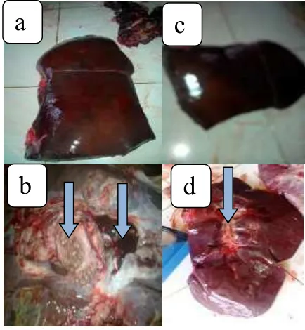
Figure 2. The condition of liver buffaloes negative (a and c) and positive (b and d) infestation fasciolosis paddy fields ( b ) and oil palm ( d ) . The arrows indicate the condition of the liver undergoing changes . In Figure 2b liver looks pale color , there are F . gigantica the bile duct and their blackish brown exudate . In Figure 2d, a thickening of the heart organ , hard consistency and widening of the bile duct .
From Figure 2 showed that the liver was not infested fasciolosis sharp edges (a and c). Then when the surgery showed a very high degree of elasticity. Instead, different looks can be observed in the liver buffalo Simeulue infested fasciolosis (b and d). At the heart organ looks intact, bile duct wall thickening and therein buried mucoid fluid and contains F. gigantica. Additionally at the time of slicing liver contained objects such as pebbles and discharge yellowish and brownish-black look their exudates found in the liver and bile ducts. Plus from the anatomic pathology changes in the gallbladder bile duct wall dilatation form, and color of exudate accumulates in the gall bladder. Clinical symptoms of liver damage are indicated as such in accordance with the fasciolosis described by Shaikh et al., (2004), and Talukder et al., (2010).
Total of the F. gigantica can affect the level of liver damage. Martindah et al (2005) reported extensive damage to the liver is affected by the number and duration
a
b
c
Ikhwan Jamil, et al. (2017) Int. J. Trop. Vet. Biomed. Res.1:8-11
10
F. gigantica infestations occur. But it is different with the results obtained Panjaitan (2012) that the number of F. gigantica in the liver are not related to changes in anatomical pathology. Meanwhile, according to Jones et al (2006) there are two factors that cause lesions in the liver damage that is the first migration of young worms and secondly because of the induction of adult worms that occur on an ongoing basis. There are several ways that F. gigantica migrated towards the liver: first through the bloodstream; second through the portal vein and the bile flow generally occurs through the intestinal perforation and penetrates the capsule of the past peritoneum and the liver parenchyma and then migrated toward the bile duct. Migration of worm the liver causing damage to the parenchym. The above process may has been experienced by the liver that is infested fasciolosis of simeulue buffalo.
The symptoms of chronic fasciolosis cases are anemia, weight loss, decreasing of milk production , and submandibular edema (Talukder et al,. 2010). The activity of sucking causes irritation of the gallbladder, inflammatory response, and blood loss to anemia (Raadsma et al., 2007). Buffaloes have a susceptibility to F. gigantica (Hambal et al., 2013). The majority of simeulue’s buffaloes which slaughtered at the abattoir of Sinabang have experienced fasciolosis, and as skin and bone of the bodies.
CONCLUSIONS
1. Infestation fasciolosis of Simeulue’s buffalo reaches 95 %
2. Changes in anatomical pathology of heart from paddy fields and palm plantations shows the different conditions . As exudate blackish brown and their objects such as pebbles.
ACKNOWLEDGMENT
Chief and Staff Graduate Studies Program of Veterinary Public Health Syiah Kuala University in Banda Aceh that has supported in the research
Head of Animal Health and Veterinary Office Simeulue and the entire staff has given permission and aid in research
To drh. Al Azhar, M. Kes, Ph.D. who has helped in the writing of this article
REFERENCE
Bhattachryya, DK. and K. Ahmed . (2005). Prevalence of helmintic infection in cattle and buffaloes.
Indian Vet. J. 8(2): 900-901.
Baker DG. (2007). Flynn’s Parasites of
Laboratory Animals. Second edition.
American College of Laboratory Animal Medicine. USA: Blackwell Publishing.
Boray JC., GW, Hutchinson., L. Stephen. (2007). Liver Fluke Disease In Sheep And Cattle. Primefact 446.
Charlier, J. DL. Meulemeester,E. Claerebout, D. Williams, J. Vercruysse.(2008). Qualitative and quantitative evaluation of coprological and serological techniques for the diagnosis of fascioliasis in cattle. Vet. Parasitol. 153: 44-51.
Hambal, M., A. Sayuti., A. Dermawan. (2013). Tingkat Kerentanan Fasciola
gigantica pada Sapi dan Kerbau di
Kecamatan Lhoong, Kabupaten Aceh Besar. Jurnal Medika Kinangop District Nyandarua District of Kenya.Bulletin Animal
Health Production Africa 43: 21-27.
Martindah, E.,S. Widjajanti, S.E Estuningsih, dan Suhardono. (2005). Meningkatkan Kesadaran dan Kepedulian Masyarakat Terhadap Fasciolosis Sebagai Penyakit Infeksius.Wartazoa, 15(3): 143-154 Marwadi, H. (2016). Pemeriksaan Feses,
Ikhwan Jamil, et al. (2017) Int. J. Trop. Vet. Biomed. Res.1:8-11
11
Akibat Infestasi Fasciola gigantica. Tesis. Program Pascasarjana, Universitas Syiah Kuala.
Panjaitan, N. (2012). Studi Kasus Fasciolosis di RPH Purwodadi Kabupaten Grobogan- Jawa Tengah: Diagnosis, Derajat Infeksi Fasciola
gigantica, dan Tingkat Kerusakan
Hati pada Sapi. Skripsi. Fakultas Kedokteran Hewan. Institut Pertanian Bogor. Bogor.
Raadsma, H.W., Kingsford, N.M., Suharyanta, Spithill, T.W., Piedrafita D. (2007). Host responses during experimental infection with Fasciola gigantica or Fasciola hepatica in Merino sheep: I. Comparative immunological and plasma biochemical changes during early infection. Vet. Par, 143(3-4):275-286.
Raunelli, F., S. Gonzales.(2009). Strategic control and prevalence of faciola
hepatica in Peru: a pilot study.
Int. J. App. Res. Vet. Med. 7(4):145-152.
Shaikh, A.A., F. M. Bilqees, and M.M. Khan. (2004). Bile Duct Hyperplasia and Associated Abnormalities in the Buffaloes Infected With Fasciola
gigantica. Pakistan J. Zool. 36(3):
231-237.
Suweta, I.G.P. 1982. Kerugian Ekonomi oleh Cacing Hati pada Sapi Bali Sebagai Implikasi Interaksi dalam Lingkungan Hidup pada Ekosistem Pertanian di Bali. Disertasi.
Universitas Padjadjaran. Bandung. Talukder, S., M.J. Bhuiyan, M.M. Hossain,
M.M. Uddin, S. Paul, M.M.R. Howlader. (2010). Pathological Investigation of Liver Fluke Infection of Slaughtered Black Bengal Goat in a Selected Area of Bangladesh. Bangl. J. Vet. Med.
8(1): 35 – 40.
Wamae, L. W. and M. K.Ihiga (1991).Fascioliasis as a limiting factor in livestock productivity, Bulletin Animal Health Production Africa 39: 257-269.