Luteinizing hormone secretion as affected by
hypophyseal stalk transection and
estradiol-17
b
in ovariectomized gilts
q
J.J. Ford
a, J.G. Berardinelli
a,1,
R.K. Christenson
a, L.L. Anderson
b,∗ aUSDA, ARS, Roman L. Hruska US Meat Animal Research Center,Agricultural Research Service, Clay Center, NE 68933, USA
bDepartment of Animal Science, Neuroscience Program, Iowa State University, Ames, IA 50011, USA
Received 20 January 2000; received in revised form 26 June 2000; accepted 30 June 2000
Abstract
The objectives were to determine hypothalamic regulation of pulsatile luteinizing hormone (LH) secretion in female pigs and the biphasic feedback actions of estradiol-17b(E2-17b). In the first study, the minimum effective dosage of E2-17bthat would induce estrus in ovariectomized gilts was determined to be 20mg/kg body weight. In the second study, ovariectomized gilts were assigned randomly on day 0 to treatments: (a) hypophyseal stalk transection (HST), (b) cranial sham-operated control (SOC), and (c) unoperated control (UOC). On day 3, gilts from each group received a sin-gle i.m. injection of either E2-17b(20mg/kg body weight) or sesame oil. Blood was collected from an indwelling jugular cannula at 15 min intervals for 3 h before (day−2) and after treat-ment (day 2) from HST, SOC and UOC gilts. On day 3, blood was collected at 2 h intervals for 12 h after E2-17bor sesame oil injection and at 4 h intervals thereafter for 108 h. Pulsatile LH secretion in all gilts 2 days after ovariectomy exhibited a frequency of 0.9±0.06 peaks/h, ampli-tude of 1.3±0.13 ng/ml, baseline of 0.8±0.07. Serum LH concentrations from SOC and UOC gilts
∗
Corresponding author. Tel.:+1-515-294-5540; fax:+1-515-294-4471. E-mail address: [email protected] (L.L. Anderson).
1Present address: Montana State University, Bozeman, MT 59717. q
Journal Paper No. J-18532 of the Iowa Agriculture and Home Economics Experiment Station, Ames, IA (Project No. 3316), and supported by the Hatch Act and State of Iowa funds, and by US Department of Agriculture, ARS, CSRS, Cooperative Agreement 58-519B-9-863. All experiments in this report were performed following standards established by the Animal Welfare Act and NIH, Guide for the Care and Use of Laboratory Animals, Publication 85-23. Mention of a trade name, proprietary product, or specific equipment does not constitute a guarantee or warranty by the US Department of Agriculture and does not imply its approval to the exclusion of other products that may be suitable.
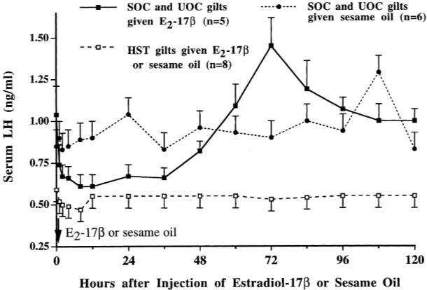
were similar on day 2 and profiles did not differ from those on day−2. In HST gilts pulsatile LH release was abolished and mean LH concentration decreased compared with controls (0 versus 0.9±0.06 peaks/h and 0.77±0.03 versus 1.07±0.07 ng/ml, respectively;P <0.05). E2-17bor sesame oil did not affect serum LH concentration in HST gilts, and LH remained constant throughout 120 h (0.7±0.07 ng/ml). In SOC and UOC control gilts, E2-17binduced a 60% decrease (P < 0.05) in LH concentration within 12 h, and LH remained low until 48 h, then increased to peak values (P < 0.05) by 72 h, followed by a gradual decline to 120 h. Although pituitary weight decreased 31% in HST gilts compared with controls (228 versus 332 mg,P <0.05), an abundance of normal basophils was evident in coronal sections of the adenohypophysis of HST comparable to that seen in control gilts. The third and fourth studies determined that hourly i.v. infusions of LHRH (2mg) and a second injection of E2-17b48 h after the first had no effect on the positive feedback action of estrogen in UOC. However, in HST gilts that received LHRH hourly, the first injection of E2-17bdecreased (P <0.05) plasma LH concentrations while the second injection of E2-17bfailed to induce a positive response to estrogen. These results indicate that both pulsatile LH secretion and the biphasic feedback action of E2-17bon LH secretion depend on hypothalamic regulatory mechanisms in the gilts. The isolated pituitary of HST gilts is capable of autonomous secretion of LH; E2-17bwill elicit direct negative feedback action on the isolated pituitary gland if the gonadotropes are supported by exogenous LHRH, but E2-17bat high concentrations will not induce positive feedback in isolated pituitaries. Thus, the direct effect of E2-17bon the pituitary of monkeys cannot be mimicked in pigs. © 2000 Elsevier Science B.V. All rights reserved.
Keywords: Luteinizing hormone; Hypothalamic regulation; Estradiol-17b; Pig
1. Introduction
Ovarian steroids modulate luteinizing hormone (LH) secretion in the rat, sheep, rhe-sus monkey, and pig. After ovariectomy, the elevated and pulsatile profiles of LH secre-tion are caused by LH-releasing hormone (LHRH) from hypothalamic secretory neurons that is secreted episodically into hypophyseal portal vessels. Hypothalamic destruction or drug-induced inhibition of secretory neurons within the hypothalamus eliminates pulsatile LH secretion in ovariectomized rats, rhesus monkeys, and ewes. The patterns of LH secretion in intact prepubertal gilts indicate few changes evident in mean serum concentration during 10–25 week of age (Diekman et al., 1983). Weekly sequential bleedings indicated that the frequency and magnitude of LH pulses remained similar during this period. Ovariectomy in gilts produces increases in LH secretion with a characteristic episodic rhythm (Berardinelli et al., 1984).
rats (Halász and Gorski, 1967; Blake, 1977). Furthermore, in rats and sheep, interruption of afferent signals from nuclei rostral to the arcuate nucleus eliminates preovulatory- as well as E2-17b-induced, surges of LH (Halász and Pupp, 1965; Blake, 1977; Jackson et al., 1978).
In contrast to rats and sheep, estrogen in monkeys can mediate its biphasic, feedback effects on LH secretion directly at the pituitary gland (Nakai et al., 1978; Ferin et al., 1979; Pavasuthipaisit et al., 1981). Thus, the two models proposed for regulation of LH secretion by estrogen include regulation primarily at either the hypothalamus or the hy-pophysis. However, the amount of estrogen required to induce positive feedback is greater in hypophyseal stalk-transected (HST) than in control monkeys (Karsch et al., 1973a; Ferin et al., 1979; Pavasuthipaisit et al., 1981). Likewise, estrogen produces positive feedback on LH secretion in gonadectomized male monkeys, but the dosage must be greater than in females (Yamaji et al., 1971; Karsch et al., 1973b). In HST gilts estrogen failed to induce a positive feedback effect on LH secretion (Kesner et al., 1989); however, this study used a single dosage of estrogen. The current study in gilts used greater dosages of estrogen to determine if the effects of E2-17bon pulsatile LH secretion are regulated primarily by (a) the pituitary gland independent of hypothalamic control or (b) the biphasic feedback of estrogen at the hypothalamus.
2. Materials and methods
2.1. Animals
2.1.1. Minimum effective dosage of E2-17β
Thirty-nine white-composite, postpubertal gilts were ovariectomized and treated with either E2-17bor estradiol benzoate (EB) at varying dosages to determine minimum effective dosages for induction of behavioral estrus. Estrogens (5, 10 or 20mg E2-17bor 1.25, 2.5 or 5mg EB per kg body weight (BW)) were given in oil as a single intramuscular injection. Gilts were observed for behavioral estrus twice daily in the presence of mature boars; proportion that exhibited estrus and latency to estrus were determined.
2.1.2. LH secretion after hypophyseal stalk transection and E2-17βtreatment
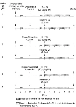
Fig. 1. Description of experimental and control groups for determination of effects of estradiol-17bon neuro-endocrine regulation of luteinizing hormone secretion in ovariectomized Yorkshire gilts.
BW) or vehicle (2.5 ml sesame oil). Fig. 1 depicts the experimental regimen and shows the number of gilts per treatment.
2.1.3. Effect of pulsatile LHRH infusion and E2-17β treatment on LH secretion
The third study evaluated the effect of exogenous, pulsatile LHRH on the biphasic effect that E2-17bhas on LH secretion. Indwelling jugular catheters were inserted into four white composite gilts that had been ovariectomized postpubertally, BW=115+2 kg. Beginning a minimum of 72 h after placement of catheters (day−2), two gilts were assigned to receive hourly i.v. infusions of 2mg LHRH for 144 h and the other two received diluent i.v. The LHRH (Sigma Chemical Co., St. Louis, MO) was diluted (1mg/ml) in 0.1% BSA and administered by infusion pumps connected to timers, 2 ml/min for 1 min each hour. All gilts received i.m. injections of E2-17b(20mg/kg BW) on days 0 and 2. Blood samples were collected at 0, 6, 12, 24, 36, 48, 53, 60, 72, 78, 84, 90, 96, 108 and 120 h (day 7 of LHRH infusion) after initiation of the first E2-17binjection. Samples were taken just before the coincident hourly infusion. Additionally, on day 0 before E2-17btreatment, blood samples were obtained from all four gilts at 0.5, 10, 20, 30, 45 and 60 min after an i.v. infusion of 2mg of LHRH. Hourly LHRH infusions stopped on day 7, and 1 h after the last infusion all four gilts received 25mg of LHRH i.v. with blood collected at 0, 7, 15, 30, 45, 60 and 90 min after this bolus treatment. Seven days after the 25mg bolus of LHRH, treatment of these gilts was reversed with respect to hourly infusions of LHRH and diluent, and the protocol repeated.
2.1.4. Effect of pulsatile LHRH infusion and E2-17βtreatment on LH secretion after
hypophyseal stalk transection
In the fourth study, postpubertal, ovariectomized, Yorkshire gilts (BW = 106±3 kg) received indwelling jugular catheters as described above. Treatments were HST (N =6) and UOC (N = 5). The HST gilts were given hourly LHRH infusion as described in the third study beginning on the morning after HST (day −2). E2-17b(20mg/kg BW) was given on days 0 and 2 to HST gilts and UOC gilts. The UOC gilts did not receive hourly infusions of LHRH. Blood samples were collected at the intervals described for the third study. Plus, all gilts received 2mg LHRH i.v. on day 0, before E2-17btreatment, with blood collected at 0, 5, 10, 20 and 30 min after LHRH. Hourly LHRH infusions of HST gilts stopped on day 7, and 1 h after the last infusion all gilts received 25mg of LHRH with blood collected at 0, 15, 30, 45, 60, 75, 90, 105 and 120 after the LHRH bolus infusion.
2.2. Hypophyseal stalk transection (HST)
2.3. Sham operation control (SOC)
Gilts were subjected to all cranial surgical procedures as HST, with the exception that the hypophyseal stalk was not severed.
2.4. Unoperated control (UOC)
Gilts were not subjected to cranial surgery.
2.5. Postoperative care and hormone treatment
During each study, all gilts were placed in individual pens. After HST or SOC, gilts were placed in a room maintained at 28–30◦C; respiration, heart rate and rectal temperature were monitored and recorded every 4 h for 48 h. Intramuscular injection of cortisone ac-etate (50 mg/ml; Merck & Co. Inc., West Point, PA) or i.v. injection of cortisone succinate (Solu-Cortif, 100 mg/ml; Merck & Co.) was administered within 24 h after surgery. Water and food intake usually returned to normal by 12–24 h postsurgery. UOC gilts were penned individually in a room separate from gilts subjected to cranial surgery.
2.6. Postmortem
Experimental and control gilts in studies two and four were sacrificed by an overdose of thiamylal sodium and decapitated. The frontal bone and calvaria were removed to expose the brain. Cranial nerves were severed, and the brain was raised to expose the hypophyseal stalk and sella turcica. The hypophyseal stalk was examined to determine completeness of stalk transection and position of the nylon disc in HST gilts. The pituitary gland was excised from the sella turcica, weighed, cut transversely into three blocks and placed into Susa’s fixative for histological evaluation. Coronal sections of the glands were cut at 6mm and stained (Heath, 1965), whereas other sections were stained with hematoxylin and eosin. Thyroid and adrenal glands were excised, connective tissue was removed, and glands were weighed. The glands were cut at 6mm and stained with hematoxylin and eosin. One set of sections from thyroid glands was stained with Mallory triple stain for identification of basophilic and acidophilic colloid.
2.7. RIA of LH and E2-17β
Serum E2-17b was assayed in 0.5 ml duplicate aliquots that had been extracted twice with diethyl ether (10 : 1 v/v; Redmer et al., 1984). The antibody was specific for E2-17b and had no cross-reactivity with estrone or estradiol-17a(<0.05%). Assay sensitivity was 10 pg/ml of serum. Interassay coefficient of variation, determined from a pool of boar serum (42±8 pg/ml) was 15% and intra-assay coefficient of variation was 12%. Extraction recoveries ranged from 91–98%.
2.8. Statistical analysis of experimental data
In the second study, characterization of pulsatile secretion of LH on day−2 and day 2 for HST and SOC gilts, or 2 and 5 days after ovariectomy in UOC gilts, were analyzed by a split-plot analysis using a one-way ANOVA, and Student’s t-tests for continuous variables was used for comparisons between groups (SAS, 1990). Fisher’s LSD test was used to determine differences among mean number of peaks, mean amplitude, baseline, and LH values with day and treatment within day as variables (Snedecor and Cochran, 1980). LH concentrations after E2-17bor sesame oil treatment were compared by analysis of variance for split-plot, with treatment as main plot and time as the subplot. Pituitary, adrenal, and thyroid gland weights were analyzed by analysis of variance using treatment as the single classification.
For the third and fourth studies, LH profiles were evaluated using Mixed Model pro-cedures for repeated measures (SAS; Littell et al., 1996). The model for the third study included treatment group, time, the interaction of these two variables and treatment within gilt. The model for the fourth study was treatment group, time and the interaction of these two variables. Specific comparisons were made with the PDIFF option.
3. Results
3.1. Minimum effective dosage of E2-17β
Proportions of ovariectomized gilts that exhibited behavioral estrus in the first study were 0/9, 5/9 and 9/9 for E2-17bdosages of 5, 10 and 20mg/kg BW, respectively. This compared to 2/4, 4/4 and 4/4 for EB dosages of 1.25, 2.5 and 5mg/kg BW. Latency to estrus was 1.9±0.5 d and did not differ (P >0.10) for the two forms of estrogen.
3.2. LH secretion after hypophyseal stalk transection and E2-17βtreatment
In the second study, three HST gilts died within 60 h after surgery from hemorrhage or elevated temperature. In the eight other HST gilts, the nylon disc was in the proper location and had prevented vascular regeneration of the hypophyseal stalk.
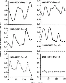
Fig. 2. Sequential profiles of luteinizing hormone concentrations in peripheral blood serum during 3 h at (a) 2 days before cranial surgery and (b) 2 days after sham operation (s) or hypophyseal stalk transection (d), as compared
with those in unoperated control (m) gilts. There was a total of eight HST, six SOC, and six UOC. All gilts were
ovariectomized 3 days before cranial surgery.
tended to be lower in HST gilts compared with controls (0.7 versus 1.0 ng/ml, respectively;
P < 0.10). Frequency and amplitude of LH pulsatile release and mean concentration in
controls 5 days after ovariectomy (day 2 of the study) did not differ significantly from those observed 2 days after ovariectomy (day 0).
Table 1
Effect of hypophysial stalk transection (HST), sham operation control (SOC), and unoperated control (UOC) on pulsatile secretion of luteinizing hormone in ovariectomized giltsa
Hormone characteristicb LH in peripheral serum (ng/ml)b
HST SOC UOC
Mean 0.77±0.03 1.07±0.13 1.07±0.07
Baseline 0.77±0.03 0.97±0.10 1.0±0.07
Amplitude NA 1.33±6.13 1.23±0.07
Frequencyc O∗ 0.9±0.06 0.9±0.07
aValues are mean±S.E.M.
bAnterior vena cava blood samples were obtained at 15 min intervals throughout a period of 180 min 2 days after cranial surgery.
cPeaks/h.
∗P <0.05.
Circulating blood concentration of E2-17bfollowed a similar sequential pattern (P > 0.05) among HST, SOC and UOC gilts. E2-17bincreased from<10 to 2500±400 pg/ml by 4 h after E2-17binjection (P < 0.01), decreased to 100±25 pg/ml by 24 h and de-clined thereafter to<10 pg/ml by 96 h. E2-17bwas<10 pg/ml after sesame oil treatment of ovariectomized HST, SOC and UOC gilts.
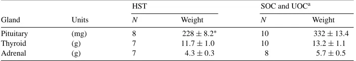
Pituitary, adrenal and thyroid gland weights remained similar (P >0.05) in SOC and UOC gilts and were pooled (Table 2). By 9 days after surgery, pituitary glands were 31% smaller (P <0.05) in HST than SOC and UOC gilts; however, thyroid and adrenal gland
Table 2
Effect of hypophyseal stalk transection on pituitary, thyroid and adrenal glands in ovariectomized gilts
HST SOC and UOCa
Gland Units N Weight N Weight
Pituitary (mg) 8 228±8.2∗ 10 332±13.4
Thyroid (g) 7 11.7±1.0 10 13.2±1.1
Adrenal (g) 7 4.3±0.3 8 5.7±0.5
aControl equals pooled values for sham-operated and unoperated control gilts because no differences were detected in ANOVA between these groups (P >0.05).
∗P <0.05.
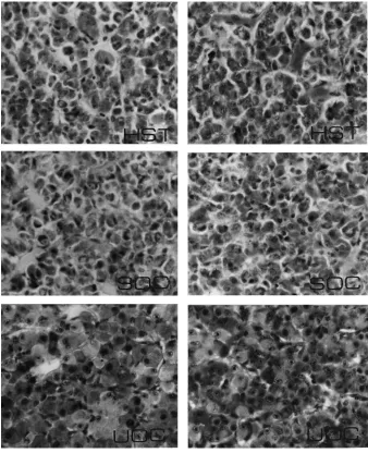
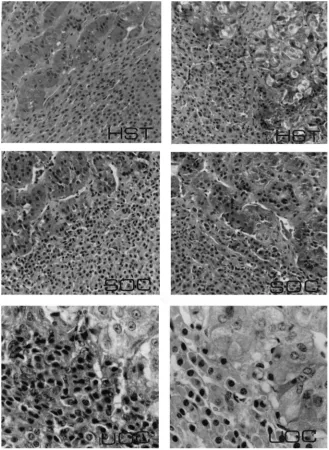
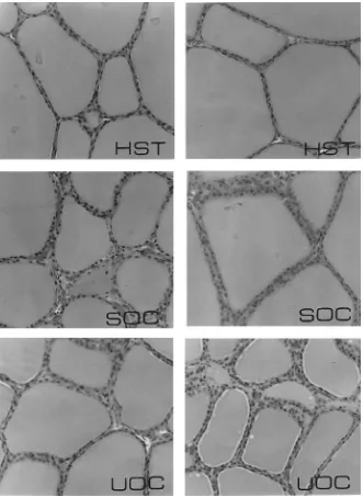
weights remained similar (P >0.05) in HST and controls. Gross examination of pituitary glands from HST gilts revealed tissue necrosis in the anterior one-third of these glands, immediately ventral to the severed end of the stalk; the remainder of the gland appeared normal. Coronal histological sections of the middle one-third of the gland from HST gilts revealed viable secretory parenchyma comparable to coronal sections of pituitary from controls (Fig. 4). Differential staining of adenohypophysis from HST gilts revealed viable appearing acidophils, basophils and chromophobes similar to SOC and UOC. Although adrenal gland weights were similar in HST and control gilts, coronal sections of adrenal revealed cortical cells with less cytoplasm compared with controls (Table 2, Fig. 5). Thyroid gland weights in HST were similar to controls (Table 2), but thyroid histology from HST gilts revealed cuboidal epithelial cells and accumulation of acidophilic colloid in large follicles compared with normal columnar epithelial cells and smaller follicles with basophilic colloid in controls (Fig. 6).
3.3. Effect of pulsatile LHRH infusion and E2-17βtreatment on LH secretion
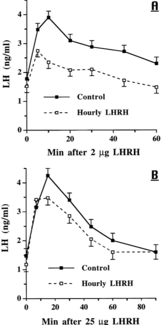
In the third study, LH secretion in response to 2mg of LHRH i.v. was greater (P <
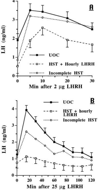
0.01) in ovariectomized, control gilts than in gilts that had received hourly LHRH for 48 h (Fig. 7A). The overall means were 2.9 versus 2.0 ng LH/ml for controls and LHRH infused gilts, respectively; the time by group interaction approached significance (P <0.08). At initiation of E2-17btreatment, LH concentrations were similar in both groups (P >0.10), and the responses to the two i.m. injections of E2-17bdid not differ (P >0.10, Fig. 9A). Clearly, the negative response to the first E2-17binjection was followed by an increase in serum LH concentrations after the second E2-17binjection. After 7 days of hourly LHRH infusions and the E2-17btreatment, LH concentrations were similar in the two groups, but the response to an i.v. bolus of 25mg LHRH was greater (P <0.01) in controls than in the infused group with the greatest difference at 15 min after LHRH (Fig. 7B).
3.4. Effect of pulsatile LHRH infusion and E2-17β treatment on LH secretion after
hypophyseal stalk transection
Fig. 7. Response to LHRH i.v. in ovariectomized gilts. Treatment groups were controls (N=4) infused hourly with diluent and gilts (N=4) infused hourly with 2mg LHRH. (A) The response to 2mg LHRH i.v. 48 h after hourly infusions began; group:P <0.01; time:P <0.01; group×time:P <0.08. (B) Response in these same gilts to 25mg LHRH i.v. on day 7 after initiation of hourly infusions combined with E2(20mg/kg BW) i.m. on days 2 and 4; group:P <0.01; time:P <0.01; group×time:P >0.10.
Fig. 8. Response to LHRH i.v. in ovariectomized gilts. Treatment groups were UOC (N = 5) and HST gilts (N=4) infused hourly with 2mg LHRH. (A) The response to 2mg LHRH i.v. 48 h after hourly infusions began; group:P <0.03; time:P <0.01; group×time:P >0.10. (B) Response in the same gilts to 25mg LHRH i.v. on day 7 after initiation of hourly infusions combined with E2(20mg/kg BW) i.m. on days 2 and 4; group:
P <0.05; time:P <0.01; group×time:P <0.01. Data from one gilt classified as an incomplete HST, due to misalignment of the nylon disc, are presented for comparison only.
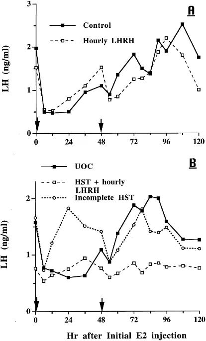
closely resemble UOC than HST gilts (Fig. 7). The response of this gilt to hourly LHRH plus the first E2-17binjection produced a much shorter interval for the negative effect of E2-17b, but after the second E2-17binjection, the LH response more closely mimicked that observed in the UOC’s (Fig. 9B).
4. Discussion
Fig. 9. Response to E2-17b(20mg/kg BW; arrows designate time of i.m. injection) in ovariectomized gilts. (A) Treatment groups were controls infused hourly with diluent and gilts infused hourly with 2mg LHRH; group:
P >0.10; time:P <0.01; group×time:P >0.10. (B) Treatment groups were HST gilts that received hourly infusion of 2mg LHRH and UOC; group:P <0.01; time:P <0.01; group×time:P <0.06. Data from one gilt classified as an incomplete HST, due to misalignment of the nylon disc, are presented for comparison only.
conclusions of Kesner et al. (1987, 1989) and Stickan et al. (1999) that estrogen manifests its positive feedback on LH secretion primarily through the central nervous system, and estrogen at excessively elevated concentrations is no more effective than concentrations observed during the estrous cycle.
These results in gilts contrast observations in female rhesus monkeys subjected to HST during the follicular phase of the menstrual cycle (Ferin et al., 1979) or in ovariectomized rhesus monkeys in which the hypophysiotropic area was destroyed and LH secretion re-established by pulsatile infusion of LHRH (Nakai et al., 1978). Although exogenous estrogen augments pituitary responsiveness and induces LH secretion presumably by hy-pothalamic regulation in rhesus monkeys, the impact of estrogen on adenohypophyseal function likely reflects action of this steroid at both of these loci (Knobil, 1980, 1981). It seems that LHRH has a permissive rather than causative role in controlling LH secretion in rhesus monkeys (Knobil et al., 1980). Estrogen inputs directed at adenohypophyseal loci are sufficient to explain a controlling influence of the steroid on LH secretion in the presence of exogenous LHRH. Exogenous estradiol caused a two-fold increase in adenohypophy-seal high-affinity LHRH receptors in ovariectomized cynomolgus macaques (Adams et al., 1981). Although LH release in response to exogenous LHRH is initially suppressed by estradiol, pituitary LHRH receptors correlate positively with estrogen-induced release of LH only during the phase of potentiated response. Thus, estrogenic augmentation (positive feedback) of pituitary responsiveness in monkeys seems to be mediated by an increase in the number of LHRH receptors on the surface of gonadotrophs.
Administration of central nervous system depressants to female pigs and rats (Kesner et al., 1987; Kimura et al., 1995), electro-lesion of the hypophysiotropic area in female rhesus monkeys (Plant et al., 1978) and ewes (Jackson et al., 1978), or interruption of hypophyseal blood circulation in female rhesus monkeys (Ferin et al., 1974), beef calves (Anderson et al., 1981) and female pigs (current study) eliminates pulsatile LH secretion. This pulsatile pattern reflects a basic rhythm of the secretory neurons of the hypophysiotropic area in the hypothalamus that secretes LHRH into hypophyseal portal vessels for transport to the adenohypophysis (Hotchkiss and Knobil, 1994; Terasawa et al., 1999).
maintained after complete deafferentation of the medial basal hypothalamus (Blake and Sawyer, 1974), as in rhesus monkeys.
In ovariectomized ewes, positive feedback action of estrogen is mediated via efferent connections of suprachiasmatic nuclei (SCh) to the medial basal hypothalamus (MBH) (Jackson et al., 1978). Interruption of neural signals from the MBH fails to alter LH secretion. In the rat, hypothalamic control of tonic LH secretion resides within the MBH, and the SCh seems necessary for the preovulatory LH surge (Halász and Pupp, 1965; Halász and Gorski, 1967; Blake, 1977). Estradiol increases the response of the adenohypophysis to exogenous LHRH (Vilchez-Martinez et al., 1974; Aiyer et al., 1974). Estradiol implants in the preoptic area (POA), but not the SCh, elicit LH release (Kalra and McCann, 1975). Estradiol also increases the amount of LHRH released after POA, but not MBH, stimulation (Sherwood et al., 1976). Thus, in the rat, LHRH can act directly on the pituitary to mediate LH secretion, whereas in the rhesus monkey, LHRH has more than a permissive action on LH secretion. In ovariectomized gilts in the present study, the positive feedback action of estrogen required mediation within the brain, and thus, pituitary secretion of LH in response to estrogen requires an increase in LHRH.
After HST, circulating LH concentration did not decrease to nondetectable values, but remained consistently lower than in control gilts indicating that the isolated pituitary gland in gilts secretes LH autonomously. The consistently low level of LH secretion in HST gilts depends upon basal function of differentiated basophils, and E2-17bmay exert its actions on these isolated gonadotrophs. In this study, E2-17bcaused a 60% decrease in serum LH concentration in ovariectomized gilts; LH decreased to a level comparable to that in HST gilts. E2-17bdid not decrease serum LH concentration in stalk-transected gilts unless they were receiving pulsatile LHRH treatment. Estrogen may affect LH secretion only when gonadotrophs function at levels above a basal rate in this species.
That E2-17bcould have affected LH secretion patterns in HST gilts because of surgical trauma and a partly interrupted blood supply to the pituitary is untenable. In gilts subjected to cranial sham operation, surgical trauma was similar to that in HST animals, with the exception of the severance of the hypophyseal stalk and insertion of the nylon disc; yet these sham-operated gilts responded to exogenous E2-17bin manner similar to that in unoperated controls. Furthermore, estrogen induced a positive increase in LH secretion in the one HST gilt in which the nylon disc allowed partial reconnection of the hypothalamic stalk. Although localized areas of necrosis of the pituitary gland were evident in the stalk-transected gilts, histological examination of coronal sections revealed an abundance of normal basophilic cells in large areas of the adenohypophysis. Previous histological evidence in sexually mature gilts indicated that these basophilic cells remain viable several weeks after HST (Anderson et al., 1967). Cellular viability of the isolated pituitary gland is indicated by a two-fold increase in serum prolactin concentration throughout the postoperative period in HST compared with SOC gilts (Anderson et al., 1982).
Acknowledgements
We thank M.E. Shell, C.R. Bohnker, W.G. McDonald, D.S. Keeney, J.R. Molina, and A. Alcivar for excellent technical assistance.
References
Adams, T.E., Norman, R.L., Spies, H.G., 1981. Gonadotropin-releasing hormone receptor binding and pituitary responsiveness in estradiol-primed monkeys. Science 213, 1388–1390.
Aiyer, M.S., Chiappa, S.A., Fink, G., 1974. A priming effect of luteinizing hormone-releasing factor on the anterior pituitary gland in the female rat. J. Endocrinol. 62, 573–588.
Anderson, L.L., Dyck, G.W., Mori, H., Henricks, D.M., Melampy, R.M., 1967. Ovarian function in pigs following hypophysial stalk transection or hypophysectomy. Am. J. Physiol. 212, 1188–1194.
Anderson, L.L., Hard, D.L., Carpenter, L.S., Awotwi, E.K., Diekman, M.A., 1981. Neuroendocrine regulation of luteinizing hormone secretion in beef calves. Biol. Reprod. 24, 795–800.
Anderson, L.L., Berardinelli, J.G., Malven, P.V., Ford, J.J., 1982. Prolactin secretion after hypophysial stalk transection in pigs. Endocrinology 111, 380–384.
Berardinelli, J.G., Ford, J.J., Christenson, R.K., Anderson, L.L., 1984. Luteinizing hormone secretion in ovariectomized gilts: effects of age, reproductive state and estrogen replacement. J. Anim. Sci. 58, 165–173. Blake, C.A., 1977. Effects of estrogen and progesterone on luteinizing hormone release in ovariectomized rats.
Endocrinology 101, 1122–1129.
Blake, C.A., Sawyer, C.H., 1974. Effects of hypothalamic deafferentation on the pulsatile rhythm in plasma concentrations of luteinizing hormone in ovariectomized rats. Endocrinology 94, 730–736.
Brinkley, H.J., 1981. Endocrine signaling and female reproduction. Biol. Reprod. 24, 22–43.
du Mesnil du Buisson, F., Léglise, P.C., Chodkiewicz, J.P., 1964. Techniques de l’hypophysectomie par voie transfrontale sus-orbitaire chez le porc. Ann. Biol. Anim. Biochim. Biophys. 4, 229–237.
Diekman, M.A., Trout, W.E., Anderson, L.L., 1983. Serum profiles of LH, FSH and prolactin from 10 weeks of age until puberty in gilts. J. Anim. Sci. 56, 139–145.
Ferin, M., Carmel, P.W., Zimmerman, E.A., Warren, M., Perez, R., Vande Wiele, R.L., 1974. Location of intrahypothalamic estrogen-responsive sites influencing LH secretion in the female rhesus monkey. Endocrinology 95, 1059–1068.
Ferin, M., Rosenblatt, H., Carmel, P.W., Antunes, J.L., Vande Wiele, R.L., 1979. Estrogen-induced gonadotropin surges in female rhesus monkeys after pituitary stalk section. Endocrinology 104, 50–52.
Ford, J.J., Maurer, R.R., 1978. Simple technique for chronic venous catheterization of swine. Lab. Anim. Sci. 28, 615–618.
Halász, B., Pupp, L., 1965. Hormone secretion of the anterior pituitary gland after physical interruption of all nervous pathways to the hypophysiotrophic area. Endocrinology 77, 553–562.
Halász, B., Gorski, R.A., 1967. Gonadotropic hormone secretion in female rats after partial or total interruption of neural afferents to the medial basal hypothalamus. Endocrinology 80, 608–622.
Heath, E.H., 1965. Application of the performic acid-Alcian blue-periodic acid-Schiff-orange G stain to sections of pituitary glands from domestic mammals. Am. J. Vet. Res. 26, 368–373.
Hotchkiss, J., Knobil, E., 1994. The menstrual cycle and its neuroendocrine control. In: Knobil, E., Neill, J.D. (Eds.), The Physiology of Reproduction, 2nd Edition. Raven Press, New York, pp. 711–749.
Jackson, G.L., Kuehl, D., McDowell, K., Zaleski, A., 1978. Effect of hypothalamic deafferentation on secretion of luteinizing hormone in the ewe. Biol. Reprod. 17, 808–819.
Kalra, S.P., McCann, S.M., 1975. The stimulatory effect on gonadotropin release of implants of estradiol or progesterone in certain sites in the central nervous system. Neuroendocrinology 19, 284–302.
Karsch, F.J., Dierschke, D.J., Weick, R.F., Yamajii, T., Hotchkiss, J., Knobil, E., 1973a. Positive and negative feedback control by estrogen of luteinizing hormone secretion in the rhesus monkey. Endocrinology 92, 799– 804.
Kesner, J.S., Kraeling, R.R., Rampacek, G.B., Johnson, B., 1987. Absence of an estradiol-induced surge of luteinizing hormone in pigs receiving unvarying pulsatile gonadotropin-releasing hormone stimulation. Endocrinology 121, 1862–1869.
Kesner, J.S., Estienne, M.J., Kraeling, R.R., Rampacek, G.B., 1989. Luteinizing hormone and prolactin secretion in hypophysial-stalk-transected pigs given estradiol and pulsatile gonadotropin-releasing hormone. Neuroendocrinology 49, 502–508.
Kimura, F., Jinnai, K., Sano, A., 1995. LHRH pulse generator is stimulated by naloxone in the pentobarbital-blocked proestrous rat. J. Neuroendocrinol. 7, 917–922.
Knobil, E., 1980. The neuroendocrine control of the menstrual cycle. Recent Prog. Horm. Res. 36, 53–88. Knobil, E., 1981. Patterns of hypophysiotropic signals and gonadotropin secretion in the rhesus monkey. Biol.
Reprod. 24, 44–49.
Knobil, E., Plant, T.M., Wildt, L., Belchetz, P., Marshall, G., 1980. Control of the rhesus monkey menstrual cycle: permissive role of hypothalamic gonadotropin-releasing hormone. Science 207, 1371–1373.
Littell, R.C., Milliken, G.A., Stroup, W.W., Wolfinger, R.D., 1996. SAS System for Mixed Models. SAS Institute Inc., Cary, NC.
Molina, J.R., Hard, D.L., Anderson, L.L., 1986. Hypothalamic deafferentation and luteinizing hormone-releasing hormone effects on secretion of luteinizing hormone in prepuberal pigs. Biol. Reprod. 35, 439–446. Nakai, Y., Plant, T.M., Hess, D.L., Keogh, E.J., Knobil, E., 1978. On the sites of the negative and positive feedback
actions of estradiol in the control of gonadotropin secretion in the rhesus monkey. Endocrinology 102, 1008– 1014.
Niswender, G.D., Reichert Jr., L.E., Zimmerman, D.R., 1970. Radioimmunoassay of serum levels of luteinizing hormone throughout the estrous cycle in pigs. Endocrinology 87, 576–580.
Pavasuthipaisit, K., Hess, D.L., Norman, R.L., Adams, T.E., Baughman, W.L., Spies, H.G., 1981. Dopamine: Effects on prolactin and luteinizing hormone secretion in ovariectomized rhesus macaques after transection of the pituitary stalk. Neuroendocrinology 32, 42–49.
Plant, T.M., Krey, L.C., Moossy, J., McCormack, J.T., Hess, D.L., Knobil, E., 1978. The arcuate nucleus and the control of gonadotropin and prolactin secretion in the female rhesus monkey (Macaca mulatta). Endocrinology 102, 52–62.
Redmer, D.A., Christenson, R.K., Ford, J.J., Day, B.N., 1984. Effect of unilateral ovariectomy on compensatory ovarian hypertrophy, peripheral concentrations of follicle-stimulating hormone and luteinizing hormone, and ovarian venous concentration of estradiol-17bin prepuberal gilts. Biol. Reprod. 31, 59–66.
SAS, 1990. SAS User’s Guide. Statistical Analysis System Institute Inc., Cary, NC.
Scaramuzzi, R.M., Tillson, S.A., Thorneycroft, I.H., Caldwell, B.V., 1971. Action of exogenous progesterone and estrogen on behavioral estrus and luteinizing hormone levels in the ovariectomized ewe. Endocrinology 88, 1184–1189.
Sherwood, N.M., Chiappa, S.A., Fink, G., 1976. Immunoreactive luteinizing hormone releasing factor in pituitary stalk blood from female rats: sex steroid modulation of response to electrical stimulation of preoptic area of median eminence. J. Endocrinol. 70, 501–511.
Snedecor, G.W., Cochran, W.G., 1980. Statistical Methods, 6th Edition. Iowa State University Press, Ames, pp. 258–298.
Stickan, F., Parvizi, N., Elsaesser, F., 1999. Compensation by pulsatile GnRH infusions for incompetence for oestradiol-induced LH surges in long-term ovariectomized gilts and castrated male pigs. J. Reprod. Fert. 115, 333–339.
Terasawa, E., Keen, K.L., Mogi, K., Claude, P., 1999. Pulsatile release of luteinizing hormone-releasing hormone (LHRH) in cultured LHRH neurons derived from the embryonic olfactory placode of the rhesus monkey. Endocrinology 140, 1432–1441.
Van de Wiel, D.F.M., Erkens, J., Koops, W., Vos, E., Van Landeghem, A.A.J., 1981. Periestrous and midluteal time courses of circulating LH, FSH, prolactin, estradiol-17band progesterone in domestic pig. Biol. Reprod. 24, 223–233.
Vilchez-Martinez, J.A., Arimura, A., Debeljuk, L., Schally, A.V., 1974. Biphasic effect of estradiol benzoate on the pituitary responsiveness to LH-RH. Endocrinology 94, 1300–1303.