Molecular characterization of IFN-T expressed in buffalo embryonic
trophoblasts and expression of recombinant BuIFN-T1a2 and BuIFN-T8
isoforms in
E. coli
Shrabani Saugandhika
a, Vishal Sharma
a, Hrudananda Malik
a, Sushil Kumar Mohapatra
a,
Vijay P. Bondre
b, Sudarshan Kumar
a, Ashok Kumar Mohanty
a, Dhruba Malakar
a,*aAnimal Biotechnology Centre, National Dairy Research Institute, Karnal, Haryana 132001, India bNational Institute of Virology, Pune, India
a r t i c l e
i n f o
Article history:
Received 21 November 2015 Received in revised form 15 January 2016 Accepted 9 February 2016 Available online 11 February 2016
Keywords:
Buffalo Cloning Isoform Sequencing Interferon tau Trophectodermal cells
a b s t r a c t
Interferon tau (IFN-T) acts as a signaling molecule for maternal recognition of pregnancy (MRP) in ru-minants. Aim of the present study was to identify various Buffalo Interferon tau (BuIFN-T) transcripts in buffalo trophoblast, phylogenetic comparison of these sequences with known mRNA sequences of buf-falo, bovine, caprine and ovine and to express and purify the recombinant BuIFN-T (rBuIFN-T) isoforms. Following RNA extraction from trophectodermal cells, RT-PCR was performed usingIfn-tgene specific primers. 13 distinct cDNA variants encoding eight different BuIFN-T proteins were identified. BuIFN-T1a2 and BuIFN-T8 were expressed in prokaryotic expression system at 37C, 25C and 16C with 1 mM IPTG for 12 h and the recombinant proteins expressed at 16
C were partially purified by Immobilised Metal Affinity Chromatography (IMAC). BuIFN-T isoforms have greater nucleotide and amino acid homology with caprine (98e100%, 96e100%), ovine (94e97%, 90e95%) and bovine (89.6e90.6%, 82e86%). These novel BuIFN-T isoforms contained pronounced nucleotide and amino acid sequence identity with one another (99.1e99.8%, 98e99%) but moderate sequence identity with previously identified buffalo IFN-T (90e92%, 82e86%). Solubility of expressed recombinant isoforms (rBuIFN-T1a2 and rBuIFN-T8) was highest at 16C. In conclusion, 13 distinctIfn-tgene variants exist in trophectoderm ofin vitrodeveloped buffalo blastocysts that encode eight different proteins. rBuIFN-T1a2 and rBuIFN-T8 were successfully expressed in soluble form inEscherichia coliexpression system at 16C with 1 mM IPTG and the resulting recombinant proteins were partially purified by IMAC.
©2016 Elsevier Inc. All rights reserved.
1. Introduction
MRP and subsequent establishment of embryo in uterus are two vital events for fruitful pregnancy. This needs maintenance of corpus luteum (CL) beyond normal estrous cycle which depends on signals received from the developing embryo. Interferon tau (IFN-T) is one such signaling molecule which is synthesized in trophecto-dermal cells of blastocyst and then secreted to act on uterine epithelial cells for further signaling resulting into diverse physio-logical actions like implantation, maternal recognition of preg-nancy, prevention of immune rejection etc.
IFN-T was initially termed as trophoblastin[1]or trophoblast
protein-1[2,3]. Thefirst report of IFN-T came in ovine[1]then it was discovered in almost all the ungulate species like cattle, goat, buffalo, red deer etc.[4,5]. IFN-T is exclusively expressed in tro-phectodermal cells of blastocyst and its expression is temporal till the implantation of blastocyst.Ifn-tbelongs to Type I IFNs[6]and
are encoded by multiple genes[7]. Conceptus-derived IFNs are
structurally distinct from other Type I IFNs though they possess many activities of Type I IFNs, such as antiviral, immunomodulatory and antiproliferative capabilities. They are collectively referred as IFN-T[8]. Unlike most Type I IFNs, IFN-T expression is not induced by viral or bacterial pathogens[9]and is expressed constitutively by the trophectoderm of blastocyst till the attachment of elongated conceptus to uterine wall[10e13].
IFN-T inhibits regression of CL by suppressing endometrial prostaglandin F2
a
(PGF2a
) release[14,15]. IFN-T prevents oxytocin receptor expression in endometrial epithelium, thereby preventing *Corresponding author.E-mail address:[email protected](D. Malakar).
Contents lists available atScienceDirect
Protein Expression and Puri
fi
cation
j o u r n a l h o m e p a g e :w w w . e l s e v i e r . c o m / l o c a t e / y p r e p
http://dx.doi.org/10.1016/j.pep.2016.02.005 1046-5928/©2016 Elsevier Inc. All rights reserved.
oxytocin from stimulating synthesis and release of PGF2
a
[16]. It has been estimated that there may be as many as 18Ifn-tgenes in cattle, all of them clustered within or in close proximity to the genetic locus of Type IIfngenes[17]. Presently, 18 distinctpoly-morphic ovine and 18 bovine alleles have been identified [18].
Multiple ovine and bovineIfntgenes are transcribed during early pregnancy which encodes proteins that can possess different bio-logical activities[7,11].
In buffalo, there is report of only one isoform ofIfn-t(ACCESSION NO: AY535404). The present work has been done with the objective to identify variousIfn-ttranscripts in the trophectodermal cells of in vitro cultured buffalo blastocyst, to compare these nucleotide sequences phylogenetically with the reported mRNA sequences of buffalo, cattle, sheep and goat, to know the relatively predominant isoform expressed at mRNA level and to clone and express it.
2. Material and methods
2.1. In vitro embryo production
Buffalo ovaries collection, oocyte aspiration,in vitromaturation, in vitrofertilisation andin vitroembryo production were performed as described earlier[19,20].
2.2. Isolation of primary trophectodermal cells from hatched blastocysts
Each of the hatched blastocysts were seeded separately on 4 well plate (Nunc, Denmark) containing standard culture medium
(DMEM/F12 supplemented with 10% FBS, 50
m
g/ml gentamycin) at38.5C under 5% CO
2. The spent medium was replaced with fresh
cultured medium in an interval of 48 h till sufficient outgrowths (10 days) from hatched blastocyst were seen. Inner cell mass was removed mechanically and the outgrowths of the trophectodermal cells were collected by trypsinisation, 0.25% trypsin for 3 min at room temperature.
2.3. RNA extraction, RT-PCR, cDNA cloning and sequence analysis
In order to avoid the variation in the gene in population, the trophoblast outgrowths of single hatched blastocysts were used in the whole experiment. Total cellular (tc) RNA was extracted from trophectodermal tissue using the RNeasy mini kit (Qiagen Corp., Carlsbad, CA) according to the manufacturer's instructions. RNA preparations were treated with RNase-free DNase enzyme (Qiagen
Corp., Madison, WI) for 1e2 min at room temp to remove genomic
DNA contamination.
2.4. Primer designing
Ifn-tsequences of different isoforms in cattle were retrieved from NCBI Genbank (Accession No: AF238611.1, AF238612.1, AF238613.1) and a consensus sequence was determined using clustalW multiple alignment program of DNAstar. The consensus sequence was used to design primers by Primer3 software (pri-mer3.ut.ee/) to amplify full lengthIfn-tin buffalo.
2.5. Synthesis of cDNA, PCR and sequence analysis
Two micrograms of tcRNA was incubated at 65C for 5 min, then reverse transcribed with M-MuLV reverse transcriptase (#K1621, Fermentas Corp, USA), oligo(dT) primer, and 10 mM each of dNTP
mix at 42 C for 60 min. PCR ampli
fication ofIfn-t genes were
performed with high fidelity dream taq DNA Polymerase
(Fer-mentas Corp, USA) and Ifn-t specific primers in 20
m
l reactionvolume. Briefly, reaction mix contained 10
m
l dream taq™greenPCR master mix (Fermentas Corp, USA), 8
m
l NFW, 0.5m
l of eachprimer and 1
m
l cDNA. The cyclic conditions used for PCR were:initial denaturation at 94
C for 3 min; 35 cycles of - 94
C for 30 s; 62C for 30s and 72C for 1 min; and
final extension at 72C for 10 min. The sequence of primers used in this study was- forward
primer: 50-AACCTACCTGAAGGTTCACCCAGA-30and reverse primer:
50-TGAGTGTACGAAGGTGATGTGGCA-30. PCR product was puri
fied on 1.5% agarose and ligated into the pJET1.2/blunt cloning vector
(Fermentas Corp. USA) and transformed in TOP10 competentE. coli
cells according to the manufacturer's instructions (Invitrogen Inc., Carlsbad, CA, USA) and plated on Luria Bertani Agar (LB) plates containing 50
m
g/ml ampicillin. Bacterial colonies were picked andpropagated in 5 ml Luria Broth containing ampicillin at 37 C
overnight. Plasmid was isolated using the Nucleospin Plasmid Miniprep Kit (Genetix Biotech Asia, New Delhi). PCR amplification using IFN-T specific primers verified presence of Buffalo IFN-T gene. Total 30 clones were sequenced twice from two different labs
(Eurofins MWG Operon, India; Xceleris genomics, Ahmedabad), in
both directions using vector primers and sequences were compiled. The identified novel buffaloIfn-tvariants and the coding sequences of bovine, ovine and caprineIfn-tvariants listed in Genbank, were compared through multiple alignment using ClustalW, BioEdit version 7.09. For phylogenetic analysis, a consensus tree was
con-structed using MEGA version 4 (http://www.megasoftware.net/)
through the Maximum Parsimony method[21].
2.6. Construction of prokaryotic expression vector of BuIFN-T
TOP10 cells harbouring isoforms BuIFNT1a2 and BuIFNT8 in
pJET vector were verified, and PCR amplified, without signal
sequence, using 50-end primers having NcoI restriction sites (50
-atcgCCATGGTGTTACCTATCTCGGAGACTCATG-30
) and 30
-end primer
having XhoI restriction site (50
-atcgCTCGAGAGGTGAGTTCA-GATTTCCACCCAT-30). The cyclic conditions used for PCR ampli
fi -cation were same as described earlier. Amplicons were cloned in pET-28b vector after double restriction digestion and transformed in TOP10 cells. Recombinant colonies having correct ORF were used to transform BL21 (DE3)E. colicells.
2.7. Expression of recombinant buffalo IFN-T (rBuIFN-T) isoforms
The colonies containing recombinant plasmid pET-28b-BuIFN-T (the positive clones) were grown to 0.6 OD at 600 nm and were initially induced with 1 mM IPTG at 37C for 4 h. To improve sol-ubility, expression was tried at lower temperature- 25
C and 16
C for overnight and 22 h, respectively with 0.5 mM IPTG. Soluble proteins from respective cell lysates were extracted using Q pro-teome Bacterial Protein prep kit (Qiagen, USA) according to man-ufacturer's instruction. The soluble protein and insoluble fraction from pellet were subjected to SDS-PAGE and western blot to analyze expression of recombinant proteins.
2.8. Purification of rBuIFN-T isoforms using His-tag affinity column
For purification of soluble rBuIFN-T1a2 and rBuIFN-T8, single colony of BL-21 DE3 cells harbouring BuIFN-T1-pET28bþ&
BuIFN-T8-pET28bþexpression constructs were cultured in 2 L LB broth
for 12 h at 16
C with 50
m
g/ml ampicillin. Cells were harvested at 6000 g for 20 min and resuspended in His binding buffer (0.3 M NaCl, 10 mM imidazole in 50 mM phosphate buffer) and sonicated (conditions- 40 amplitude, 5 s pulse, 5 s on/off for 30 min). Soluble and insoluble fractions were then separated at 15,000 g for 30 min at 4C and the cleared lysates were used for puriHisTrap HP cartridge (GE Healthcare) for purification of His-tag eluted rBuIFN-T protein through IMAC. After equilibration of col-umn with His binding buffer, protein was allowed to bind at aflow rate of 1.0 ml/min and the bound protein was washed with wash buffer (50 mM sodium phosphate buffer, 0.3 M NaCl, 20 mM imidazole, pH 8.0). Adsorbed His-tag-BuIFN-T was eluted with imidazole step wise gradient. A gradient of 0e30% was run in 50 ml of elution buffer (1 M imidazole, 50 mM sodium phosphate buffer, 0.3 M NaCl, pH 8.0). The purified fractions (i.e. the His elutes con-taining the recombinant fusion protein), washing fraction (i.e. fraction collected during washing with 20 mM imidazole wash buffer), unbound fractions along with cell pellet and cell lysate (positive control) were subjected to SDS-PAGE.
2.9. Western blot
The purified recombinant BuIFN-T proteins- rBuIFN-T1a2 and
rBuIFN-T8 were confirmed by western blot analysis. For western
blot, the membrane was incubated with primary antibody, anti
6x-His Epitope Tag mice Antibody (Pierce, Thermo scientific), at
1:1000 dilutions and secondary antibody, horseradish
peroxidase-conjugated Goat Anti-Mouse IgG (Abnova, Thermoscientific), at
1:2000 dilution.
2.10. Antiviral assay
The antiviral assay for the target protein was performed by using Madin-Darby bovine kidney cell line (MDBK) and Chandipura virus (CHPV). The MDBK cell monolayer for assay was developed as mentioned earlier[22]. Initially, to produce working stock, 1108 plaque forming units (PFU) of CHPV was propagated in BHK-21 cells to attain approximately 1 multiplicity of infection (moi). Following development of virus induced cytopathic effects (CPE) in 80e90% cells of the monolayer, the PFU of CHPV to be used for antiviral
assay was standardized as described earlier [22]. The Standard
bovine IFN-T (My biosource, MBS836704) was diluted to 10-fold dilution and its activity against CHPV was estimated. To analyze the potency of test rBuIFN-T1a2, it was diluted with broad range of dilutions i.e. 10-fold dilution, increment starting from 104e1015.
2.10.1. Assay protocol
4104MDBK cells suspension in MEMþ10% FCS was seeded in
each well of 96-wellflat bottom plate (Corning) and incubated at
37C in CO
2 incubator. After 4 h of seeding cells were fed with respective standard bovine IFNT and test rBuIFN-T1a2 dilution, the virus control and cell control was fed with 100
m
l of MEMþ10% FCS and incubated further. After 18 h of IFNT treatment, cells wereinfected with 0.1 ml of CHPV suspension containing 1105PFU
and incubated further in CO2incubator at 37 C. 30 h post virus infection (when virus control showed 100% CPE infection) the assay was terminated and the plates were washed and stained with 0.1% Amido Black stain as described earlier[22].
3. Results
3.1. Production of hatched blastocysts
In the present study, 182 cleaved zygotes were obtained and finally 15 blastocysts were hatched. These hatched blastocysts were
culturedin vitroand expanded trophectodermal outgrowths were
observed within 10 days of culture.
3.2. Sequence analysis, identification and nomenclature of various isoforms
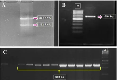
The integrity of RNA isolated from trophectodermal cells was
confirmed by two distinct bands corresponding to 18S and 28S
rRNAs on 1.5% agarose gel (Fig. 1A). PCR amplification of Ifnt resulted in product of 694 bp length (Fig. 1B). Following cloning, sequence analysis yielded 13 different cDNA sequences (Fig. 1C). All these 13 cDNA sequences (defined as buffalo IFN-T: 1a1, 1a2, 1a3, 1b1, 1b2, 2a, 2b, 3, 4, 5, 6, 7 and 8 in supplementaryfileFig. 1) represent an ORF (determined by DNAstar and DNA to PROTEIN software) of 588 bp coding for 195 amino acids. Out of these 13 cDNA sequences, eight cDNA sequences upon predicted translation (by DNA star and DNA to PROTEIN software) encoded eight distinct proteins differing from each other by at least one amino acid
(supplementaryfileTable 1). The cDNA sequences of these eight
distinct proteins have been submitted to NCBI Genbank data base (Accession numbers: JX481982, JX481987, JX481989, JX481990, JX481991, JX481992, JX481993 and JX481994. Interestingly, the newly identified buffaloIfn-tisoforms contain pronounced nucle-otide and amino acid sequence identity with each other (99.1e99.8%, 98e99%) but have only moderate sequence identity with reported buffaloIfn-t(90e92%, 82e86%) (supplementaryfile Figs. 1 and 2). Restfive sequences were differing at nucleotide level
without any change in amino acid sequence (supplementaryfile
Table 1).
Numeric values 1 to 8 represent distinct isoforms where dif-ference in nucleotide sequence results in change in amino acids. Letters “a” or “b” following first numerical represents variants under specific isoform, which differ in nucleotide sequence without changing amino acid. The third numerical denotes different posi-tional variants within same isoform (supplementaryfileTable 1).
The sequences of eight distinct isoforms of IFN-T in buffalo were compared to different isoforms of IFN-T in sheep, goat and cattle.
The similarity among all these buffalo IFN-T isoforms and five
bovine isoforms (Accession no: M31557, AF196320, AF196324, EU828780, EU828775), four sheep IFN-T (Accession no: M26386,
NM_001123400.1, NM_001101735, NM_001123401) andfive goat
IFN-T (Accession no: AY357327, AY357329, AY357332, AY357335,
AY357336) available in NCBI was 89.6e90.6%, 94.7e96.9% and
98.3e100%, respectively. Amino acid sequence analysis
(supple-mentaryfileFig. 2) and phylogenetic tree analysis (supplementary fileFig. 3) indicated high similarity of buffalo IFN-T sequences with
cattle, goat, and sheep IFN-T. This suggests that the BuIFN-T gene is closely related to IFN-T sequence of ruminants. However, among all ruminants, buffalo IFN-T group together with caprine IFN-T while
ovine and bovine IFN-T make separate groups (supplementaryfile
Fig. 3).
3.3. In silico analysis of predicted protein
The novel BuIFN-T proteins are predicted to contain structural features indicative of IFN-T and other Type I IFNs, includingfive
long helices (helixes AeE) by PSIPRED v3.3 Secondary Structure
Prediction server (supplementaryfileFigs. 1 and 4). In addition, the novel buffalo IFN-T proteins are expected to possess cysteine
resi-dues required for disulfide bridging (Cys24/Cys52 and Cys122/
Cys162) (DISULFIND). Sequence analysis of predicted translation product of all isoforms reveals 23 amino acids as signal peptide at N terminal (Signal IP 4.1 Server). It seems these isoforms do not have O-linked glycosylation, but some of the buffalo IFN-T isoforms contain a putative N-linked glycosylation site (Asn78) (netOGlyc 3.1 and netNGlyc 3.1 prediction program). Similarly, Netphos 2.0
al-gorithm predicted three ser (25th, 28th and 74th position), five
threonine (70th, 98th, 119th, 153rd and 163rd position) and two tyrosine (58th and 136th position) residues with potential phos-phorylation sites.
3.4. Expression of recombinant BuIFN-T isoforms
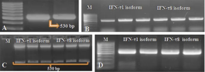
PCR products (Fig. 2A) of TOP10 cells, harbouring
isoforms-BuIFNT1a2 and BuIFNT8 in the pJET cloning vector, were subcloned into pET28 vector. Transformed recombinant plasmid pET28b-BuIFN-T1a2 and pET28b-BuIFN-T8 in TOP10 cells were screened by colony PCR (Fig. 2B). The positive colonies were cross-checked
by restriction digestion with Nco1 and Xho1 enzymes (Fig. 2C).
Thefinal positive clones transformed intoE. coliBL21 (DE3) strain were analysed for correct ORF of the insert sequence (Fig. 2D). On
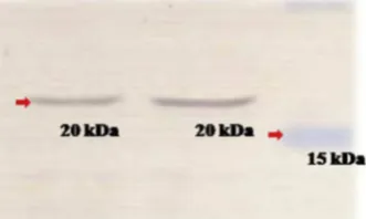
induction at 37C with 1 mM IPTG for 4 h, these recombinant
colonies expressed only insoluble aggregates of recombinant pro-tein (rBuIFN-T1a2 and rBuIFN-T8) of about 20 kDa as confirmed by SDS PAGE and mass spectroscopy (Fig.3A andsupplementaryfile). To improve the solubility of recombinant proteins, cultured cells
were induced for overnight at 25
C and for 22 h at 16
C with 0.5 mM IPTG. On the basis of band intensity in SDS-PAGE, it was observed that solubility of recombinant proteins improved at 16C (Fig. 3B).
3.5. Purification of recombinant BuIFN-T isoforms
Purification of the rBuIFN-T proteins through IMAC yielded two broad fractions- eluted fraction (during gradient of 300 mM Imid-azole) and washing fraction (i.e. fraction collected during washing with 20 mM Imidazole wash buffer). SDS PAGE analysis (of each isoform) showed that both the fractions contained purified protein as major band (Fig. 3C and D). Interestingly, the washing fraction was having very low level of contaminating proteins as compared to the eluted fraction. Finally, the purified T1a2 and rBuIFN-T8 were confirmed by western blotting (Fig. 4).
3.6. Antiviral activity of rBuIFN-T protein
After 30 h of virus infection, the test rBuIFN-T1a2 exhibited IFN activity corresponding to 10 IU/ml of standard bovine IFNT at 107 dilution. On estimation of total protein content in the lyophilized stock of test IFNT, it was found that 0.067107mg of rBuIFN-T1a2 resulted in 50% reduction in CPE after 30 h of viral infection exposure. The specific antiviral activity exhibited by rBuIFN-T1a2 was found to be 1.5108antiviral units/mg of protein parallel to the standard bovine IFN-T protein.
4. Discussion
Previously, variety of Ifn-ttranscripts have been identified in caprine[23], ovine[7]and bovine[11]conceptus. From this study it can be said that at least eight distinct IFN-T proteins encoded by 13 Ifn-tvariants are expressed in the buffalo conceptus. A single set of primers, derived from the consensus sequences of bovine and caprine, were used for PCR amplification of Ifn-t transcripts in buffalo trophectoderm. Due to high degree of homology between
the buffalo IFN-T isoforms, single set of primers amplified all
possibleIfn-tvariant transcripts. But some isoforms having varia-tion at the primer annealing site (because of degenerate sequences) might have been missed in this study. Owing to small size ofIfn-t, PCR generated errors were expected to be least, still the sequence obtained from two different labs were compared. The PCR sequence from two different labs were same and also the sequence readings of both the forward and reverse strands were same, so we can say with high confidence that there was no artefact in the sequence of the gene. Analysis of sequences gave rise to eight isoforms of buf-falo IFN-T encoded by 13Ifn-tcDNA variants. As per earlier reports Ifn-tevolved fromIfn-
u
by gene duplication about the timerumi-nants diverged from other artiodactyls [4]. PCR amplified and
sequencedIfn-tusing same primers on genomic DNA isolated from
blood, following BLAST revealedIfn-tto be closer toIfn-
u
. Hence,we advise a word of caution that probably because of similarity between Ifn-tand Ifn-
u
, Ifn-u
genes are preferentially amplified overIfn-t. Since, IFN-T expression is exclusive to conceptus, whichleads to relatively more mRNA transcripts of Ifn-t, reverse
transcriptase-PCR(RT-PCR) based approach is better than genomic (gDNA) based PCR.
Comparison of the nucleotide sequence of buffalo IFN-T iso-forms with other species IFN-T isoiso-forms followed by phylogenetic analysis revealed that BuIFN-T gene sequence is closely related to goat (96e100%), sheep (90e95%) and cattle (82e86%) IFN-T se-quences at amino acid level (supplementary file Fig. 2). Greater homology of buffalo IFN-T gene with caprine IFN-T gene may be correlated with similar reproductive physiology involved in em-bryo implantation, placentation and/or maternal recognition of pregnancy. A similarfinding has been reported in IFN-T gene ofBos frontalis,whereB. frontalisIFN-T gene was having more than 85% homology with IFN-T gene of other ruminants but in phylogenetic tree it made a clade with IFN-T gene of Red Deer[26]. Nevertheless,
about 89e90% homology of IFN-T gene among all ruminants
sug-gests that IFN-T gene is conserved across the species. Similar to the findings of Walker et al. (2009), in our study the overall similarity
among the thirteen buffaloIfn-tvariants was quite high ranging
between 99.1 and 99.8% at nucleotide level and 98e99% at amino
acid level.
Historically, different isoforms of IFN-T have been named and symbolized differently in many species. In our study the different isoforms and variants have been named in a very simple way. The sequences varying only in their amino acid sequence were considered distinct isoforms which resulted in eight main groups of isoforms namely 1, 2, 3, 4, 5, 6, 7 and 8. Under each group, se-quences which varied at nucleotides without altering the amino acids were considered as distinct variants ofIfn-tand denoted with isoform group number followed by letters a, b etc. Within such variants according to the position of variation these variants were
further distinguished as 1, 2, or 3. Sequence analysis of 30 clones resulting from two independent PCR products also revealed infor-mation about the relative abundance of various isoforms. The real time PCR based quantification of relative copy number would have been more informative. But as these isoforms were similar in se-quences, distinct primers amplifying each isoform coding mRNA transcripts could not be designed. Thus in our study, based upon frequency of obtaining a particular type of sequence, it could be said that particular variant (i.e. IFN-T1a2) is probably transcribed in more copy and hence, represents more clones in the pool of randomly selected large number of recombinant colonies. IFN-T expression is exclusive to blastocyst and its expression increases from morula till hatched blastocyst and decreases post implanta-tion[24]. Regarding role of different isoforms, it has been suggested that either specific isoform is unique to function during
implanta-tion[23] or to maintain enough molecules of IFN-T during
im-plantation, IFN-T genes might have been duplicated [11,25].
Functional implication of different isoforms of IFN-T suggests that they differ in their biological potency- like all the isoforms have antiluteolytic function and antiviral activity but some have high antiviral activity while others have stronger antiluteolytic activity [7,10].
It is interesting to note that none of the buffalo IFN-T contain O-linked glycosylation. But majority of the buffalo IFN-T isoforms contain a potential N-linked glycosylation site at amino acid posi-tion 78 (Asn78 in buffalo IFN-T1, T3, T4, T5, T6, T7 and T8) where as buIFN-T2 isoform does not contain this site (Asp78). This suggests that both N-glycosylated and non-glycosylated forms of IFN-T proteins are produced by the buffalo conceptus, like caprine
spe-cies [23]. In the ovine conceptus, some IFN-T isoforms contain
Asn78 residue but none of these proteins are glycosylated[7,27]. In contrast to buffalo or goat, all known boIFN-T isoforms contain Asn78 and all IFN-T produced by bovine trophectoderm are gly-cosylated[11,27]. The functional importance of glycosylated moi-eties on IFN-T remains mystery. Non-glycosylated recombinant boIFN-T and native boIFN-T have similar biological activities [11,28]. In addition, glycosylated and non-glycosylated caprine IFN-T proteins purified from goat conceptuses also have identical bio-logical activity[29]. Whether glycosylation enhances the functional lifespan of IFN-T activity in utero or not is unknown. Glycosylation of recombinant human IFN-B increased protein stability and bio-logical longevity[30]. IFN-T is a secretory protein which after its translation is secreted out of cell and acts on uterine epithelial cells [31]. Initial 23 amino acids are predicted to be signal peptide which is conserved across ovine, caprine and bovine except BuIFNT8 in which leucine has been replaced by proline at 7th position. In our study, out of total 10 potential ser, thr and tyr phosphorylation sites, most of them seem to be present in 2/3rd C-terminal region. Mol-ecules like IFN-T are involved in a cascade of signal transduction Fig. 3.SDS PAGE analyses of expressed and purified rBuIFN-T1a2 and rBuIFN-T8. Expression at 25C and 37C with 1 mM concentration of IPTG: lane M- Marker, lane 1- Uninduced (control), lane 2 and 3- BuIFN-T1a2 at 25C in pellet and soluble fraction, respectively, lane 4 and 5- BuIFN-T8 at 25C in pellet and soluble fraction, respectively, lane 6 and 7-BuIFN-T1a2 at 37C in pellet and soluble fraction, respectively and lane 8 and 9- BuIFN-T8 at 37C in pellet and soluble fraction, respectively (A). Expression at 16C with 1 mM IPTG: lane M- Marker, lane 1 and 6- Uninduced (control), lane 2 and 3- BuIFN-T1a2 pellet and soluble fraction, respectively, lane 4 and 5- BuIFN-T8 soluble and pellet fraction, respectively) (B). Purified rBuIFN-T1a2: lane 1, 2 and 3- Washing fraction, lane 4- Unbound, lane 5- Eluted fraction, lane 6- Cell lysate, lane 7- Cell pellet, lane M- marker (C). Purified rBuIFN-T8: lane 1- Unbound, lane 2- Eluted fraction, lane 3- Washing fraction, lane 4- Cell lysate, lane 5- Cell pellet, lane M- marker (D).
reaction[31]. The catalytic activity of IFN-T may be attributed to the C-terminal region as was revealed by mutagenesis study[32].
In our study, from sequence analysis it was found that IFN-T1a2 was the major isoform and IFN-T8 was the isoform which diverged maximally from other isoforms. So the expression of these two isomers (IFN-T1a2 and IFN-T8) as recombinant protein was tried. Now, despite the advances in eukaryotic expression systems,E. coli
remains the first host of choice for most researchers, as
over-production and purification of recombinant protein fromE. coliis faster, cheaper and easier than from other organisms. For these reasons, of the 58 biopharmaceuticals products approved from
2006 to 2010, 17 are produced in E. coli, 32 are produced in
mammalian cell lines (mainly Chinese hamster ovary; CHO) and four are produced inSaccharomyces cerevisiae, transgenic animals, or via direct synthesis[33]. However, the production of proteins to high yields remains a process of trial and error. Initially, as we tried
to express the recombinant proteins at 37
C with 1 mM IPTG for 4 h, it led to expression in the form of insoluble aggregates. To limit the in vivo aggregation of recombinant proteins cultivation of bacteria at reduced temperature, with reduced concentration of
inducer[34,35] has been proved to be effective. So, we tried to
express these proteins at 25C and 16C for overnight and 22 h, respectively with 0.5 mM IPTG. The solubility of these recombinant
proteins increased significantly at 16 C. Strong temperature
dependent hydrophobic interactions that determine the aggrega-tion reacaggrega-tions are favored at higher temperature[36]. At reduced temperature the heat shock proteases that are induced under over expression conditions are partially eliminated[37]. In E. colithe
activity and expression of chaperones are increased at 30 C
[38,39]. All these factors partially explain increased stability and correct folding at low temperature. Our result was in contrast to a reported literature[40]which reported that bovine IFN-T expressed as inclusion bodies at low temperatures. Finally, the recombinant proteins expressed at 16C were puri
fied by IMAC chromatography.
Affinity chromatography by nickelenitrilotriacetic acid (Ni-NTA) column resulted in two broader fractions, one was eluted fraction at 300 mM imidazole and other was the fraction collected during washing by 20 mM imidazole wash buffer. Recombinant protein was present as major band in both the fractions, as evident from
SDS PAGE analysis (Fig. 3C and D). Surprisingly, number of
contaminating bands in washing fraction was very less. Taking into account the greater yield of rBuIFN-T1a2, obtained in a single step
purification and its purification level (in terms of number of
contaminating bands) compared to rBuIFN-T8, we tried to examine its functional activity by antiviral assay. Interestingly, the antiviral
assay on CHPV revealed that 0.067 107 mg of rBuIFN-T1a2
protein was able to inhibit CPE up to 50% exhibiting a specific
antiviral activity of 1.5 108 antiviral units/mg of protein.
Compared to our previous work[41]the antiviral activity in this study was more, which may be due to the different cell line and virus used.
5. Summary/conclusion
In buffalo at least 13 distinctifntgene variants exist which are transcribed into mRNA. Theseifntgene variants are supposed to be encoding eight different proteins during early pregnancy by the buffalo conceptus. IFN-T seems to be a multicopy gene with small variations. Based upon frequency of obtaining a particular type of sequence, IFN-T1 may be the major isoform expressed at mRNA level in the buffalo trophectodermal cells. All BuIFN-T are closely related to bovine, caprine and ovine IFN-T. IFN-T in buffalo has 23 N-terminal amino acids as signal peptide. Majority of the isoforms seem to be N-glycosylated and heavily phosphorylated in 2/3rd C-terminal region. Relative abundance of individual isoforms can be
further validated for its functional significance. Out of 8 isoforms, IFN-T1a2 and IFN-T8 were successfully expressed inE. coli
expres-sion system and purified through IMAC chromatography. However,
both yield and purification of protein may be enhanced further.
Acknowledgement
We would like to thank Council of Scientific and Industrial
Research (CSIR), India for providing financial assistance to the
student under file number 09/130(59)/12-EMR-I in the form of
senior research fellowship and Indian Council of Agricultural Research (ICAR) for providing funds for the research. The authors declare that there is no conflict of interest that would prejudice the impartiality of this scientific work.
Appendix A. Supplementary data
Supplementary data related to this article can be found athttp:// dx.doi.org/10.1016/j.pep.2016.02.005.
References
[1] J. Martal, M.C. Lacroix, C. Loudes, M. Saunier, S. Wintenberger-Torres, Troph-oblastin, an antiluteolytic protein present in early pregnancy in sheep, J. Reprod. Fertil. 56 (1979) 63e73. F.F.
[2] R.M. Bartol, F.W. Roberts, G.S. Bazer, J.D. Lewis, W.W. Godkin, Thatcher, characterization of proteins produced in vitro by periattachment bovine conceptuses, Biol. Reprod. 32 (1985) 681e693.
[3] J.D. Godkin, F.W. Bazer, W.W. Thatcher, R.M. Roberts, Proteins released by cultured day 15e16 conceptuses prolong luteal maintenance when intro-duced into the uterine lumen of cyclic ewes, J. Reprod. Fertil. 71 (1984) 57e64. [4] R.M. Roberts, L. Liu, Q. Guo, D. Leaman, J. Bixby, The evolution of the type I
interferons, J. Interf. Cytok. Res. 18 (1998) 805e816.
[5] R.M. Roberts, A.D. Ealy, A.P. Alexenko, C.S. Han, T. Ezashi, Trophoblast in-terferons, Placenta 20 (1999) 259e264.
[6] K. Imakawa, T.R. Hansen, P.V. Malathy, R.V. Anthony, H.G. Polites, K.R. Marotti, Molecular cloning and characterization of complementary deoxyribonucleic acids corresponding to bovine trophoblast protein-1: a comparison with ovine trophoblast protein-1 and bovine interferon-alpha II, Mol. Endocrinol. 3 (1989) 127e139.
[7] G.L. Winkelman, R.M. Roberts, P.A. James, A.P. Alexenko, A.D. Ealy, Identifi -cation of the expressed forms of ovine interferon-tau in the periimplantation conceptus: sequence relationships and comparative biological activities, Biol. Reprod. 61 (1999) 1592e1600.
[8] R.M. Roberts, J.C. Cross, D.W. Leaman, Interferons as hormones of pregnancy, Endocrinol. Rev. 13 (1992) 432e452.
[9] J.C. Cross, R.M. Roberts, Constitutive and trophoblast-specific expression of a class of bovine interferon genes, Proc. Natl. Acad. Sci. U. S. A. 88 (1991) 3817e3821.
[10] A.D. Ealy, J.A. Green, A.P. Alexenko, D.H. Keisler, R.M. Roberts, Different ovine interferon-tau genes are not expressed identically and their protein products display different activities, Biol. Reprod. 58 (1998) 566e573.
[11] A.D. Ealy, S.F. Larson, L. Liu, A.P. Alexenko, G.L. Winkelman, H.M. Kubisch, Polymorphic forms of expressed bovine interferon-tau genes: relative tran-script abundance during early placental development, promoter sequences of genes and biological activity of protein products, Endocrinology 142 (2001) 2906e2915.
[12] T.R. Hansen, K. Imakawa, H.G. Polites, K.R. Marotti, R.V. Anthony, R.M. Roberts, Interferon RNA of embryonic origin is expressed transiently during early pregnancy in the ewe, J. Biol. Chem. 263 (1988) 12801e12804.
[13] J.J. Hernandez-Ledezma, J.D. Sikes, C.N. Murphy, A.J. Watson, G.A. Schultz, R.M. Roberts, Expression of bovine trophoblast interferon in conceptuses derived by in vitro techniques, Biol. Reprod. 47 (1992) 374e380.
[14] J. Parent, C. Villeneuve, A.P. Alexenko, A.D. Ealy, M.A. Fortier, Influence of different isoforms of recombinant trophoblastic interferons on prostaglandin production in cultured bovine endometrial cells, Biol. Reprod. 68 (2003) 1035e1043.
[15] J.L. Vallet, F.W. Bazer, M.F. Fliss, W.W. Thatcher, Effect of ovine conceptus secretory proteins and purified ovine trophoblast protein-1 on interoestrous interval and plasma concentrations of prostaglandins F-2 alpha and E and of 13,14-dihydro-15-keto prostaglandin F-2 alpha in cyclic ewes, J. Reprod. Fertil. 84 (1988) 493e504.
[16] K.J. Demmers, K. Derecka, A. Flint, Trophoblast interferon and pregnancy, Reprod 121 (2001) 41e49.
[17] A.M. Walker, K. Kimura, R.M. Roberts, Expression of bovine interferon-tau variants according to sex and age of conceptuses, Theriogenology 72 (2009) 44e53.
interferon-tau, J. Interf. Cytok. Res. 20 (2000) 817e822.
[19] M.S. Chauhan, S.K. Singla, P. Palta, R.S. Manik, M.L. Madan, In vitro maturation and fertilization, and subsequent development of buffalo (Bubalus bubalis) embryos: effects of oocyte quality and type of serum, Reprod. Fertil. Dev. 10 (1998) 173e177.
[20] S. Saugandhika, D. Kumar, M.K. Singh, R. Shah, T. Anand, M.S. Chauhan, R.S. Manik, S.K. Singla, P. Palta, Effect of sodium nitroprusside, a nitric oxide donor, and aminoguanidine, a nitric oxide synthase inhibitor, on in vitro development of buffalo (Bubalus bubalis) embryos, Reprod. Dom. Anim. 4 (2010) 931e933.
[21] R. Erck, M. Dayhoff, Atlas of Protein Sequence and Structure, Natl Biomed Res Found, Silver Springs MD, 1966.
[22] V.P. Bondre, R.S. Jadi, A.C. Mishra, P.N. Yergolkar, V.A. Arankalle, West Nile virus isolates from India: evidence for a distinct genetic lineage, J. Gen. Virol. 88 (2007) 875e884.
[23] A.D. Ealy, S. Wagner, A. Sheils, N. Whitley, D. Kiesling, S. Johnson, G. Barbato, Identification of interferon-T isoforms expressed by the peri-implantation goat (Capra hircus) conceptus, Dom. Anim. Endocrinol. 27 (2004) 39e49. [24] M. Takahashi, H. Takahashi, S. Hamano, S. Watanabe, S. Inumaru, M. Geshi,
K. Okuda, Y. Yokomizo, A. Okano, Possible role of interferon tau on in vitro development of bovine embryos, J. Reprod. Dev. 49 (4) (2003) 297e305. [25] F.W. Bazer, T. Spencer, T. Ott, Interferon tau: a novel pregnancy recognition
signal, Am. J. Reprod. Immunol. 37 (6) (1997) 412e420.
[26] K. Rajaravindra, A. Mitra, A. Sharma, M. Sitangshu, A. Sharma, Molecular characterization of the interferon-tau gene of the mithun (bos frontalis), Zool. Sci. 23 (7) (2007) 607e611.
[27] R.V. Anthony, S.D. Helmer, S.F. Sharif, R.M. Roberts, P.J. Hansen, W.W. Thatcher, Synthesis and processing of ovine trophoblast protein-1 and bovine trophoblast protein-1, conceptus secretory proteins involved in the maternal recognition of pregnancy, Endocrinology 123 (1988) 1274e1280. [28] S.W. Klemann, J.Z. Li, K. Imakawa, J.C. Cross, H. Francis, R.M. Roberts, The
production, purification, and bioactivity of recombinant bovine trophoblast protein-1 (bovine trophoblast interferon), Mol. Endocrinol. 4 (1990) 1506e1514.
[29] M. Guillomot, P. Reinaud, B.C. La, G. Charpigny, Characterization of conceptus-produced goat interferon tau and analysis of its temporal and cellular
distribution during early pregnancy, J Reprod. Fertil. 112 (1998) 149e156. [30] L. Runkel, W. Meier, R.B. Pepinsky, M. Karpusas, A. Whitty, K. Kimball,
Structural and functional differences between glycosylated and non-glycosylated forms of human interferon-beta (IFN-beta), Pharmcol. Res. 15 (1998) 641e649.
[31] F.W. Bazer, Pregnancy recognition signaling mechanisms in ruminants and pigs, J. Anim. Sci. Biotech. 4 (1) (2013) 23.
[32] C.H. Pontzer, T.L. Ott, F.W. Bazer, H.M. Johnson, Structure/function studies with interferon Tau: evidence for multiple active sites, J. Interf. Cytok. Res. 14 (3) (1994) 133e141.
[33] G. Walsh, Biopharmaceutical benchmarks 2010, Nat. Biotechnol. 28 (2010) 917e924.
[34] C.H. Schein, Production of soluble recombinant proteins in bacteria, BioTechnology 7 (1989) 1141e1148.
[35] J.A. Vasina, F. Baneyx, Expression of aggregation prone recombinant proteins at low temperatures: a comparative study of the Escherichia coli cspA and tac promoter systems, Protein Expr. Purif. 9 (1997) 211e218.
[36] T. Kiefhaber, R. Rudolph, H.H. Kohler, J. Buchner, Protein Aggregation in Vitro and in Vivo: a Quantitative Model of the Kinetic Competition between Folding and Aggregation, 1991, pp. 825e829. Biotechnology (NY) 9.
[37] J.A. Chesshyre, A.R. Hipkiss, Low temperatures stabilize interferon a-2 against proteolysis in methylophilus methylotrophus and Escherichia coli, Appl. Microbiol. Biotechnol. 31 (1989) 158e162.
[38] A. Mogk, M.P. Mayer, E. Deuerling, Mechanisms of protein folding: molecular chaperones and their application in biotechnology, Chembiochem 3 (2002) 807e814.
[39] M. Ferrer, T.N. Chernikova, M.M. Yakimov, P.N. Golyshin, K.N. Timmis, Chap-eronins govern growth of Escherichia coli at low temperature, Nat. Biotechnol. 21 (2003) 1266e1267.
[40] G.F. Fang, W.Z. Yi, Z.S. Ming, Bovine interferon-tau expression in Escherichia coli and identification of its biological activities, J. Agric. Biotech. 16 (2) (2008) 208e213.