THE JOURNAL OF TROPICAL LIFE SCIENCE OPEN ACCESS Freely available online
VOL. 7, NO. 1, pp. 1 - 7, January 2017 Submitted March 2016; Revised September 2016; Accepted January 2017
Cross Reaction among Antibody Pili subunit Hemagglutinin Proteins and Outer Membrane subunit
Hemagglutinin Proteins of Shigella flexneri
Avin Ainur Fitrianingsih 1,2
*, Lailia Nur Rachma 1,2
, Alvi Milliana 1,2
, Tinny Endang Hernowati 3
, Aulanni’am 4
, Sanarto Santoso 5 , Sumarno Reto Prawiro 5
1 Biomedical Magister Program, Medical Faculty, Brawijaya University, Malang, Indonesia
2 Science and Technology Faculty, Islamic State University, Malang, Indonesia
3
Department of Clinical Pathology Medical Faculty, Brawijaya University, Malang, Indonesia 4
Departement of Biology Natural Science Faculty, Brawijaya University, Malang, Indonesia 5
Department of Microbiology, Medical Faculty, Brawijaya University, Malang, Indonesia
ABSTRACT
Shigella flexneri is the most common causal agent of shigellosis. Its pili are composed of pili protein subunits. Adhesion molecules can be found on the pili and outer membrane proteins (Omp). A hemagglutination reaction can be used for screening of adhesion molecules. Objectives: The purpose of this study was to determine the molecular weight of the pili protein subunits and outer membrane proteins of S. flexneri that act as hemagglutinin proteins, and to prove whether there is a crossreaction between antibodies against hemagglutinin pili protein sub -units and outer membrane proteins of S. flexneri. Methods: Pili protein sub-units were isolated using pili bacteria cutters, and the outer membrane proteins were solubilized and obtained using sodium dodecyl sulfate 0.05% as detergent for Omp isolation. The hemagglutination reaction used mice erythrocytes. The cross reactions between subunit pili proteins were conducted by Western blot and Dot blot. Results:. Antibodies against hemagglutinin sub unit pili protein 18 kDa responded to pili protein subunits 18 kDa; 23 kDa; 34 kDa; and 53 kDa; and Omp 23 kDa and 27 kDa. Omp and subunit pili proteins S. flexneri consists of several identical epitopes that were respon-sible for the similarity of the response profile in the cross-reactions of antibodies.
Keywords: Pili, Omp, hemagglutinin, cross reaction, Shigella flexneri
Shigellosis is a type of diarrhea caused by Shigella spp. Shigellosis is an endemic disease that occurs mainly in developing countries and is the most com-mon cause of bloody diarrhea. These diseases are en-demic and cause 120 million cases each year with se-vere dysentery. In developing countries endemic shigel-losis is a significant cause of morbidity and mortality especially in children under five [1].
Shigella is Gram-negative bacteria in the family En-terobacteriaceae, consisting of non-motile bacilli. Shigella genus consists of four species: S. dysenteriae, S. flexneri, S. sonnei and S. boydii [2]. From the four species of Shigella, S. flexneri most commonly causes
dysentery and is recorded as causing 60 % of cases of shigellosis in developing countries [3].
The outer membrane protein (Omp), is known as an adhesion molecule in many species of bacteria and as well as Shigella, allowing bacteria to attach to cell receptors. [4]. Many pathogenic bacteria use a two-stage process of attachment: the first two-stage, where the bacteria is loosely attached with pili, and then the sec-ond stage, a stronger bsec-ond with the surface cell recep-tors using Omp. Hemagglutination activity is directly correlated with the colonization ability of Shigella spp. in a suckling mouse model [5]. Both pili and Omp hemagglutinin are virulence factors for infection-caus-ing bacteria colonization [6].
INTRODUCTION
*Corresponding author: Avin Ainur Fitrianingsih
Medical Faculty, Brawijaya University Jalan Veteran, Malang, Indonesia 65145 E-mail: avinainur@yahoo.com
How to cite:
Developing vaccines against bacterial adhesion mol-ecules may have an advantage in that the immune re-sponse may better eliminate bacteria and will not gen-erate other side effects when compared with a whole cell–based vaccine or a bacterial lipopolysaccharide. Antibodies produced against Omp S. flexneri 34 kDa also may react with other Shigella species [4]. Recent research has shown that the Omp S. flexneri 2a is one of the most immunodominant antigens in the outer membrane of gram-negative bacteria and has many de-sirable characteristics for a candidate vaccine. Omp S. flexneri 2a is cross-reactive to antigens and common among Shigella spp. and is a widely recognized epitope on the cell surface and able to generate protective im-munity in mice. Protective imim-munity involves the par-ticipation of both humoral and cellular immune re-sponses, as OmpA increased rapidly induced IgG and IgA in both systemic and mucosal compartments and also activated Th1 cells [7, 8].
Recent studies have clarified that the anti-subunit hemagglutinin pili protein 7.9 kDa and subunit hemag-glutinin pili protein 49.8 kDa S. dysenteriae are adhe-sion molecules [9, 10]. Subsequent studies have con-firmed that there are similar epitopes between subunit pili protein 7.9 kDa S. dysenteriae and subunit pili tein 7.9 kDa S. boydii, and between subunit pili pro-tein 49.8 kDa S. dysenteriae and pili subunit propro-tein 49.8 S. boydii as well, but differences in epitopes than with sub unit pili 7.9 kDa and 49.8 kDa protein in S. flexneri and S. sonnei [11].
This research was conducted in order to clarify whether there is any cross-reaction among hemagglu-tinin subunit pili proteins and Omp S. flexneri. The re-sult may be very important in designing a vaccine for shigellosis using an adhesion molecule–based vaccine.
Culture of Shigella flexneri
Cultures of S. flexneri used in this research were derived from the Health Research Laboratory in DI Yogyakarta Indonesia. S. flexneri were grown in Mac-Conkey’s or salmonella-shigella agars. A Carbonate Thiaproline Glutamate (TCG) medium was used to en-rich the growth of S. flexneri pili. Bacterial culture on TCBS media were harvested and put in a bottle con-tained 1000 mL of brain-heart infusion broth (BHI). The bottles were then shaken for 30 minutes in a water bath at a temperature of 37°C. Furthermore, from the bottle of 10 mL bacterial suspension included in each bottle that already contained media TCG, then the bac-teria in media TCG incubation was conducted at a
temperature of 37°C for 2 × 24 hours.
Method of harvesting subunit pili protein and isolating Omp from Shigella flexneri
In order to harvest the pili, we applied the method using a pili cutter as described in Sumarno [12], which was a slight modification of Evan’s method. Modifica-tions that were made included precipitating the free pili and flagella from the bacteria after the last round of cutting and further separation using column chro-matography was not used. Pellets were suspended with PBS at pH 7.4 until the volume reached 5 times the volume of the pellet, then SDS was added until the concentration reached 0.05%. Then the mixture was homogenized using a vortex mixer at full speed for 1 minute. The mixture was then centrifuged at 12,000 rpm at 4°C for 15 minutes. The supernatant was col-lected and stored at 4°C [19].
Sodium Dodecyl Sulfate Polyacrylamide Electrophoresis (SDS-PAGE)
For protein identification and characterization on the electrophoresis gels, the protein stain comassie bril-liant blue was used along with standard low-range molecular markers from Sigma Chemical. After calcu-lating the molecular weight of the protein samples, SDS-PAGE was replicated for times to get a sufficient amount of protein for further analyses [13].
Subunit pili protein and Omp purification.
Purification of subunit pili protein and Omp used the method of electroelution [9]. Bands of interest in the SDS-PAGE gels were cut perpendicularly so that each piece contained one protein band. The cut bands were collected and inserted into a piece of membrane tape that was filled with an electrophoresis running buffer. The membrane was put in a horizontal elec-trophoresis apparatus, and the protein from each eluted for 90 minutes under 120 mV of current. Fol-lowing this, the membrane tape was dialyzed in a PBS pH 7.4 fluid buffer for 28 h with the replacement of the buffer 4 times in between (replaced 4 times after 7 minute intervals).
Hemagglutination test methods
Hemagglutination assays were performed according to Hanne and Finkelstein [14]. Sample dilutions were made at half-concentration in micro plates with well volumes of 50 µL. In every well, mouse red blood cell suspensions at a concentration of 0.5% was added in the same volume. These were then shaken using a
tator plate for 1 minute. Subsequently plates were incu-bated at room temperature for 1 hour. The titer was determined by observing the agglutination of red blood cells at the lowest dilution.
Production of polyclonal antibodies
Mice were acclimatized for 4 days before immu-nization. The antigen used was subunit pili proteins from S. flexneri. Mice were injected with antigen emul-sified with Complete Freud's Adjuvant (CFA), in-traperitoneal, 100 µg/µL doses. Booster injections were performed in weeks two to four using antigens emulsi-fied with Incomplete Freud's Adjuvant (IFA). A booster dose of 0.1 mL was used in a intraperitoneal injection. Serum was taken 1 week after the last booster [15].
Western Blotting method
Western blots were performed according to the Bio-rad technical protocol [16]. The SDS-PAGE gels con-taining protein bands were transferred onto nitrocellu-lose (NC) membranes using a semi-dry blotter (Bio-rad). After that, the NC membrane was incubated overnight and washed with TBE plus 0.05% tween-20 twice. Next, the primary antibody of mice IgG at con-centrations of 1/1000 in TBE pH 7.4 containing 1% so-lution of BSA was applied to the NC. The NC was washed again using the same solution and then an anti-mouse secondary antibody IgG at a concentration of 1/1000 in TBE pH 7.4 and a 1% solution of BSA were added. Subsequently the NC was washed twice for 5 minutes using TBE pH 7.4 with tween-20 at a concentration of 0.05%. For staining, Cip β tablets dis-solved in 10 mL H2O were applied after this washing.
Dot Blot method of examination
Dot blots are used to detect reactions with serologi-cal specificity between antigens and antibodies. The NC were trimmed into measuring 7.5 × 11 cm rectan-gles and inserted between two pieces of metal blotter apparatus. Then they were mounted on the dot blot, incubated overnight at a temperature of 4°C and de-gassed until the antigen was completely absorbed into the NC membrane. Further TBS blocking was carried out with blocking buffer (containing 50 mM Tris Base, 0.2 M NaCl, 5% skim milk, pH 7.4). Next, 50 mL of primary antibody was added, incubated for 2 h at room temperature and then placed in a shaker. The solution was removed, and then washed 3 times with TBS-0.05% Tween-20. Next, the secondary antibody with 1 : 2500 dilution was added. Again the NC was washed
3 times with TBS 0.05% Tween-20 and chromogenic substrate (BCIP-NBT) was added [17].
Data analysis
The data obtained from the results of antigen-anti-body reaction by Dot Blot method were transformed in Corel Photo Paint. The results of the interpretation of data on Corel Photo Paint were then analyzed using ANOVA statistical test and correlation test.
The isolated sub unit pili protein and Omp of Shigella flexneri
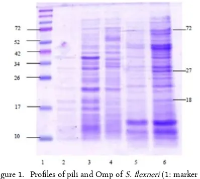
The subunit pili protein S. flexneri and Omp pro-files are shown in Figure 1. Propro-files and the calculation of molecular weight (MW) of the subunit pili and Omp of S. flexneri showed a similar picture. Our result showed that the isolated protein derived from pili and Omp with a molecular weight of 72 kDa.
Figure 1. Profiles of pili and Omp of S. flexneri (1: marker pro-tein; 2 and 3: pili slices 1 and 2 of S. flexneri; 3, 4, and 5 Omp isolation 1 and 2 of S. flexneri)
The aim of the erythrocyte agglutination test was to see whether purified subunit pili protein and Omp have the ability to perform mouse erythrocyte aggluti-nation (hemagglutiaggluti-nation). The results hem agglutina-tion sub unit pili protein and Omp S. flexneri shown in Figure 2. The hemagglutination test results showed that the subunit pili protein 18 kDa S. flexneri showed the highest titer (1/128), therefore this protein was se-lected for further research.
The hemagglutination test carried out on Omp S. flexneri from three 3 band purification results are shown in Figure 3. The results showed that the Omp 72 kDa, 27 kDa and 18 kDa proteins can agglutinate erythrocyte at a titer of 1/128.
Figure 2. Hemagglutination test of subunit pili protein S. flexneri shows negative agglutination (red arrow) and positive agglutination (blue arrow). (A) Hemaggluti-nation result of sub unit pili protein 72 kDa S. flexneri. (B) Hemagglutination result of sub unit pili protein 27 kDa S. flexneri. (C) Hemagglutination re-sult of sub unit pili protein 18 kDa S. flexneri. (CT) Control (PBS + erythrocyte)
Figure 3. Hemagglutination test of purified Omp S. flexneri shows negative agglutination (red arrow) and posi-tive agglutination (blue arrow). (A) Hemagglutina-tion result of Omp 72 kDa S. flexneri. (B) Hemag-glutination result of Omp 27 kDa S. flexneri. (C) Hemagglutination result of Omp 18 kDa S. flexneri. (CT) Control (PBS + erythrocyte)
Antigen-antibody reaction by the method of Western Blotting
The Western blot test was used to assess the anti-gen-antibody reaction using antibodies against subunit pili protein 18 kDa S. flexneri (the highest titer) to its subunit pili 18 kDa S. flexneri antigen, and with others sub unit pili protein and Omp S. flexneri.
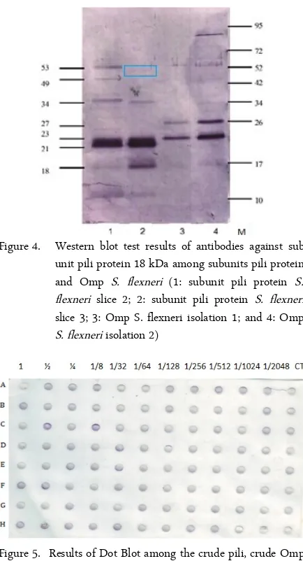
Figure 4 shows the results of the expression of anti-gen-antibody reaction of subunit pili protein and Omp S. flexneri with antibodies against subunit pili protein 18 kDa S. flexneri. Subunits of the pili protein with molecular weights of 18 kDa; 23 kDa; 34 kDa; 49 kDa and 53 kDa S. flexneri responded to antibodies against sub unit pili protein 18 kDa S. flexneri. Meanwhile an-tibodies against sub unit pili protein 18 kDa S. flexneri responded to OMP S. flexneri with a molecular weight of 23 kDa and 27 kDa.
Antigen-antibody reaction by the method of Dot Blot The results of the antigen-antibody reaction using the dot blot method can be seen in Figure 5; the results
Figure 4. Western blot test results of antibodies against sub unit pili protein 18 kDa among subunits pili protein and Omp S. flexneri (1: subunit pili protein S. flexneri slice 2; 2: subunit pili protein S. flexneri slice 3; 3: Omp S. flexneri isolation 1; and 4: Omp S. flexneri isolation 2)
Figure 5. Results of Dot Blot among the crude pili, crude Omp and sub unit pili protein of S. flexneri with the anti-bodies against sub unit pili protein 18 kDa S. flexneri (1/500, 1/1000, 1/2000……..1/1024000 dilutions). (A) The crude sub unit pili protein, (B) the sub unit pili protein 72 kDa, (C) the sub unit pili protein 27 kDa, (D) the sub unit pili protein 18 kDa, (E) the crude Omp, (E) the Omp 72 kDa (F) the Omp 27 kDa Omp, and (G) the Omp 18 kDa.
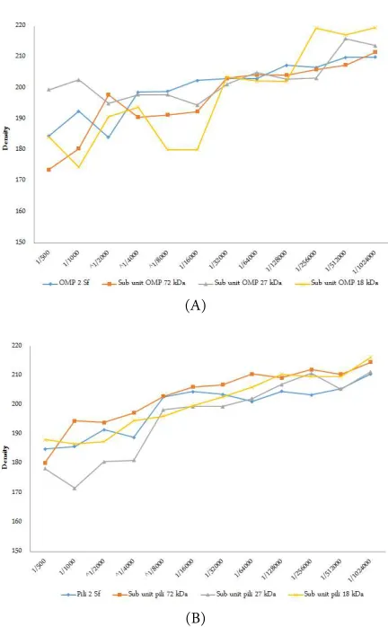
of calculation of these antigen and antibody reactions using the semi-quantitative Dot Blot method is de-picted in Figure 6.
There were significant differences (p = 0.000 < α)
on all 8 different types of S. flexneri antigens, i.e., pieces of pili 2 S. flexneri; subunit pili protein purified 72 kDa, 27 kDa, 18 kDa; crude Omp S. flexneri; and subunit Omp 72 kDa, 27 kDa, 18 kDa.
relationship between treatment pili protein 2 S. flexneri with antibody concentrations (p = 0.000 < α)
with a coefficient of correlation r = 0.895. For subunit pili protein 72 kDa S. flexneri, there was a meaningful relationship with pili protein antibody concentrations (p = 0.000 < α) with a correlation coefficient r = 0.924.
There was also a significant relationship between sub-unit pili protein 27 kDa, 18 kDa, Omp S. flexneri, sub unit Omp 72 KDa, 27 kDa and 18 kDa with pili pro-tein antibody concentrations (p = 0.000 < α) with each
of the correlation coefficient r = 0.932; 0.979; 0.922; 0.913; 0.731; and 0.879.
The attachment of fimbriae or pili and other surface molecules to the surface of a host cell is mediated by specific receptors called adhesion molecules. The bond between the adhesion molecules with the receptor will activate signal transduction in host cells for the initial activation in the pathogenesis process and increased bacterial colonization [18].
The observation and calculation of molecular weight profiles for the pili protein subunits and Omp S. flexneri shows the same picture as depicted in Fig 1. The first piece pili subunit band seem thinner than the second and third pieces. Similarly, the same finding was found in the first and second Omp isolation. First isolated produce images that are thinner than in isola-tion to second. These differences in Omp band thick-nesses could be caused by the location of the protein on the cell wall. The thickess differences in the pili protein subunits, however, could be caused by differ-ences in the speeds used in the pili cutter.
During pili formation, sub unit pili (pilins) secreted into the periplasmic space through the secretory path-way and binds to chaperon (companion) which assist the folding process and prevent the formation of pre-mature sub-units. Then the complex pili/ chaperones brought into outer membrane usher which serves as a platform for the creation of pili. Then, the complex proteins form pores in the outer membrane that allows impassable helical strands [20, 21].
Three bands of both protein sub units pili and Omp with each molecular weight 72 kDa; 27 kDa; and 18 kDa were taken as the hemagglutinin protein candi-date. This selection was based on the relative thickness of the band protein.
The hemagglutination test aims is to find protein hemagglutinin activity of S. flexneri pili proteins. The hemagglutinin protein is identical to the adhesion mol-ecule that is responsible for the adhesion of bacteria to the host cell [10]. Therefore, the hemagglutination re-action is a screening tool for bacterial adhesion
mole-cules, which allowed these adhesion molecules to be used as a tool for diagnosis candidates and vaccine component [12, 22].
At times, precipitation of erythrocyte cells occurred in the bottom of the well showing no agglutination. This was caused by the absence of the bond between erythrocyte cells with pili protein subunits S. flexneri. Hemagglutinin protein is considered as one of the vir-ulence factors of pathogenic bacteria. Bacteria that are able to perform agglutination and attach to the ery-throcyte cell also have the ability to attach to receptors found on host mucosal cells. This is because the recep-tors that exist in the erythrocyte cell membrane are be-lieved to have similarities with receptors on host cell mucosa [23].
The Western blot was results showed a maximum yield of antigen dilution pili sub units BM 18 kDa pro-tein S. flexneri at a titer of 1/20, whereas the maximum results for antibody against pili sub units S. flexneri 18 kDa protein was at 1/500.
The Marrack Lattice theory states that if an antigen and antibody molecule react to form a precipitate, it will produce a different concentration ratio depending on the reaction zone in which the precipitin is formed. If the antibody-antigen ratio is above 1, it will settle. Whereas when the ratio is below 1, will remain in complex formed in the supernatant. The results of anti-gen-antibody reaction can be described in terms of a rainfall curve, which describes a rising curve shape as an excess antibody zone, which means a lot of free an-tibody molecules are in the supernatant, while a de-scending curve illustrates an excess antigen zone, which means a lot of free antigen molecules in the su-pernatant. Maximum precipitation is in the zone where there is no equivalent antigen or antibody detected in the supernatant [24].
From the results of the check board above it can be concluded that the equivalent zone lies in antibody and antigen titers between 1/500 and 1/20. Antigen-body bond formation requires the interaction of anti-body bivalent and multivalent antigens that produces a complex bond. The more epitopes recognized by anti-bodies, the more and increasingly complex the bonds formed [25].
(A)
(B)
Figure 6. (A) The result of dot blot protein crude pili and sub unit pili protein purified with antibodies against sub unit pili protein 18 kDa S. flexneri. (B) The result of dot blot protein crude Omp and Omp purified with antibodies against sub unit pili protein 18 kDa S. flexneri
antibody against Omp S. flexneri was a protein with a molecular weight of 23 kDa and 27 kDa. This result seemed to be caused by a common epitopes which found on the sub unit pili protein and Omp S. flexneri. This study results are related to the character of pili proteins in gram-negative bacteria as a group of media adhesion and self-defense, in order to recognize and bind to at least one epitope molecule. The results also showed that there are similarities epitopes of the pro-tein constituent subunits pili and Omp S. flexneri, so both can function as adhesin proteins.
Based on Anam (2015), it was found that there are predicted epitope similarities between the 7.9 kDa pro-tein pili subunits of S. dysenteriae with 7.9 kDa propro-tein pili subunits of S. boydii and the 49.8 kDa protein pili subunits of S. dysenteriae with 49.8 protein pili sub-units of S. boydii as well, but differences in the
epi-topes between the 7.9 kDa and 49.8 kDa protein pili subunits of S. flexneri and S. sonnei. [11].
The statistical analysis found that there were signifi-cant differences in treatment between antibody reac-tion with the antigen pili proteins (p = 0.000 < α). The
value of the lowest mean reaction between antibodies contained in the pili with an antigen protein is 27 kDa subunit. This means that the antigen protein 27 kDa subunit of pili reacted most stably/strongly with the pili protein antibodies. In the treatments with Omp antigens and antibodies to the subunit pili protein 18 kDa there were also significant differences (p = 0.000 <
α). This seems to mean that the antibody sub unit pili
protein when reacted with Omp antigen with different molecular weights will produce different antibody-anti-gen bond strengths.
Furthermore, the correlation of test results also showed significant correlation (p = 0.000 < α) between
the treatment of pili proteins and antibody concentra-tion Omp against pili proteins 18 kDa S. flexneri. It can be concluded that the smaller concentration of an-tibody gave a smaller density value of the antigen anti-body reaction. This may indicate an antigen excess.
The results of this study can be summarized as fol-lows: hemagglutinin subunit pili protein and Omp 72 kDa, 27 kDa and 18 kDa is present in S. flexneri. An-tibodies against hemagglutinin subunit pili protein 18 kDa can responded to the sub unit pili proteins 18 kDa, 23 kDa, 34 kDa, and 53 kDa, as well as to Omp 23 kDa and 27 kDa. This likely indicates that the sub unit of pili and Omp S. flexneri consist of several iden-tical epitopes and is thus responsible for the similarity of the response profile in the cross-reactions of anti-bodies.
This research was supported by the Ministry of Health of Indonesia in the scheme of Iptekdok 2014.
1. Niyogi SK (2005) Shigellosis. Journal of microbiology (Seoul, Korea) 43 (2): 133–143.
2. van der Ploeg C, Vinas M, Terragno R et al. (2010) Title: Laboratory protocol: Serotyping of Shigella spp. protocol number: 2010GFNLAB003 Effective. http://www.antimi-crobialresistance.dk/. Accessed: February 2017.
3. WHO (2005) Shigellosis: Disease burden, epidemiology and case management. Weekly Epidemiological Record 80 (11): 94–99.
CONCLUSION
ACKNOWLEDGMENT
4. Pore D, Mahata N, Pal A, Chakrabarti MK (2010) 34kDa MOMP of Shigella flexneri promotes TLR2 mediated macrophage activation with the engagement of NF- Bκ and p38 MAP kinase signaling. Molecular Immunology 47 (9): 1739–1746. doi: 10.1016/j.molimm.2010.03.001. 5. Mitra S, Saha DR, Pal A et al. (2012) Hemagglutinating
activity is directly correlated with colonization ability of Shigellae in suckling mouse model. Canadian Journal of Microbiology 58 (10): 1159–1166. doi: 10.1139/w2012-095.
6. Salyers AA, Whitt DD (2002) Bacterial pathogenesis : A molecular approach. Washington DC, ASM Press. 7. Pore D, Chakrabarti MK (2013) Outer membrane protein
A (OmpA) from Shigella flexneri 2a: A promising subunit vaccine candidate. Vaccine 31 (36): 3644–3650. doi: 10.1016/j.vaccine.2013.05.100.
8. Chakrabarti MK (2010) A simple approach towards the development of a Shigella vaccine. Al Ameen Journal of Medical Sciences 3 (4): 263–264.
9. Agustina W, Fitri LE, Yudani T et al. (2012) Antibody protein hemagglutinin subunit pili with MW 49,8 kDa Shigella dysenteriae can inhibit Shigella dysenteriae adhe-sion on mice enterocyte. IOSR Journal of Pharmacy 2 (5): 13–20.
10. Sumarno RP, Avanita AS, Winarsih S et al. (2015) Haemaglutination of Shigella dysenteriae subunit pili pro-tein with anti-haemaglutination of S. dysenteriae subunit pili protein as a molecule adhesion in mouse enterocytes. African Journal of Microbiology Research 9 (11): 781– 787. doi: 10.5897/AJMR2014.7273.
11. Anam K, Nurdiana, Hernowati TE et al. (2016) Cross im-munity among the 49.8 KDa pili subunit hemagglutinin proteins and 7.9 KDa pili subunit anti hemagglutinin pro-teins of Shigella spp. |. International Journal of Pharmacy and Pharmaceutical Research 7 (2): 19–30.
12. Sumarno RP, Yanuhar U, Winarsih S, Islam S, Santoso S (2012) Detection of molecule adhesion sub-unit pili 48 kDa Salmonella typhi by immunochemistry method using sera patients suffering from typhoid fever. Journal of Basic Application Science Research 2 (9): 8527–8532.
13. Laemmli UK (1970) Cleavage of structural proteins dur-ing the assembly of the head of bacteriophage T4. Nature
227 (5259): 680–685. doi: 10.1038/227680a0.
14. Hanne LF, Finkelstein RA (1982) Characterization and distribution of the hemagglutinins produced by Vibrio cholerae. Infection and immunity 36 (1): 209–214. 15. IACUC (2013) Guidelines for the use of complete
Fre-und’s adjuvant in rabbits: Institutional animal care and use committee. http://iacuc.utk.edu/. Accessed: February 2017.
16. Fisher Scientific (1997) Protein blotting applications guide, technical protocol TP001. https://beta-static.fisher-sci.com/. Accessed: February 2017.
17. Harlow E, Lane D (1988) Antibodies: A laboratory man-ual. New York, Cold Spring Harbor Laboratory.
18. Forest C, Faucher SP, Poirier K et al. (2007) Contribution of the stg fimbrial operon of Salmonella enterica serovar Typhi during interaction with human cells. Infection and immunity 75 (11): 5264–71. doi: 10.1128/IAI.00674-07. 19. G-Biosciences (2010) Detergents: Handbook &
Selec-tion guide to detergents and detergent removal. https://www.gbiosciences.com/. Accessed: February 2017. 20. Soto GE, Hultgren SJ (1999) Minireview: Bacterial
ad-hesins: Common themes and variations in architecture and assembly. Journal of Bacteriology 181 (4): 1059–1071. 21. Proft T, Baker EN (2009) Pili in Gram-negative and gram-positive bacteria — structure, assembly and their role in disease. Cellular and Molecular Life Sciences 66 (4): 613– 635. doi: 10.1007/s00018-008-8477-4.
22. Inatsuka CS, Julio SM, Cotter PA (2005) Bordetella fila-mentous hemagglutinin plays a critical role in im-munomodulation, suggesting a mechanism for host speci-ficity. Proceedings of the National Academy of Sciences 102 (51): 18578–18583. doi: 10.1073/pnas.0507910102. 23. Chmiela M, Lawnik M, Czkwianianc E et al. (1997)
At-tachment of Helicobacter pylori strains to human epithe-lial cells. Journal of Physiology and Pharmacology: An Of-ficial Journal of the Polish Physiological Society 48 (3): 393–404.
24. Cruse JM, Lewis RE (2004) Atlas of immunology: Second edition. Boca Raton, CRC Press.