140
Science Letters
2016 | Volume 4 | Issue 2 | Pages 140-146
Ameliorative effects of
Costus
speciosus
on biochemical and
histopathological changes in alloxan-induced diabetic mice
Ana Triana Maiyah1, Endang Linirin Widiastuti1, Sajid Umar2
1Department of Biology, Faculty of Mathematics and Sciences, University of Lampung, Indonesia
2Department of Pathobiology, Faculty of Veterinary and Animal Sciences, Arid Agriculture University, Rawalpindi, Pakistan
Abstract
The present study evaluated the ameliorative effects of Costus speciosus (CS) on biochemical and histopathological changes in alloxanized mice. Diabetes was induced in mice by administration of alloxan monohydrate (0.65mg/100g BW). Root extract of CS (10, 20 and 50 mg/100g BW) and metformin (19.5 mg/100g BW) as the standard drug were administered orally using a gavage to alloxanized mice daily for 14 days.Our study showed that oral administration of CS significantly decreased the blood glucose and total serum cholesterol levels. Moreover, CS restored the altered plasma enzyme (AST, ALT, LDH, ALP and ACP) levels. The reversal in liver and pancreatic histopathology further supports the protective effect of the CS extract towards diabetic damage. Extract of CS is effective in controlling blood glucose in diabetes and protecting liver and pancreatic tissues from diabetic damage. However, further studies are indeed required to prove the safety and efficacy of CS extracts as a potential anti-diabetic agent in clinical practices.
Keywords Alloxan, biochemical changes, Costus speciosus, Diabetes mellitus, histopathology, mice.
Received May 15, 2016 Accepted July 15, 2016 Published August 15, 2016 *Corresponding author Sajid Umar E-mail [email protected] Tel +782316692
To cite this manuscript: Maiyah AT, Widiastuti EL, Umar S. Ameliorative effects of Costus speciosus on biochemical and histopathological changes in alloxan-induced diabetic mice. Sci Lett 2016; 4(2):140-146.
Introduction
Diabetes mellitus (DM) is one of the most common metabolic disorders worldwide, affects the life quality of patients by bringing huge pressure to society and public health [1]. The global prevalence of DM has shown an upward trend over the past few decades. Nearly 2.2% of the total deaths in the world are caused by DM [2]. Every ten seconds a person dies from DM related causes. DM, therefore, has become a very serious public health problem with a heavy socioeconomic burden to most of the South Asian countries including Indonesia. Current numbers of DM patients are approximately 150 million to 300 million, which is predicted to be double by the year 2025[2]. Persistent hyperglycemia in DM leads to the development of secondary complications including neuropathy, nephropathy, and retinopathy [3, 4]. Increased production of free radicals and reactive oxygen species (ROS) have been observed during DM leading to increased lipid peroxidation and degradation of DNA and proteins. Autoimmune reactions and inflammatory cytokines initiate ROS
production in type 1 diabetes, which cause 𝛽-cell
dysfunction [5]. While in type 2 diabetes, 𝛽-cell
apoptotic pathways impair insulin synthesis and are activated by ROS leading to insulin resistance [3, 6].
Despite the great strides made in the
understanding and management of DM, related complications are increasing vigorously unabatedly
[7]. Currently, there are no therapeutic regimens available which can fully cure DM, although most of them can reduce blood glucose and fat levels in blood circulation [1]. Traditional treatment aims to restore blood glucose levels and pancreatic islet function only. Additionally, some oral antihyperglycemic agents display various adverse effects such as
indigestion, constipation, insulin resistance,
hypoglycemia and edema. Metformin, a biguanide antihyperglycaemic agent, is widely used to lower blood glucose in type 2 DM and do not cause
hypoglycaemia [8].However, many researchers
reported unsatisfactory therapeutic results for current medicines. The basic reasons for limiting compliance for therapeutic regimen are lack of confidence, patient education/ belief, side effects of medicine and medicine taste. These issues have led to the selection of alternative therapeutic regimen for the treatment of DM. Searching for alternative treatment of DM and related complications are highly demanded.
Plants are considered an important source for novel drugs due to the potent efficacy with few side effects. Plants with anti-diabetic activities could provide useful ingredients for the management and treatment of DM [9]. Active compounds found in plants are being evaluated and used for the treatment of various diseases, including high blood pressure,
cancer, heart problems and gastrointestinal
disturbances [10]. Recently, antidiabetic agents of
plant origin, such as Costus speciosus (CS) have
Science Letters 2016; 4(2):140-146
141
gained the attention of scientist due to unwanted side effects with other pharmacological antidiabetic drugs.
CS (Family: Costaceae) is a valuable medicinal plant
which is being used for the treatment of various possessing a polymodal protective action are under the intensive research in this context [11, 12]. However, the data on the mechanisms of action of these drugs are insufficient. Keeping this in view, the present study was designed to study the antidiabetic and antilipidemic effects of CS along with
ameliorative effects on pancreas and liver
histopathology in alloxan induced diabetic mice.
Materials and Methods
Plant material
The plant material for the present investigation was collected from the field areas of Bandar Lampung, Lampung Indonesia in June 2014. Plant material was verified by a botanist and a voucher specimen deposited at Department of Botany, Lampung University Indonesia.
Preparation of the extract
Soxhlet apparatus is a method to extract a soluble fraction from a solid medium. This apparatus consists of a condenser, an extraction unit and a round bottom flask. It has a standard paper round glass joint. The extraction unit and control unit plays a vital role in this apparatus. The sun-dried coarsely ground rhizome of CS (100g) was weighed and placed inside the extraction unit. The flask was filled with solvent100% ethanol (250ml) and apparatus was switched on. After a few hours (approximately four cycles), it was switched off and the extract was collected from the flask. The evaporated final content was used for the phytochemical work and animal treatment [6].
Animals and diabetes induction
All animal experiments were approved by the University Ethics Committee on the use of laboratory animals and experiments were performed according
to the Committee guidelines. Thirty male mice (Mus
musculus) were purchased from the Veterinary
Investigation Center (BPPV), Lampung, Indonesia (age 4 months and weight 30-40 g). Mice were conditioned in polyurethane boxes, placed in an air-conditioned environment at 25°C with controlled
lighting and exhaust, and received standard laboratory diet (protein, 16.04%; fat, 3.63%; fiber, 4.1%; and
metabolic energy, 0.012 MJ) and water ad libitum.
Prior to the inoculation of alloxan monohydrate, no feed was offered to mice for 12 hours. Type 2 DM was induced using intravenous alloxan (Sigma- Aldrich 3050 Spruce St., St. Louis, MO 63103, USA) at a dose of 0.65mg/100g of body weight into tail veins. Mice with a fasting plasma glucose range >200 mg/dl were considered diabetic and used for the investigation. The control animals were administered with phosphate buffer saline (PBS; pH 7.0).Diabetic animals that died during the post-induction period or at follow-up were replaced to avoid compromising the final number of mice in the sample. Animals were randomly assigned to 6 experimental treatments as shown in Table 1.Hyperglycemia mice were treated using an ethanol extract of CS dissolved in Tween 40, through oral administration daily upto 14 days.
Table 1 Experimental design.
Treatments
BW= body weight; CS= Costus speciosus; DM= Diabetes mellitus
Collection of blood sample
Mice were euthanized using pentobarbital sodium after 14 days of treatment. Blood samples were taken and harvested sera stored at -20°C for biochemical kits (Perkin Elmer, Massachusetts, USA). Alanine aminotransferase (ALT), aspartate aminotransferase (AST), lactate dehydrogenase (LDH), alkaline phosphatase (ALP) and acid phosphatase (ACP) activities were measured using Bayer Express plus
Clinical Chemistry Auto-analyzer (Bayer®
Germany).
Histopathological investigations
Science Letters 2016; 4(2):140-146
142
dehydration. The minimum time for dehydration between two different concentrations was 1 h. The fixed tissues were then processed for routine
histological examination. The sections (5 𝜇m) from
each of the tissues were examined using a light
microscope (×40) after staining with hematoxylin and
eosin dye [13].
Statistical analysis
Biochemical and histological lesion data were compared using one-way analysis of variance
(ANOVA) followed by Post Hoc Dunnett’s test(SPSS
13.0 for Windows program; SPSS Inc., Chicago,
Illinois, USA). The P value of <0.05 was considered
statistically significant and values were expressed as the mean ±standard deviation of the mean (SDM).
Results
Biochemical analysis
The anti-hyperglycemic effect of the C Son the fasting serum glucose levels in diabetic mice was determined. Diabetic mice showed significantly higher levels of glucose than mice in the negative control group. Our data revealed a drop in serum glucose levels with CS in a dose dependent manner (Table 2). It was revealed that CS can significantly
decrease (P<0.05) serum glucose levels at a dose rate
of 50mg/100 g BW/day compared with the diabetic mice. Similarly, mice treated with metformin also
showed a significant reduction (P<0.05) in glucose
level than mice in the negative control group.
Moreover, a significant reduction in (P<0.05)
insulin and C-peptide levels in alloxanized mice was observed. After treatment with CS, the level of insulin and C-peptide was increased in a dose dependent manner. At the end of the study, values of insulin and C-peptide were similar to that of negative control mice in CS (50 mg/100 g BW/day) and metformin treated mice. In addition, a significant
increase (P<0.05) in the levels of total cholesterol in
diabetic mice than in normal mice was observed. controls. However, oral administration of CS (50mg/100g BW) for 14 days significantly (P<0.05) decreased the levels of these enzymes in CS and metformin treated mice. However, a decrease in the level of these enzymes was more prominent in those mice treated with higher doses of CS (Table 3).
Histopathologic observations
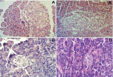
Non diabetic control mice showed normal liver parenchyma with general structures preserved, including hepatic lobules with normal hepatocytes surrounded by sinusoids and distributed radially towards the centrilobular veins, and containing kupffer and red blood cells in the capillary lumen. Portal spaces were also normal, with no observed inflammatory infiltration, fatty degeneration or abnormal distribution of fibroblasts or collagen in mice sacrificed at 14 days of follow-up (Fig. 1A). More prominent pathological lesions were observed commonly in the liver and pancreas due to toxicity of alloxan. Severe micro-vesicular fatty degeneration was observed in the livers of diabetic untreated mice associated with the presence of dilated sinusoids and a progressive loss of general organ structure (Fig.
1B). Inflammatory changes consistent with
steatohepatitis, which were represented by
mononuclear inflammatory infiltrates of moderate intensity of analyzed liver histological sections. CS nullified the toxic effects of alloxan on liver tissue in a dose dependent manner. Interestingly, metformin showed better ameliorative effects than CS at dose rate of 10 and 20 mg/100g BW, but less than 50 mg/kg BW CS (Fig.1C and 1D).
The sections of the pancreas from untreated diabetic mice showed an extensive destruction of islet cells as compared with that of healthy control mice (Fig. 2Aand2B). Further, there was a definite reduction in the number of islets in diabetic mice than in the healthy mice. However, hemorrhages were not observed and acinar cells were intact in the pancreatic tissues of alloxan induced diabetic control mice. Further, severe inflammatory cell infiltrations in islets were also observed in diabetic control mice. The best amelioration was seen with CS 50mg/100g BW in the islets of Langerhans compared to CS 10 and 20 mg/100g BW (Fig. 2C and 2D). The pancreas sections from CS treated diabetic mice revealed a statistically significant regeneration of islet cells with some hyperplastic islets (P< 0.05). Further, the CS extract produced a significant increase in the mean profile diameter in large islets compared to the alloxan induced diabetic control mice. Severity of histopathologic lesions was significantly reduced in CS treated group (50mg/100g BW) (Table 4).
Discussion
Science Letters 2016; 4(2):140-146
143
Table 2 Effect of oral administration of Costus speciosus on blood glucose, total cholesterol, plasma insulin and C-peptide levels (mean + SD) in normal and alloxan-induced diabetic male mice.
Treatments Glucose
(mg/dL)
Cholesterol (mg/dL)
Plasma Insulin
(μU/ml)
C-Peptide (ng/ml) Control negative 153.50± 52.00c 115.00 ± 42.04c 18.15±0.33a 6.38±0.11a Control positive (DM) 309.75±194.76a 210.50 ± 25.94a 6.9±0.1.2d 3.44±0.29d
DM+ CS (10mg/100g BW) 172.20±30.80b 141.60 ± 24.94b 9.33±0.6c 3.75±0.66c
DM+ CS (20mg/100 g BW) 166.25±35.83b 139.25 ± 42.92b 10.31±0.73c 3.89±0.42c
DM+ CS (50mg/100 g BW) 158.00±36.31c 129.75 ± 32.99b 14.47±1.2b 5.35±0.15b
DM + Metformin (19.5mg /100 g BW) 170.25±4.84b 133.25 ± 29.35b 11.28±1.4b 4.53±0.48b
a-dMean values in the same column that do not share a common letter differ significantly (P < 0.05)
Table 3Effect of oral administration of Costus speciosus on plasma AST, ALT, LDH, ALP and ACP levels in normal and alloxan induced diabetic male mice.
Treatments AST (U/dl) ALT (U/dl) LDH (U/dl) ALP (U/dl) ACP (U/dl) Control negative 32.6 ± 1.7d 53.0± 1.2c 1089.2 ± 12.3d 47.3± 4.7c 13.8± 0.2c
Control positive (DM) 66.4± 3.6 a 97.3± 2.6a 1727.2 ± 22.2a 86.2 ±3.9a 27.0± 6.3a
DM+ CS (10mg/100 g BW) 56.7 ±1.2b 71.8± 6.3b 1356.2 ± 25.9b 67.8± 3.7b 18.9± 1.7b
DM+ CS (20mg/100 g BW) 47.8 ±3.7b 69.5± 3.9b 1266.4 ± 23.5c 64.5± 3.2c 17.2± 2.7b
DM+ CS (50mg/100 g BW) 42.2 ±6.5c 68.2± 6.7b 1203.2 ± 12.3c 61.4± 7.5c 17.44± 1.4b
DM + Metformin (19.5mg /100 g BW) 49.9 ±2.2b 65.8± 5.7b 1262.2 ± 17.6c 67.6± 2.0b 18.67± 2.2b
CS: Costus spiosus, DM: Diabetes mellitus, AST: Aspartate aminotransferase, ALT: Alanine aminotrasferase, LDH: Lactate dehydrogenase, ALP: Alkaline phosphatase, ACP: Acid phosphatase. a-dMean values in the same column that do not share a common letter differ significantly (P < 0.05)
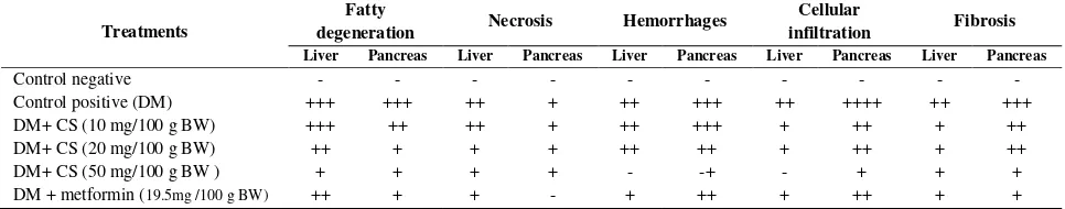
Table 4: Severity of histopathological lesions in liver and pancreas in normal and alloxan induced diabetic male mice.
Treatments
Fatty
degeneration Necrosis Hemorrhages
Cellular
infiltration Fibrosis
Liver Pancreas Liver Pancreas Liver Pancreas Liver Pancreas Liver Pancreas
Control negative - - - -
Control positive (DM) +++ +++ ++ + ++ +++ ++ ++++ ++ +++ DM+ CS (10 mg/100 g BW) +++ ++ ++ + ++ +++ + ++ + ++ DM+ CS (20 mg/100 g BW) ++ + + + ++ ++ + ++ + ++ DM+ CS (50 mg/100 g BW ) + + + + - -+ - + + + DM + metformin (19.5mg /100 g BW) ++ + + - + ++ + ++ + +
- normal ; + very mild ; ++ mild; +++ moderate; ++++ severe
currently available antidiabetic agents. The FDA approved anti-diabetic agents are used for the therapy to control blood glucose level [14, 15]. Incorporation of natural antidiabetic preparation is given the privilege to make use of it, as it holds less toxicity and negligible side effects. Almost all the flavonoids have potential for antidiabetic activity, but are limited in usage due to lack of studies on their antidiabetic activities.
In the present study, low dose of alloxan monohydrate (0.65 mg/100g BW) induced type II DM by partial destruction of pancreatic beta cells leading to insufficient insulin production [16]. Dose dependent decrease in the glucose level was observed during the present study in CS treated diabetic mice. This decrease in the level of glucose might be due to insulin like effects of CS or its ingredients which lead either to increase in glucose uptake mechanism possibly by inhibiting the process of gluconeogenesis [17].Moreover, the CS might also enhance the
regeneration process of 𝛽-cells in pancreas [18-21].
Further, CS might inhibit the expression of nitric oxide synthase leading to increase insulin secretion [22].
It has been reported that hypoglycemic activity of herbal medicines might be due to improvement in the effect of insulin on the target cells, stimulating glucose dependent insulin secretion or by enhancing
the recovery process for the damaged 𝛽-cells of islets
Science Letters 2016; 4(2):140-146
144
Fig 1 Histological pattern of liver from nondiabetic control mice showing normal appearing of hepatocytes, portal space, sinusoids, and Kuppfer cells (A); Histological pattern of liver in untreated diabetic showing severe fatty degeneration, sinusoidal enlargement and interlobular mononuclear inflammatory infiltrate (B);liver of metformin-treated diabetic group showing mild fatty degeneration and mononuclear inflammatory infiltrates (C); liver of CS (50mg/100g BW) treated diabetic group showed normal hepatic architecture which was almost similar to that of the control group (D), (×400).
Science Letters 2016; 4(2):140-146
145
leakage of these enzymes from the damaged liver
tissue to blood circulation indicating the
hepatotoxic effect of alloxan. However, we rats.Moreover, CS treated mice showed increased levels of insulin and C-peptide, indicating the effect of CS on the regeneration of pancreatic tissue. C-peptides have been reported to have insulin-mimetic effects and can stimulate insulin receptors; leading to increased glycogen synthesis from blood glucose [30-31].Phytochemical compounds in plants may
regulate the enzymes of glycolysis and
gluconeogenesis [25]. However, we have not studied the mode of action of these phytochemical agents in the present study; therefore our analysis is based on assumptions.
Abnormalities in lipid profile are considered one of the leading complications in most of the DM cases [26].Low levels of insulin during DM can result in increased lipase secretion leading to enhanced mobilization of fatty acids [27]. Fatty degeneration was seen during histopathological examination of liver that might be the result of increase fatty acid mobilization in diabetic mice [32, 33]. Regeneration of pancreatic and liver tissue with minimal lesions was observed in CS treated mice. Similarly, the regeneration of pancreatic tissues was observed by Dhanavathy [34] in streptozotocin induced diabetic mice after treatment with plant extract.
In conclusion, oral administration of ethanolic extract of CS to alloxan-induced diabetic mice improved their glucose tolerance, an important finding in the control of diabetes. This suggested that CS is useful in the protection and amelioration of diabetic complications through the enhancement
of regeneration of 𝛽-cells of the islets of
Langerhans. Moreover, the present results showed that CS consumption reversed most of the histological changes in the liver and pancreas of the diabetic rats in a dose dependent manner. In
addition, the plant extract exerts
anti-hyperlipidemic activities in diabetic mice. So, we can say that CS had a significant hepatoprotective role in diabetic rats and offers promising perspectives deserve further investigation. Further studies are needed to define the active agents present and their mode of activity.
Acknowledgement
The authors wish to thank Mahmud Rudini, Dr. Sutyarso and Dr. Nugroho Susanto for their technical help in data analysis and interpretation of results.
References
[1] Wang J, Teng L, Liu Y, Hu W, Chen W, Hu X, et al. Studies on the antidiabetic and antinephritic activities of Paecilomyces hepiali water extract in diet-streptozotocin-induced diabetic Sprague Dawley rats.J Diabetes Res 2016; e4368380.
[2] World Health Organization. Global report on diabetes [Internet]. 2016 [cited 2016 Apr 29]. Available from: http://apps.who.int/iris/bitstream/10665/204871/1/97892415652 57eng.pdf.
[3] Alamoudi EF, Khalil WKB, Ghaly IS, Hassan NHA, Ahmed ES. Nanoparticles from of Costus speciosus extract improves the antidiabetic and antilipidemic effects against STZ-induced
Diabetes mellitus in albino rats. Int J Pharm Sci Rev Res 2014; 29(1):279–288.
[4] Taha MA, Ahmed SJ, Rashid KI. Effect of arabidopsis extracts on the status of liver histology of alloxan-induced diabetic mice. J Nat Sci Res 2016; 6(6):111–121.
[5] Attanayake AP, Jayatilaka KAPW, Pathirana C, Mudduwa LKB, Attanayake AP, Jayatilaka KAPW, et al. Gmelina arborea Roxb. (Family: Verbenaceae) extract upregulates the β-cell regeneration in STZ induced diabetic rats. J Diabetes Res 2016; e4513871.
[6] Revathy J, Abdullah SS, Kumar PS. Antidiabetic Effect of
Costus Speciosus rhizome extract in alloxan induced albino rats. J Chem Biochem 2014; 2(1):13–22.
[7] Agwaya MS, Vuzi PC, Nandutu AM, Agwaya MS, Vuzi PC, Nandutu AM. Hypoglycemic activity of aqueous root bark extract Zanthoxylum chalybeum in alloxan-induced diabetic rats. J Diabetes Res 2016; 2016:8727590.
[8] Obi BC, Okoye TC, Okpashi VE, Igwe CN, Alumanah EO, Obi BC, et al. Comparative study of the antioxidant effects of metformin, glibenclamide, and repaglinide in alloxan-induced diabetic rats. J Diabetes Res 2016; 2016:1635361.
[9] Alagammal M, Agnel RA, Mohan VR. Antidiabetic and antihyperlipidaemic effect of Polygala javana DC on alloxan induced diabetic rats. Int Res J Pharm 2012; 3:231–234. [10] Rani AS, Sulakshana G, Patnaik S. Costus speciosus,
anantidiabetic plant - Review. FS J Pharm Res 2012;1:52–53. [11] Nehete J, Bhatia M, Narkhede M. In-vitro Evaluation of
antioxidant activity and phenolic content of costus speciosus
(Koen) J.E. Sm. Iran J Pharm Res IJPR 2010; 9(3):271–277. [12] Jha MK, Alam MB, Hossain MS, Islam A. In vitro antioxidant
and cytotoxic potential of Costuss peciosus (Koen.) Smith rhizome. Int J Pharm Sci Res 2012; 1:138–144.
[13] Drury RAB, Wallington EA. Preparation and fixation of tissues. In: Carleton HM, Drury RAB, Wallington EA, editors.
Carleton’s Histological technique. Oxford University Press;
1980, p. 41–45.
[14] Akiyama S, Katsumata S-I, Suzuki K, Ishimi Y, Wu J, Uehara M. Dietary hesperidin exerts hypoglycemic and hypolipidemic effects in streptozotocin-induced marginal type 1 diabetic rats. J Clin Biochem Nutr 2010; 46(1):87–92.
[15] Barwal I, Sood A, Sharma M, Singh B, Yadav SC. Development of stevioside Pluronic-F-68 copolymer based PLA-nanoparticles as an antidiabetic nanomedicine. Coll Surf B Biointer 2013; 101:510–516.
[16] Aybar M, Sanchez-Riera AN, Grau A, Sanchez SS. Hypoglycemic effect of the water extract of Smallanthus soncifolius (yacon) leaves in normal and diabetic rats. J Ethnopharmacol 2002; 74:125–132.
Science Letters 2016; 4(2):140-146
146
seed, and whole plant of Momordica charantia on normal and diabetic model rats. Planta Med 1993; 59(5):408–412. [18] Shanmugasundaram ER, Gopinath KL, Radha
Shanmugasundaram K, Rajendran VM. Possible regeneration of the islets of langerhans in streptozotocin-diabetic rats given
Gymnema sylvestre leaf extracts. J Ethnopharmacol 1990; 30(3):265–279.
[19] Li WL, Zheng HC, Bukuru J, De Kimpe N. Natural medicines used in the traditional Chinese medical system for therapy of diabetes mellitus. J Ethnopharmacol 2004; 92(1):1–21. [20] Achrekar S, Kaklij GS, Pote MS, Kelkar SM. Hypoglycemic
activity of Eugenia jambolana and Ficus bengalensis: mechanism of action. Vivo Athens Greece 1991; 5(2):143–147. [21] Bansal R, Ahmad N, Kidwani JR. Effect of oral administration of Eugenia jambolana seeds and chlororopamide on blood glucose level and pancreatic cathapsin B in rats. Indian J Biochem Biophys 1981; 18:377–381.
[22] Gunawardana SC, Head WS, Piston DW. Dimethyl amiloride improves glucose homeostasis in mouse models of type 2 diabetes. Am J Physiol Endocrinol Metab 2008; 294(6):E1097-1108.
[23] Mao-Ying Q-L, Kavelaars A, Krukowski K, Huo X-J, Zhou W, Price TJ, et al. The anti-diabetic drug metformin protects against chemotherapy-induced peripheral neuropathy in a mouse model. PLOS One 2014; 9(6):e100701.
[24] Ayodhya S, Kusum S, Anjali S. Hypoglycemic activity of different extracts of various herbal plants. International J Res Ayurveda Pharm 2010; 1(1):212–224.
[25] Arya A, Cheah SC, Looi CY, Taha H, Mustafa MR, Mohd MA. The methanolic fraction of Centratherum anthelminticum seed downregulates pro-inflammatory cytokines, oxidative stress, and hyperglycemia in STZ-nicotinamide-induced type 2 diabetic rats. Food Chem Toxicol 2012; 50(11):4209–4220. [26] Ravi K, Rajasekaran S, Subramanian S. Antihyperlipidemic
effect of Eugenia jambolana seed kernel on
streptozotocin-induced diabetes in rats. Food Chem Toxicol 2005; 43(9):1433– 1439.
[27] Soltani N, Keshavarz M, Dehpour AR. Effect of oral magnesium sulfate administration on blood pressure and lipid profile in streptozocin diabetic rat. Eur J Pharmacol 2007; 560(2–3):201–205.
[28] Murali B, Upadhyaya UM, Goyal RK. Effect of chronic treatment with Enicostemma littorale in non-insulin-dependent diabetic (NIDDM) rats. J Ethnopharmacol 2002; 81(2):199– 204.
[29] El-Demerdash FM, Yousef MI, El-Naga NIA. Biochemical study on the hypoglycemic effects of onion and garlic in alloxan-induced diabetic rats. Food Chem Toxicol 2005; 43(1):57–63.
[30] Gomathi, Ganesan Ravikumar, Manokaran Kalaiselvi, Kanakasabapathi Devaki and Chandrasekar Uma, Efficacy of
Evolvulus alsinoides (L.) on insulin and antioxidants activity in pancreas of streptozotocin induced diabetic rats. J Diabet Metab Disord 2013; 12:39.
[31] Senthilkumar G, Subramanian SP, Biochemical studies on the effect of Terminalia chebula on the levels of glycoproteins in streptozotocin induced experimental diabetes in rats. J Appl Biomed2008; 6:105–115.
[32] Eliza J, Rajalakshmi M, Ignacimuthu SJ, Daisy P. Normalizing effects of Costus speciosus rhizome crude extracts and its fractions on diabetic complications in STZ-induced diabetic rats. Med Chem Res 2010; 20(7):1111–1118.
[33] Girgis S, Shoman T, Kassem S, Ezz El-Din A, Abdel-Aziz K. Potential Protective Effect of Costus speciosus or its nanoparticles on streptozotocin-induced genotoxicity and histopathological alterations in rats. J Nutr Food Sci 2015; S3(002).