review article
Thenew england journalofmedicine
medical progress
The Pathophysiology and Treatment of Sepsis
Richard S. Hotchkiss, M.D., and Irene E. Karl, Ph.D.
From the Departments of Anesthesiology (R.S.H.), Medicine (R.S.H., I.E.K.), and Surgery (R.S.H.), Washington University School of Medicine, St. Louis. Address re-print requests to Dr. Hotchkiss at the De-partment of Anesthesiology, Washington University School of Medicine, Campus Box 8054, St. Louis, MO 63110, or at hotch@morpheus.wustl.edu.
epsis is the leading cause of death in critically ill patients in the United States. Sepsis develops in
750,000 people annually, and more than 210,000 of them die.1,2 After numerous
unsuccessful trials of antiinflammatory agents in patients with sepsis, investigators doubted that mortality could be decreased. Advances in unraveling the pathophysiolo-gy and genetic basis for the host response to sepsis have changed the prevailing under-standing of the syndrome, and several therapies have demonstrated surprising effica-cy. In this article, we examine evolving concepts of sepsis and discuss new and potential therapies.
The prevailing theory has been that sepsis represents an uncontrolled inflammatory response.3-5 Lewis Thomas popularized this notion when he wrote that “the
micro-organisms that seem to have it in for us . . . turn out . . . to be rather more like bystanders. . . . It is our response to their presence that makes the disease. Our arse-nals for fighting off bacteria are so powerful . . . that we are more in danger from them than the invaders.”6 A consensus conference defined sepsis as “the systemic
in-flammatory response syndrome that occurs during infection.”3 Numerous trials were
conducted of agents that block the inflammatory cascade — corticosteroids,7
antien-dotoxin antibodies,8 tumor necrosis factor (TNF) antagonists,9,10
interleukin-1–recep-tor antagonists,11 and other agents.12 The failure of antiinflammatory agents led
inves-tigators to question whether death in patients with sepsis results from uncontrolled inflammation.4,13-15 Clinical trials of treatments for sepsis are difficult because of the
heterogeneity of patients and the high rates of culture-negative sepsis. Interpretation is complicated, because the analysis of outcomes generates post hoc stratifications that have not been prospectively defined.
The theory that death from sepsis was attributable to an overstimulated immune system was based on studies in animals that do not seem to reflect the clinical picture in humans.16-18 These studies used large doses of endotoxin or bacteria; consequently,
levels of circulating cytokines such as tumor necrosis factor a (TNF-a) were exponen-tially higher in animals than they are in patients with sepsis.17 In these studies, the
an-imals died from “cytokine storm,” and compounds and macromolecules that block these mediators improved survival.16-18
In certain forms of sepsis — for example, meningococcemia — circulating TNF-a
levels are high and correlate with mortality.19,20 Of 55 children with severe infectious
purpura (32 of them with Neisseria meningitidis infection), 91 percent had elevated levels of circulating TNF-a.19 Nevertheless, studies have shown that the frequency of an
ex-aggerated systemic inflammatory response is lower than it was originally thought to be.21-24 Debets et al. reported that only 11 of 43 patients with sepsis had detectable
cir-culating TNF (limit of detection, 5 to 10 pg per milliliter).21 In another study of 87
pa-s
medical progress
tients with sepsis, fewer than 10 percent had meas-urable TNF-a or interleukin-1b.22,23
Although cytokines are considered to be cul-prits, they also have beneficial effects in sepsis. Studies in an animal model of peritonitis demon-strated that blocking TNF-a worsens
surviv-al.25,26 Combination immunotherapy against
TNF-a and interleukin-1 receptors was fatal in a neutropenic model of sepsis.27 In clinical trials, a
TNF antagonist increased mortality.9 The role of
TNF-a in combating infection has recently been underscored by the finding that sepsis and other infectious complications developed in patients with rheumatoid arthritis who were treated with TNF antagonists.28
The debate about the merits of inhibiting cyto-kines in patients with sepsis has been rekindled by a recent trial that indicated that a subgroup of pa-tients with sepsis who had therapy directed against TNF-a had improved survival.29 Also, a
meta-analy-sis of clinical trials of antiinflammatory agents in patients with sepsis showed that although high doses of antiinflammatory agents were generally harmful in such patients, a subgroup of patients (approximately 10 percent) benefited.13
Advances in our understanding of cell-signal-ing pathways that mediate the response to microbes have demonstrated that the concept of blocking endotoxin in order to prevent septic complications may be simplistic. Cells of the innate immune sys-tem recognize microorganisms and initiate respons-es through pattern-recognition receptors called toll-like receptors (TLRs).30-32 Insight into the role of
TLRs in combating infection has been provided by studies in C3H/HeJ mice,30 which are resistant to
endotoxin because of a mutation in the toll-like re-ceptor 4 gene (TLR4). Despite their resistance to endotoxin, these mice have increased mortality with authentic sepsis.33,34 TLR4 mutations have been
identified in humans and may make persons more susceptible to infection.35 Therefore, although
dotoxin has deleterious effects, total blockade of en-dotoxin may be detrimental. Reasons for the failure of monoclonal antiendotoxin antibodies to improve outcomes in trials involving patients with sepsis are complex.36
Patients with sepsis have features consistent with immunosuppression, including a loss of delayed hypersensitivity, an inability to clear infection, and
a predisposition to nosocomial infections.37-39 One
reason for the failure of antiinflammatory strate-gies in patients with sepsis may be a change in the syndrome over time. Initially, sepsis may be charac-terized by increases in inflammatory mediators; but as sepsis persists, there is a shift toward an antiin-flammatory immunosuppressive state.38,39 There
is evidence of immunosuppression in sepsis from studies showing that lipopolysaccharide-stimulat-ed whole blood from patients with sepsis releases markedly smaller quantities of the inflammatory cy-tokines TNF-a and interleukin-1b than does that of control patients.40 The adverse sequelae of
sepsis-induced immunosuppression were reversed with the administration of interferon-g in patients with sepsis.41 This immune stimulant restored
macro-phage TNF-a production and improved survival.41
a shift to antiinflammatory cytokines
Activated CD4 T cells are programmed to secrete cytokines with either of two distinct and antago-nistic profiles.42,43 They secrete either cytokines
with inflammatory (type 1 helper T-cell [Th1]) prop-erties, including TNF-a, interferon-g, and interleu-kin-2, or cytokines with antiinflammatory (type 2 helper T-cell [Th2]) properties — for example, in-terleukin-4 and interleukin-10 (Fig. 1). The factors that determine whether CD4 T cells have Th1 or Th2 responses are unknown but may be influenced by the type of pathogen, the size of the bacterial inocu-lum, and the site of infection.42 Mononuclear cells
from patients with burns or trauma have reduced levels of Th1 cytokines but increased levels of the Th2 cytokines interleukin-4 and interleukin-10, and reversal of the Th2 response improves survival among patients with sepsis.38,44 Other studies have
demonstrated that the level of interleuk10 is in-creased in patients with sepsis and that this level predicts mortality.43,45
anergy
Anergy is a state of nonresponsiveness to antigen. T cells are anergic when they fail to proliferate or secrete cytokines in response to their specific an-tigens. Heidecke et al. examined T-cell function in patients with peritonitis and found that they had de-creased Th1 function without inde-creased Th2 cyto-kine production, which is consistent with anergy.46
Defective T-cell proliferation and cytokine secretion f a i l u r e o f t h e i m m u n e s y s t e m ?
Thenew england journalofmedicine
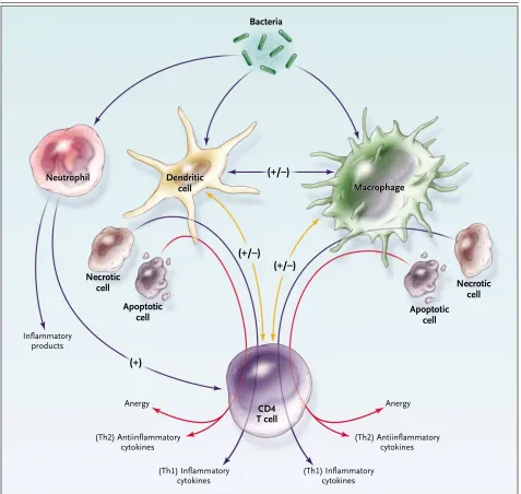
Figure 1. The Response to Pathogens, Involving “Cross-Talk” among Many Immune Cells, Including Macrophages, Dendritic Cells, and CD4 T Cells.
Macrophages and dendritic cells are activated by the ingestion of bacteria and by stimulation through cytokines (e.g., interferon-g) secreted by CD4 T cells. Alternatively, CD4 T cells that have an antiinflammatory profile (type 2 helper T cells [Th2]) secrete interleukin-10, which suppresses macrophage activation. CD4 T cells become activated by stimulation through macrophages or dendritic cells. For example, macrophages and dendritic cells secrete interleukin-12, which activates CD4 T cells to secrete inflammatory (type 1 helper T-cell [Th1]) cytokines. Depending on numerous factors (e.g., the type of organism and the site of infection), macrophages and dendritic cells will respond by inducing either in-flammatory or antiinin-flammatory cytokines or causing a global reduction in cytokine production (anergy). Macrophages or dendritic cells that have previously ingested necrotic cells will induce an inflammatory cytokine profile (Th1). Ingestion of apoptotic cells can induce either an anti-inflammatory cytokine profile or anergy. A plus sign indicates up-regulation, and a minus sign indicates down-regulation; in cases where both a plus sign and a minus sign appear, either up-regulation or down-regulation may occur, depending on a variety of factors.
Bacteria
Dendritic Dendritic
cell cell Neutrophil
Neutrophil
Macrophage Macrophage
Dendritic cell Neutrophil
Macrophage
Necrotic cell
T cellT cell
CD4CD4
Necrotic cell Apoptotic
cell Apoptotic
cell
Inflammatory products
AnergyAnergy
(Th2) Antiinflammatory cytokines
(Th2) Antiinflammatory cytokines
(Th1) Inflammatory cytokines
(Th1) Inflammatory cytokines (+)
(+/–)
medical progress
correlated with mortality.46 Patients with trauma or
burns have reduced levels of circulating T cells, and their surviving T cells are anergic.47
Apoptotic cell death may trigger sepsis-induced anergy. Although the conventional belief was that cells die by necrosis, recent work has shown that cells can die by apoptosis — genetically pro-grammed cell death. In apoptosis, cells “commit suicide” by the activation of proteases that disas-semble the cell.48,49 Large numbers of lymphocytes
and gastrointestinal epithelial cells die by apopto-sis during sepapopto-sis.50-52 A potential mechanism of
lymphocyte apoptosis may be stress-induced en-dogenous release of glucocorticoids.53,54 The type
of cell death determines the immunologic function of surviving immune cells (Fig. 1).55-57 Apoptotic
cells induce anergy or antiinflammatory cytokines that impair the response to pathogens, whereas ne-crotic cells cause immune stimulation and enhance antimicrobial defenses (Fig. 1).55-57
death of immune cells
Autopsy studies in persons who had died of sep-sis disclosed a profound, progressive, apoptosep-sis- apoptosis-induced loss of cells of the adaptive immune sys-tem.50-52 Although no loss of CD8 T cells, natural
killer cells, or macrophages occurred, sepsis mark-edly decreased the levels of B cells (Fig. 2), CD4 T cells (Fig. 3), and follicular dendritic cells (Fig. 3). The loss of lymphocytes and dendritic cells was es-pecially important, because it occurred during life-threatening infection, when clonal expansion of lymphocytes might have been expected.
The magnitude of the apoptosis-induced loss in lymphocytes during sepsis was apparent in exami-nations of the circulating lymphocyte count in pa-tients.50 In one study, 15 of 19 patients with sepsis
had absolute lymphocyte counts below the lower limit of normal (a mean [±SD] of 500±270 per cubic millimeter vs. the lower limit of 1200 per cubic mil-limeter). Losses of B cells, CD4 T cells, and
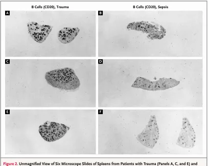
dendrit-Figure 2. Unmagnified View of Six Microscope Slides of Spleens from Patients with Trauma (Panels A, C, and E) and Patients Who Died of Sepsis (Panels B, D, and F), with Staining for B Cells (CD20).
The dark stained regions are concentrations of B cells in lymphoid follicles that are visible to the naked eye. The patients with sepsis have dramatically smaller and fewer lymphoid follicles than the patients with trauma.
B Cells (CD20), Trauma B Cells (CD20), Sepsis
A B
C D
Thenew england journalofmedicine
ic cells decrease antibody production, macrophage activation, and antigen presentation, respectively. The potential importance of the depletion of lym-phocytes is illustrated by studies in animals show-ing that prevention of lymphocyte apoptosis im-proves the likelihood of survival.58-61 Immune
defects identified in patients with sepsis, including monocyte dysfunction,41,62,63 are listed in Table 1.
Investigators are challenging Lewis Thomas’s theory6 that the body’s primary response to
infec-tion and injury is uncontrolled hyperinflamma-tion.4,13,14,64 Munford and Pugin maintain that the
body’s normal stress response is activation of anti-inflammatory mechanisms and that, outside of af-fected tissues, the body’s systemic antiinflamma-tory responses predominate.64 They postulate that
immune cells and cytokines have both pathogenic and protective roles and that blocking these media-tors may worsen the outcome.64 Heidecke et al.
ex-amined T-cell function in patients with sepsis and reported that immunosuppression was evident at the onset of sepsis, suggesting a primary hypo-immune response.46
Weighardt and associates examined lipopoly-r e a p p lipopoly-r a i s a l o f
l e w i s t h o m a s ’ s t h e o r y
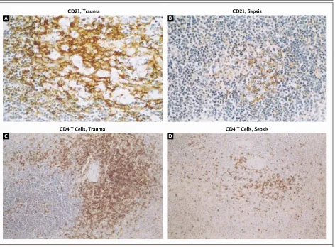
Figure 3. Immunohistochemical Staining for Follicular Dendritic Cells (CD21) (Top Panels, ¬600) and CD4 T Cells (Bottom Panels, ¬600) in Spleens from Patients with Trauma (Panels A and C) or Patients Who Died of Sepsis (Panels B and D).
The patients with sepsis have dramatically fewer follicular dendritic cells and CD4 T cells (located in the T-cell–rich periarteriolar zone) than patients with trauma.
CD21, Trauma
CD4 T Cells, Trauma CD4 T Cells, Sepsis
CD21, Sepsis
A B
medical progress
saccharide-stimulated production of cytokines by monocytes in patients with sepsis that occurred af-ter major visceral surgery.65 Postoperative sepsis
was associated with the immediate onset of defects in the production of both inflammatory and antiin-flammatory cytokines by monocytes, and survival among patients with sepsis correlated with the re-covery of the inflammatory but not the antiinflam-matory response.65 These investigators concluded
that immunosuppression was a primary rather than a compensatory response to sepsis.65 Others
pos-tulate a sequential response to sepsis, with initial marked inflammation followed by immunosup-pression.14,38,39
On the basis of studies in identical twins and adop-tees, genetic factors are known to be major deter-minants of susceptibility to death from infectious disease.66 Some persons have single base-pair
alter-ations (single-nucleotide polymorphisms) in genes controlling the host response to microbes.67-69
Identified alterations include polymorphisms in TNF receptors, interleukin-1 receptors, Fcg recep-tors, and TLRs.67-69 Polymorphisms in cytokine
genes may determine the concentrations of inflam-matory and antiinflaminflam-matory cytokines produced and may influence whether persons have marked hyperinflammatory or hypoinflammatory respons-es to infection. The risk of death among patients with sepsis has been linked to genetic polymor-phisms for TNF-a and TNF-b.69 Trials examining
the effect of polymorphisms in patients with pneu-monia and sepsis are under way; such polymor-phisms may ultimately be used to identify patients
at high risk for the development of sepsis and organ dysfunction during infection. Thus, physicians may, in the future, be able to use genetic information to dictate immune-based therapy to modulate the re-sponse in a given patient.
Neutrophils have been regarded as double-edged swords in sepsis. Although neutrophils were thought to be essential for the eradication of pathogens, ex-cessive release of oxidants and proteases by neutro-phils was also believed to be responsible for injury to organs. Because of the intrapulmonary seques-tration of neutrophils and the frequent complica-tion of the acute respiratory distress syndrome in patients with sepsis, this link between overly exu-berant neutrophil activation and organ injury was thought to affect the lungs in particular.70 Although
findings from studies in animals implicated neutro-phil-mediated injury, other studies in which granu-locyte colony-stimulating factor (G-CSF) was used — to increase the number of neutrophils and en-hance their function — demonstrated improved survival among patients with sepsis.
Two randomized trials of G-CSF were conduct-ed in patients with community-acquirconduct-ed and hos-pital-acquired pneumonia.71,72 Despite an increase
in the white-cell count to 70,000 per cubic milli-meter, there was no evidence of adverse effects on lung function in patients with community-acquired pneumonia.71 Although a subgroup of patients
with multilobar pneumonia had fewer complica-tions and shorter stays in the intensive care unit with G-CSF, there was no improvement in survival. Similarly, hospitalized patients with community-acquired or nosocomial pneumonia who were treat-ed with G-CSF had no survival benefit, no decrease in organ dysfunction, and no decrease in the num-ber of days in intensive care.72
Although marked leukocytosis resulting from G-CSF was not injurious, it is not necessarily possi-ble to extrapolate from such data whether marked leukocytosis would be harmful in patients with se-vere sepsis. However, these two clinical studies im-ply that blocking neutrophil function to prevent complications of sepsis would be unlikely to be ben-eficial. Furthermore, therapies aimed at enhancing the number or function of neutrophils in patients with sepsis are also unlikely to be efficacious. h o s t g e n e t i c f a c t o r s
s u r p r i s i n g i n s i g h t s a b o u t n e u t r o p h i l s
* Th1 denotes type 1 helper T cell, and Th2 type 2 helper T cell. Table 1. Potential Mechanisms of Immune Suppression in Patients with Sepsis.*
Shift from an inflammatory (Th1) to an antiinflammatory (Th2) response
Anergy
Apoptosis-induced loss of CD4 T cells, B cells, and dendritic cells
Loss of macrophage expression of major-histocompatibility-complex class II and costimulatory molecules
Thenew england journalofmedicine
Autopsy studies in persons who died in the inten-sive care unit show that the failure to diagnose and appropriately treat infections with antibiotics or surgical drainage is the most common avoidable error.73,74 Our laboratory conducted an autopsy
study of 20 patients who died in intensive care units50; consent was obtained immediately after
each patient’s death, so that tissues were usually acquired within 30 to 90 minutes after death, there-by permitting tissue morphology to be assessed be-fore autolytic changes occurred. Autopsies were also performed in a control group consisting of patients who had died while critically ill but who did not have clinical sepsis. Immunohistochemical analy-sis showed that in the majority of patients with sep-sis, only two types of cells — lymphocytes and gas-trointestinal epithelial cells — were dying; this finding parallels those of studies in animals.39,54,75
As had been noted previously, there was a profound loss of cells of the adaptive immune system. Lym-phocytes and gastrointestinal epithelial cells nor-mally undergo rapid turnover through apoptosis, and sepsis most likely accelerates these physiologic processes. Focal necrosis occurred in hepatocytes in the region of the central vein (presumably be-cause this region is vulnerable to hypoxia) in 7 of 20 patients, as well as in the brain and heart in 3 pa-tients who had evidence of infarction before death.
Another intriguing finding from the autopsy study was a discordance between histologic findings and the degree of organ dysfunction seen in patients who died of sepsis.50 Cell death in the heart,
kid-ney, liver, and lung was relatively minor and did not reflect the clinical evidence of more profound or-gan dysfunction. There was no evidence of injury to cardiac myocytes in patients with sepsis who had myocardial depression. (No patient had meningo-coccemia, which causes myocarditis with infiltra-tion of organisms and granulocytes.) Histologic findings in patients with sepsis and acute renal failure showed only focal injury with preservation of normal glomeruli and renal tubules.50 These
re-sults are similar to those of studies in patients with acute renal failure in which microscopy showed a dissociation between the degree of tubular
necro-sis and the level of renal dysfunction.76,77 Most
patients who survive sepsis and acute renal failure recover base-line renal function, suggesting that re-nal-cell death is not overwhelming during sepsis.78
We speculate that much organ dysfunction in patients with sepsis can be explained by “cell hiber-nation” or “cell stunning,” as occurs during myo-cardial ischemia.79 Presumably, sepsis activates
de-fense mechanisms that cause cellular processes to be reduced to basic “housekeeping” roles. A possi-ble molecular basis for cellular stunning was sug-gested by work from the laboratory of Fink et al.,80
who showed that immunostimulated enterocytes have diminished oxygen consumption as a result of depletion of nicotinamide adenine dinucleotide secondary to activation of the nuclear enzyme poly– adenosine diphosphate (ADP)–ribose polymerase by peroxinitrite or other oxidants.
No autopsy studies have revealed why patients with sepsis die. Occasionally, a patient with sepsis may die of refractory shock, but this is exception-al. Although patients with sepsis have profound myocardial depression, cardiac output is usually maintained because of cardiac dilatation and tach-ycardia.81 Although the acute respiratory distress
syndrome frequently develops in patients with sep-sis, such patients rarely die of hypoxemia or hyper-carbia.82 Renal failure is common, but that alone is
not fatal, because dialysis may be used. Liver dys-function rarely progresses to hepatic encephalop-athy. Thus, the exact cause of death in patients with sepsis remains elusive. Many patients die when care is withdrawn or not escalated when families, in con-sultation with physicians, decide that continued therapy is futile.
Physicians caring for patients in intensive care units need a thorough knowledge of common infectious and noninfectious causes of fever in this popula-tion of patients (Table 2). Many patients in whom sepsis develops — for example, elderly patients or patients with uremia — do not become febrile.83
The lack of an apparent acute-phase response in pa-tients with sepsis is associated with high mortality and may reflect the immunosuppressive phase of sepsis. Early manifestations of sepsis include sub-l e s s o n s f r o m a u t o p s y s t u d i e s
c e l l u l a r h i b e r n a t i o n a s a m e c h a n i s m o f o r g a n d y s f u n c t i o n
d e a t h o f p a t i e n t s w i t h s e p s i s
medical progress
tle changes in mental status, minor increases or decreases in white-cell count or neutrophil percent-age, or elevated blood glucose levels. Early recogni-tion of sepsis is a key to successful treatment.
activated protein c
Recombinant human activated protein C, an anti-coagulant, is the first antiinflammatory agent that has proved effective in the treatment of sepsis.84,85
In patients with sepsis, the administration of acti-vated protein C resulted in a 19.4 percent reduction in the relative risk of death and an absolute risk re-duction of 6.1 percent.84 Activated protein C
inacti-vates factors Va and VIIIa, thereby preventing the generation of thrombin.85 The efficacy of an
anti-coagulant agent in patients with sepsis has been at-tributed to feedback between the coagulation sys-tem and the inflammatory cascade.85 Inhibition of
thrombin generation by activated protein C decreas-es inflammation by inhibiting platelet activation, neutrophil recruitment, and mast-cell degranula-tion. Activated protein C has direct antiinflamma-tory properties, including blocking of the produc-tion of cytokines by monocytes and blocking cell adhesion.
A puzzling issue is why activated protein C was successful whereas two other anticoagulants — antithrombin III86 and tissue factor–pathway
in-hibitor — failed as treatments of sepsis. A possible explanation for the failure of these two anticoagu-lant agents is that they work at different sites in the coagulation cascade. Also, activated protein C has
antiapoptotic actions that may contribute to its ef-ficacy.87
The debate regarding the appropriate use of ac-tivated protein C, as well as its potential adverse ef-fects, particularly bleeding, has been discussed in recent articles.88-90 A major risk associated with
ac-tivated protein C is hemorrhage; in a study of acti-vated protein C, 3.5 percent of patients had serious bleeding (intracranial hemorrhage, a life-threaten-ing bleedlife-threaten-ing episode, or a requirement for 3 or more units of blood), as compared with 2 percent of pa-tients who received placebo (P<0.06). With open-label use of activated protein C after the trial, 13 of 520 patients (2.5 percent) had intracranial hemor-rhage.88 Caution is advised in the use of activated
protein C in patients with an international normal-ized ratio greater than 3.0 or a platelet count of less than 30,000 per cubic millimeter. Currently, activat-ed protein C is approvactivat-ed only for use in patients with sepsis who have the most severe organ com-promise and the highest likelihood of death. Use of activated protein C is restricted in many hospitals to the more seriously ill patients who meet the cri-teria for sepsis specified by the Acute Physiology and Chronic Health Evaluation (APACHE II) scor-ing system.
intensive insulin therapy
for hyperglycemia
Van den Berghe et al. demonstrated that intensive insulin therapy that maintained the blood glucose level at 80 to 110 mg per deciliter (4.4 to 6.1 mmol per liter) resulted in lower morbidity and mortality among critically ill patients than did conventional therapy that maintained the blood glucose level at 180 to 200 mg per deciliter (10.0 to 11.1 mmol per liter).91 Intensive insulin therapy reduced the
fre-quency of episodes of sepsis by 46 percent. Patients with bacteremia who were treated with intensive insulin therapy had lower mortality than those who received conventional therapy (12.5 percent vs. 29.5 percent). Insulin therapy reduced the rate of death from multiple-organ failure among patients with sepsis, regardless of whether they had a history of diabetes.
The protective mechanism of insulin in sepsis is unknown. The phagocytic function of neutrophils is impaired in patients with hyperglycemia, and correcting hyperglycemia may improve bacterial phagocytosis. Another potential mechanism in-volves the antiapoptotic effect of insulin.92 Insulin
prevents apoptotic cell death from numerous stimu-Table 2. Infectious and Noninfectious Causes of Fever
in the Intensive Care Unit.
Infected intravascular catheters
Sinusitis or otitis media (in patients with intranasal devices such as nasogastric tubes or nasal endotracheal tubes)
Acalculous cholecystitis
Drug fever
Pulmonary emboli
Deep venous thrombosis
Central fever (in patients with head trauma)
Clostridium difficile colitis
Postcardiotomy syndrome
Secondary infection by resistant organisms
Thenew england journalofmedicine
li by activating the phosphatidylinositol 3-kinase– Akt pathway.90 Regardless of mechanism, it seems
reasonable to control blood glucose more tightly in critically ill patients. Clinicians must avert hy-poglycemic brain injury in attempting to maintain the blood glucose level at 80 to 110 mg per deciliter. Frequent monitoring of blood glucose is impera-tive, and studies are needed to determine whether less tight control of blood glucose — for example, a blood glucose level of 120 to 160 mg per decili-ter (6.7 to 8.9 mmol per lidecili-ter) — provides similar benefits.
volume resuscitation
Another recent study by Rivers et al. showed that early aggressive therapy that optimized cardiac pre-load, afterpre-load, and contractility in patients with severe sepsis and septic shock improved the likeli-hood of survival.93 Rivers et al. used infusions of
colloid or crystalloid, vasoactive agents, and trans-fusions of red cells to increase oxygen delivery. Re-suscitation end points chosen for assessment of the adequacy of oxygen delivery were the normalization of values for mixed venous oxygen saturation, lac-tate concentration, base deficit, and pH. Patients in the group that received early goal-directed therapy received more fluid, inotropic support, and blood transfusions during the first six hours than did con-trol patients, who received standard resuscitation therapy. During the interval from 7 to 72 hours, pa-tients in the group receiving early goal-directed treatment had a higher mean central venous oxygen concentration, a lower mean lactate concentration, a lower mean base deficit, and a higher mean pH than the control group. Mortality was 30.5 percent in the group receiving early goal-directed treat-ment, as compared with 46.5 percent in the control group (P=0.009). Thus, early therapeutic interven-tion to restore balance between oxygen delivery and oxygen demand improved survival among patients presenting with severe sepsis. The use of objective measures, including lactate concentration, base def-icit, pH, and possibly central venous oxygen satura-tion, in the follow-up of patients who are receiving resuscitation therapy is advisable.
corticosteroids
Administration of high doses of corticosteroids (e.g., 30 mg of methylprednisolone per kilogram of body weight) does not improve survival among
patients with sepsis and may worsen outcomes by increasing the frequency of secondary infections.94
Despite the negative effects of high-dose cortico-steroids, a 2001 study by Annane indicated that pa-tients with sepsis who are extremely ill and have persistent shock requiring vasopressors and pro-longed mechanical ventilation may benefit from “physiologic” doses of corticosteroids.95 It is
pos-tulated that such patients may have “relative” adre-nal insufficiency despite elevated levels of circulat-ing cortisol.96
The proposed explanation for the physiological response to corticosteroids (despite normal or ele-vated plasma cortisol levels) is desensitization of corticosteroid responsiveness with down-regula-tion of adrenergic receptors.96 Catecholamines
in-crease arterial pressure through effects on adrener-gic receptors of the vasculature; corticosteroids increase the expression of adrenergic receptors. Testing involving stimulation by adrenocortico-tropic hormones may not be useful in identifying patients with relative adrenal insufficiency. Such patients may have markedly elevated base-line plas-ma cortisol levels and a blunted response to stimu-lation by adrenocorticotropic hormones. A random plasma cortisol concentration of less than 20 µg per deciliter suggests an inadequate adrenal response to stress.96
A recent study, also by Annane and colleagues, in which hydrocortisone (a 50-mg intravenous bo-lus four times per day) and fludrocortisone (50 µg per day) were administered for seven days to patients in septic shock showed improved survival in com-parison with controls.97 Combination therapy was
beneficial even in patients with elevated base-line plasma cortisol levels if their serum cortisol level did not increase by more than 9 µg per deciliter when stimulated by adrenocorticotropic hormone. Somewhat worrisome was the fact that patients who did not have adrenal insufficiency and who re-ceived corticosteroids had a slight, albeit not statis-tically significant, trend toward increased mortali-ty.98 A second issue that has been raised is the high
medical progress
Our current hypothesis regarding the activity of the immune system during sepsis is illustrated in Fig-ure 4, which depicts the responses of three hypo-thetical patients. The type of response is determined by many factors, including the virulence of the or-ganism, the size of the inoculum, and the patient’s coexisting conditions, nutritional status, age, and polymorphisms in cytokine genes or other immune-effector molecules or their receptors.
Our evaluation of spleens removed after the death of patients with sepsis demonstrated that the more prolonged the sepsis, the more profound was the loss of B cells and CD4 T cells.51 Most deaths
occurred during the prolonged hypoimmune state, and reversal or prevention of this immune deficien-cy should be a major focus of research. Antiinflam-matory strategies applied early in patients with a hyperinflammatory immune response may be life-saving.13,14,29,78,84 In addition to TNF-a and
inter-leukin-1b, other inflammatory mediators may have critical roles in mediating cell injury in sepsis. Re-cently, high-mobility group 1 protein was identified as a late mediator of the lethality of endotoxin in mice and has correlated with outcome in patients with sepsis.99,100
Measurement of circulating concentrations of inflammatory mediators may prove to be useful in evaluating the stage of sepsis and in tailoring the administration of antiinflammatory agents. Alter-natively, antiinflammatory agents used during the hypoimmune phase may worsen outcome.9,13,39
When patients are determined to be in a hypo-immune state, inflammatory strategies that en-hance the function of the innate or adaptive im-mune system may be found to be efficacious.15,39,43
The ability of interferon-g, a potent macrophage ac-tivator, to improve survival in a subgroup of patients with sepsis may be the first example of immune-enhancing therapy for sepsis.41 Interferon-g was
found to restore macrophage HLA-DR expression and TNF-a production in patients with sepsis.
Diverse new agents have shown efficacy in clinical-ly relevant animal models and offer hope as well as new insight into sepsis. O’Suilleabhain et al. noted that interleukin-12, a potent immune stimulant and
Th1 inducer, reduced mortality from subsequent sepsis when administered after burn injury.101
Ad-ministration of antibodies against complement-activation product C5a decreased the frequency of bacteremia, prevented apoptosis, and improved survival.102-104 Calandra and associates reported
that high concentrations of macrophage inhibitory factor were present in patients with sepsis and that the administration of antibodies against macro-phage migration inhibitory factor protected mice from peritonitis.105 Strategies that block apoptosis
of lymphocytes or gastrointestinal epithelial cells have improved survival in experimental models of a n e m e r g i n g c o n c e p t
o f t h e n a t u r e o f t h e i m m u n e r e s p o n s e i n s e p s i s
p o t e n t i a l t h e r a p i e s f o r s e p s i s
Figure 4. Immunologic Response of Three Hypothetical Patients with Sepsis. The individual response is determined by many factors, including the viru-lence of the organism, the size of the inoculum, and the patient’s coexisting conditions, age, and polymorphisms in genes for cytokines. The initial im-mune response is hyperinflammatory, but the response rapidly progresses to hypoinflammatory. A secondary bump in the hyperimmune state can occur during the hospital course with secondary infections. In the hypothetical healthy person who has contracted a serious meningococcal infection, there is an initial robust hyperinflammatory response. This patient would have ex-tremely high plasma concentrations of TNF-a and other inflammatory cyto-kines. Death may occur due to a hyperinflammatory state, and
antiinflammatory treatments may improve the likelihood of survival. If infec-tion resolves rapidly, there is only a minimal hypoimmune state. In the hypo-thetical elderly malnourished person with diverticulitis, the initial response is limited, and, if infection persists, a prolonged hypoinflammatory response develops, followed by either recovery or death. In the hypothetical patient with diabetes, chronic renal failure, and pneumonia, the initial response is blunt-ed, and there is prolonged depression of immune function, culminating in death.
1 3 5 7
Recovery
Death
Immune Status
Days
Healthy person with meningococcemia Elderly patient with malnutrition and diverticulitis Patient with diabetes, chronic renal failure, and pneumonia
Hyperimmune
Normal
Hypoimmune
Thenew england journalofmedicine
sepsis.58-61,106,107 Mice with sepsis that are
defi-cient in poly–ADP–ribose polymerase 1 (PARP) have improved survival, and administration of a PARP inhibitor was beneficial in pig models.108,109 The
central nervous system is an important modulator of inflammation; electrical stimulation of the vagus nerve protects against endotoxic shock.110 Thus, a
variety of agents hold promise as effective new ther-apies for sepsis.
A major shift has occurred in the way investigators view the problem of sepsis. Sepsis may not be attrib-utable solely to an “immune system gone haywire” but may indicate an immune system that is severely compromised and unable to eradicate pathogens. Mechanisms of organ failure and death in patients with sepsis remain unknown, and autopsy studies do not reveal widespread necrosis. Current clinical
advances in the treatment of sepsis include therapy with activated protein C, tight control of blood glu-cose, and early goal-directed therapy to treat the cellular oxygen deficit. Future therapy may be direct-ed at enhancing or inhibiting the patient’s immune response, depending on genetic polymorphisms, the duration of disease, and the characteristics of the particular pathogen.
Supported by grants (GM 44118 and GM 55194) from the Na-tional Institutes of Health and by the Alan A. and Edith L. Wolff Foundation.
Dr. Hotchkiss has reported receiving grant support from Merck Frosst.
We are indebted to Kevin W. Tinsley, Katherine C. Chang, Dale F. Osborne, Christopher G. Davis, Thomas Howard, and Nemani Rateri for their dedication and skills; to our colleagues Paul E. Swanson, Timothy G. Buchman, J. Perren Cobb, Craig Coopersmith, Bradley D. Freeman, and Robert E. Schmieg for their contributions to the for-mulation of the ideas expressed in this review; to Gordon R. Bernard for helpful discussions regarding the manuscript; and to the nurses and staff members of the 8400 surgical intensive care unit at Barnes Jewish Hospital.
c o n c l u s i o n s
r e f e r e n c e s
1. Angus DC, Linde-Zwirble WT, Lidicker J, Clermont G, Carcillo J, Pinsky MR. Epide-miology of severe sepsis in the United States: analysis of incidence, outcome, and associated costs of care. Crit Care Med 2001;29:1303-10.
2. Murphy SL. Deaths: final data for 1998. National vital statistics report. Vol. 48. No. 11. Hyattsville, Md.: National Center for Health Statistics, 2000. (DHHS publication no. (PHS) 2000-1120 0-0487.)
3. Bone RC, Balk RA, Cerra FB, et al. Def-initions for sepsis and organ failure and guidelines for the use of innovative thera-pies in sepsis. Chest 1992;101:1644-55.
4. Warren HS. Strategies for the treat-ment of sepsis. N Engl J Med 1997;336: 952-3.
5. Stone R. Search for sepsis drugs goes on despite past failures. Science 1994;264: 365-7.
6. Thomas L. Germs. N Engl J Med 1972; 287:553-5.
7. Bone RC, Fisher CJ Jr, Clemmer TP, et al. A controlled clinical trial of high-dose methylprednisolone in the treatment of severe sepsis and septic shock. N Engl J Med 1987;317:653-8.
8. Ziegler EJ, Fisher CJ Jr, Sprung CL, et al. Treatment of gram-negative bacteremia and septic shock with HA-1A human monoclonal antibody against endotoxin: a randomized, double-blind, placebo-con-trolled trial. N Engl J Med 1991;324:429-36.
9. Fisher CJ Jr, Agosti JM, Opal SM, et al. Treatment of septic shock with the tumor necrosis factor receptor:Fc fusion protein. N Engl J Med 1996;334:1697-702.
10.Abraham E, Wunderink R, Silverman H, et al. Efficacy and safety of monoclonal
anti-body to human tumor necrosis factor alpha in patients with sepsis syndrome: a random-ized, controlled, double-blind, multicenter clinical trial. JAMA 1995;273:934-41.
11.Fisher CJ Jr, Slotman GJ, Opal SM, et al. Initial evaluation of human recombinant interleukin-1 receptor antagonist in the treatment of sepsis syndrome: a random-ized, open-label, placebo-controlled multi-center trial. Crit Care Med 1994;22:12-21.
12.Bernard GR, Wheeler AP, Russell JA, et al. The effects of ibuprofen on the physi-ology and survival of patients with sepsis. N Engl J Med 1997;336:912-8.
13.Zeni F, Freeman BF, Natanson C. Anti-inflammatory therapies to treat sepsis and septic shock: a reassessment. Crit Care Med 1997;25:1095-100.
14.Natanson C, Hoffman WD, Suffredini AF, Eichacker PQ, Danner RL. Selected treatment strategies for septic shock based on proposed mechanisms of pathogenesis. Ann Intern Med 1994;120:771-83.
15.Nelson S. A question of balance. Am J Respir Crit Care Med 1999;159:1365-7.
16.Fink MP, Heard SO. Laboratory models of sepsis and septic shock. J Surg Res 1990; 49:186-96.
17.Deitch EA. Animal models of sepsis and shock: a review and lessons learned. Shock 1998;9:1-11.
18.O’Reilly M, Newcomb DE, Remick D. Endotoxin, sepsis, and the primrose path. Shock 1999;12:411-20.
19.Girardin E, Grau GE, Dayer J-M, Roux-Lombard P, J5 Study Group, Lambert PH. Tumor necrosis factor and interleukin-1 in the serum of children with severe infec-tious purpura. N Engl J Med 1988;319:397-400.
20.Hatherill M, Tibby SM, Turner C, Rat-navel N, Murdoch IA. Procalcitonin and cytokine levels: relationship to organ fail-ure and mortality in pediatric septic shock. Crit Care Med 2000;28:2591-4.
21.Debets JMH, Kampmeijer R, van der Linden MPMH, Buurman WA, van der Lin-den CJ. Plasma tumor necrosis factor and mortality in critically ill septic patients. Crit Care Med 1989;17:489-94.
22.Oberholzer A, Oberholzer C, Mold-awer LL. Cytokine signaling — regulation of the immune response in normal and critically ill states. Crit Care Med 2000;28: Suppl:N3-N12.
23.Pruitt JH, Welborn MB, Edwards PD, et al. Increased soluble interleukin-1 type II receptor concentrations in postoperative patients and in patients with sepsis syn-drome. Blood 1996;87:3282-8.
24.Rogy MA, Coyle SM, Oldenburg HS, et al. Persistently elevated soluble tumor necro-sis factor receptor and interleukin-1 recep-tor antagonist levels in critically ill patients. J Am Coll Surg 1994;178:132-8.
25.Eskandari MK, Bolgos G, Miller C, Nguyen DT, DeForge LE, Remick DG. Anti-tumor necrosis factor antibody therapy fails to prevent lethality after cecal ligation and puncture or endotoxemia. J Immunol 1992; 148:2724-30.
26.Echtenacher B, Weigl K, Lehn N, Man-nel DN. Tumor necrosis factor-dependent adhesions as a major protective mechanism early in septic peritonitis in mice. Infect Immun 2001;69:3550-5.
medical progress
28.Keane J, Gershon S, Wise RP, et al. Tuberculosis associated with infliximab, a tumor necrosis factor a–neutralizing agent. N Engl J Med 2001;345:1098-104.
29.Reinhart K, Karzai W. Anti-tumor necrosis factor therapy in sepsis: update on clinical trials and lessons learned. Crit Care Med 2001;29:Suppl:S121-S125.
30.Modlin RL, Brightbill HD, Godowski PJ. The toll of innate immunity on microbial pathogens. N Engl J Med 1999;340:1834-5.
31.Vasselon T, Detmers PA. Toll receptors: a central element in innate immune responses. Infect Immun 2002;70:1033-41.
32.Underhill DM, Ozinsky A. Toll-like receptors: key mediators of microbe detec-tion. Curr Opin Immunol 2002;14:103-10.
33.Hagberg L, Briles DE, Eden CS. Evi-dence for separate genetic defects in C3H/ HeJ and C3HeB/FeJ mice, that affect suscep-tibility to gram-negative infections. J Immu-nol 1985;134:4118-22.
34.Hotchkiss RS, Swanson PE, Knudson CM, et al. Overexpression of Bcl-2 in trans-genic mice decreases apoptosis and improves survival in sepsis. J Immunol 1999;162: 4148-56.
35.Arbour NC, Lorenz E, Schutte BC, et al. TLR4 mutations are associated with endo-toxin hyporesponsiveness in humans. Nat Genet 2000;25:187-91.
36.Warren HS, Amato SF, Fitting C, et al. Assessment of ability of murine and human anti-lipid A monoclonal antibodies to bind and neutralize lipopolysaccharide. J Exp Med 1993;177:89-97.
37.Meakins JL, Pietsch JB, Bubenick O, et al. Delayed hypersensitivity: indicator of acquired failure of host defenses in sepsis and trauma. Ann Surg 1977;186:241-50.
38.Lederer JA, Rodrick ML, Mannick JA. The effects of injury on the adaptive immune response. Shock 1999;11:153-9.
39.Oberholzer A, Oberholzer C, Moldawer LL. Sepsis syndromes: understanding the role of innate and acquired immunity. Shock 2001;16:83-96.
40.Ertel W, Kremer J-P, Kenney J, et al. Downregulation of proinflammatory cyto-kine release in whole blood from septic patients. Blood 1995;85:1341-7.
41.Docke WD, Randow F, Syrbe U, et al. Monocyte deactivation in septic patients: restoration by IFN-gamma treatment. Nat Med 1997;3:678-81.
42.Abbas AK, Murphy KM, Sher A. Func-tional diversity of helper T lymphocytes. Nature 1996;383:787-93.
43.Opal SM, DePalo VA. Anti-inflamma-tory cytokines. Chest 2000;117:1162-72.
44.O’Sullivan ST, Lederer JA, Horgan AF, Chin DHL, Mannick JA, Rodrick ML. Major injury leads to predominance of the T helper-2 lymphocyte phenotype and diminished interleukin-12 production associated with decreased resistance to infection. Ann Surg 1995;222:482-92.
45.Gogos CA, Drosou E, Bassaris HP, Skoutelis A. Pro- versus anti-inflammatory
cytokine profile in patients with severe sep-sis: a marker for prognosis and future thera-peutic options. J Infect Dis 2000;181:176-80.
46.Heidecke C-D, Hensler T, Weighardt H, et al. Selective defects of T lymphocyte func-tion in patients with lethal intraabdominal infection. Am J Surg 1999;178:288-92.
47.Pellegrini JD, De AK, Kodys K, Puyana JC, Furse RK, Miller-Graziano C. Relation-ships between T lymphocyte apoptosis and anergy following trauma. J Surg Res 2000; 88:200-6.
48.Raff M. Cell suicide for beginners. Nature 1998;396:119-22.
49.Haslett C, Savill J. Why is apoptosis important to clinicians? BMJ 2001;322: 1499-500.
50.Hotchkiss RS, Swanson PE, Freeman BD, et al. Apoptotic cell death in patients with sepsis, shock, and multiple organ dys-function. Crit Care Med 1999;27:1230-51.
51.Hotchkiss RS, Tinsley KW, Swanson PE, et al. Sepsis-induced apoptosis causes pro-gressive profound depletion of B and CD4+ T lymphocytes in humans. J Immunol 2001; 166:6952-63.
52.Hotchkiss RS, Tinsley KW, Swanson PE, et al. Depletion of dendritic cells, but not macrophages, in patients with sepsis. J Immunol 2002;168:2493-500.
53.Fukuzuka K, Edwards CK III, Clare-Salzler M, Copeland EM III, Moldawer LL, Mozingo DW. Glucocorticoid-induced, caspase-dependent organ apoptosis early after burn injury. Am J Physiol Regul Integr Comp Physiol 2000;278:R1005-R1018.
54.Ayala A, Herdon CD, Lehman DL, DeMaso CM, Ayala CA, Chaudry IH. The induction of accelerated thymic pro-grammed cell death during polymicrobial sepsis: control by corticosteroids but not tumor necrosis factor. Shock 1995;3:259-67.
55.Green DR, Beere HM. Apoptosis: gone but not forgotten. Nature 2000;405:28-9.
56.Voll RE, Herrmann M, Roth EA, Stach C, Kalden JR, Girkontaite I. Immunosup-pressive effects of apoptotic cells. Nature 1997;390:350-1.
57.Fadok VA, Bratton DL, Rose DM, Pear-son A, Ezekewitz RA, HenPear-son PM. A recep-tor for phosphatidylserine-specific clear-ance of apoptotic cells. Nature 2000;405:85-90.
58.Chung CS, Xu YX, Wang W, Chaudry IH, Ayala H. Is Fas ligand or endotoxin respon-sible for mucosal lymphocyte apoptosis in sepsis? Arch Surg 1998;133:1213-20.
59.Oberholzer C, Oberholzer A, Bahjat FR, et al. Targeted adenovirus-induced expres-sion of IL-10 decreases thymic apoptosis and improves survival in murine sepsis. Proc Natl Acad Sci U S A 2001;98:11503-8.
60.Hotchkiss RS, Tinsley KW, Swanson PE, et al. Prevention of lymphocyte cell death in sepsis improves survival in mice. Proc Natl Acad Sci U S A 1999;96:14541-6.
61.Hotchkiss RS, Chang KC, Swanson PE,
et al. Caspase inhibitors improve survival in sepsis: a critical role of the lymphocyte. Nat Immunol 2000;1:496-501.
62.Manjuck J, Saha DC, Astiz M, Eales LJ, Rackow EC. Decreased response to recall antigens is associated with depressed costimulatory receptor expression in septic critically ill patients. J Lab Clin Med 2000; 135:153-60.
63.Ertel W, Morrison MH, Ayala A, Dean RE, Chaudry IH. Interferon-gamma attenu-ates hemorrhage-induced suppression of macrophage and splenocyte functions and decreases susceptibility to sepsis. Surgery 1992;111:177-87.
64.Munford RS, Pugin J. Normal responses to injury prevent systemic inflammation and can be immunosuppressive. Am J Respir Crit Care Med 2001;163:316-21.
65.Weighardt H, Heidecke C-D, Emmanui-lidis K, et al. Sepsis after major visceral sur-gery is associated with sustained and inter-feron-g-resistant defects of monocyte cytokine production. Surgery 2000;127: 309-15.
66.Sørensen TIA, Nielsen GG, Andersen PK, Teasdale TW. Genetic and environmen-tal influences on premature death in adult adoptees. N Engl J Med 1988;318:727-32.
67.van Deventer SJ. Cytokine and cytokine receptor polymorphisms in infectious dis-ease. Intensive Care Med 2000;26:Suppl 1: S98-S102.
68.van der Pol WL, Huizinga TW, Vidars-son G, et al. Relevance of Fcgamma receptor and interleukin-10 polymorphisms for meningococcal disease. J Infect Dis 2001; 184:1548-55.
69.Freeman BD, Buchman TG. Gene in a haystack: tumor necrosis factor polymor-phisms and outcome in sepsis. Crit Care Med 2000;28:3090-1.
70.Kollef MH, Schuster DP. The acute res-piratory distress syndrome. N Engl J Med 1995;332:27-37.
71.Nelson S, Belknap SM, Carlson RW, et al. A randomized controlled trial of filgras-tim as an adjunct to antibiotics for treatment of hospitalized patients with community-acquired pneumonia. J Infect Dis 1998;178: 1075-80.
72.Root RK, Marrie TJ, Lodato RF, et al. A multicenter, double-blind, placebo-con-trolled study of the use of filgrastim in patients hospitalized with pneumonia and severe sepsis. Crit Care Med (in press).
73.Mort T, Yeston NS. The relationship of pre mortem diagnoses and post mortem findings in a surgical intensive care unit. Crit Care Med 1999;27:299-303.
74.Blosser SA, Zimmerman HE, Stauffer JL. Do autopsies of critically ill patients reveal important findings that were clini-cally undetected? Crit Care Med 1998;26: 1332-6.
medical progress
B-cell-deficient mice. Crit Care Med 1997; 25:1298-307.
76.Weinberg JM, Venkatachalam MA. Guanine nucleotides and acute renal failure. J Clin Invest 2001;108:1279-81.
77.Solez K, Morel-Maroger L, Sraer JD. The morphology of “acute tubular necrosis” in man: analysis of 57 renal biopsies and a comparison with the glycerol model. Medi-cine (Baltimore) 1979;58:362-76.
78.Wheeler AP, Bernard GR. Treating patients with severe sepsis. N Engl J Med 1999;340:207-14.
79.Sawyer DB, Loscalzo J. Myocardial hiber-nation: restorative or preterminal sleep? Cir-culation 2002;105:1517-9.
80.Khan AU, Delude RL, Han YY, et al. Lip-osomal NAD(+) prevents diminished O(2) consumption by immunostimulated Caco-2 cells. Am J Physiol Lung Cell Mol Physiol 2002;282:L1082-L1091.
81.Parrillo JE. Pathogenetic mechanisms of septic shock. N Engl J Med 1993;328:1471-7.
82.Bell RC, Coalson JJ, Smith JD, Johanson WG Jr. Multiple organ system failure and infection in adult respiratory distress syn-drome. Ann Intern Med 1983;99:293-8.
83.Gleckman R, Hibert D. Afebrile bactere-mia: a phenomenon in geriatric patients. JAMA 1982;248:1478-81.
84.Bernard GR, Vincent J-L, Laterre P-F, et al. Efficacy and safety of recombinant human activated protein C for severe sepsis. N Engl J Med 2001;344:699-709.
85.Matthay MA. Severe sepsis — a new treatment with both anticoagulant and anti-inflammatory properties. N Engl J Med 2001;344:759-62.
86.Warren BL, Eid A, Singer P, et al. High-dose antithrombin III in severe sepsis: a ran-domized controlled trial. JAMA 2001;286: 1869-78. [Erratum, JAMA 2002;287:192.]
87.Joyce DE, Gelbert L, Ciaccia A, DeHoff B, Grinnell BW. Gene expression profile of antithrombotic protein C defines new mechanisms modulating inflammation and apoptosis. J Biol Chem 2001;276:11199-203.
88.Warren HS, Suffredini AF, Eichacker PQ, Munford RS. Risks and benefits of acti-vated protein C treatment for severe sepsis. N Engl J Med 2002;347:1027-30.
89.Manns BJ, Lee H, Doig CJ, Johnson D, Donaldson C. An economic evaluation of activated protein C treatment for severe sep-sis. N Engl J Med 2002;347:993-1000.
90.Siegel JP. Assessing the use of activated protein C in the treatment of severe sepsis. N Engl J Med 2002;347:1030-4.
91.Van den Berghe G, Wouters P, Weekers F, et al. Intensive insulin therapy in critically ill patients. N Engl J Med 2001;345:1359-67.
92.Gao F, Gao E, Yue TL, et al. Nitric oxide mediates the antiapoptotic effect of insulin in myocardial ischemia-reperfusion: the roles of PI3-kinase, Akt, and endothelial nitric oxide synthase phosphorylation. Cir-culation 2002;105:1497-502.
93.Rivers E, Nguyen B, Havstad S, et al. Early goal-directed therapy in the treatment of severe sepsis and septic shock. N Engl J Med 2001;345:1368-77.
94.Cronin L, Cook DJ, Carlet J, et al. Corti-costeroid treatment for sepsis: a critical appraisal and meta-analysis of the litera-ture. Crit Care Med 1995;23:1430-9.
95.Annane D. Corticosteroids for septic shock. Crit Care Med 2001;29:Suppl:S117-S120.
96.Shenker Y, Skatrud JB. Adrenal insuffi-ciency in critically ill patients. Am J Respir Crit Care Med 2001;163:1520-3.
97.Annane D, Sebille V, Charpentier C, et al. Effect of treatment with low doses of hydrocortisone and fludrocortisone on mortality in patients with septic shock. JAMA 2002;288:862-71.
98.Abraham E, Evans T. Corticosteroids and septic shock. JAMA 2002;288:886-7.
99.Wang H, Bloom O, Zhang M, et al. HMG-1 as a late mediator of endotoxin lethality in mice. Science 1999;285:248-51.
100. Andersson U, Wang H, Palmblad K, et al. High mobility group 1 protein (HMG-1) stimulates proinflammatory cytokine
syn-thesis in human monocytes. J Exp Med 2000;192:565-70.
101. O’Suilleabhain C, O’Sullivan ST, Kelly JL, Lederer J, Mannick JA, Rodrick ML. Inter-leukin-12 treatment restores normal resist-ance to bacterial challenge after burn injury. Surgery 1996;120:290-6.
102. Czermak BJ, Sarma V, Pierson CL, et al. Protective effects of C5a blockade in sepsis. Nat Med 1999;5:788-92.
103. Guo RF, Huber-Lang M, Wang X, et al. Protective effects of anti-C5a in sepsis-induced thymocyte apoptosis. J Clin Invest 2000;106:1271-80.
104. Huber-Lang MS, Sarma JV, McGuire SR, et al. Protective effects of anti-C5a pep-tide antibodies in experimental sepsis. FASEB J 2001;15:568-70.
105. Calandra T, Echtenacher B, Roy DL, et al. Protection from septic shock by neutral-ization of macrophage migration inhibitory factor. Nat Med 2000;6:164-70.
106. Coopersmith C, Stromberg PR, Dunne WM, et al. Inhibition of intestinal epithelial apoptosis and survival in a murine model of pneumonia-induced sepsis. JAMA 2002; 287:1716-21.
107. Oberholzer C, Oberholzer A, Clare-Salzler M, Moldawer LL. Apoptosis in sep-sis: a new target for therapeutic exploration. FASEB J 2001;15:879-92.
108. Soriano FG, Liaudet L, Szabo E, et al. Resistance to acute septic peritonitis in poly(ADP-ribose) polymerase-1-deficient mice. Shock 2002;17:286-92.
109. Goldfarb RD, Marton A, Szabo E, et al. Protective effect of a novel, potent inhibitor of poly(adenosine 5'-diphosphate-ribose) synthetase in a porcine model of severe bac-terial sepsis. Crit Care Med 2002;30:974-80.
110. Borovikova LV, Ivanova S, Zhang M, et al. Vagus nerve stimulation attenuates the systemic inflammatory response to endo-toxin. Nature 2000;405:458-62.