Hemorrhagic Stroke: Any Difference
Between Acute Spontaneous
Intracerebral Hemorrhage and
Acute Non-traumatic Subarachnoid
Hemorrhage?
Osama Shukir Muhammed Amin1, Sarwer Jamal Al-Bajalan2, Alaa Mubarak2
ABSTRACT
Background: A variety of ECG changes occur as an aftermath of stroke. Prolongation of the QTc interval is a well-documented change. We analyzed QTc interval prolongation among pa-tients with acute hemorrhagic strokes. Methods: This observational study was conducted at the Emergency Department of Sulaymaniyah General Teaching Hospital and Shar Hospital from September 1st, 2014 to August 31st, 2015. Fifty patients who developed acute
sponta-neous hypertensive intracerebral hemorrhage (ICH) and 50 patients who developed acute non-traumatic subarachnoid hemorrhage (SAH) were included in the study. All patients un-derwent resting 12-lead ECG within half an hour of admission. The QTc interval was calculated and analyzed in those 100 patients. Results: Females (62%) outnumbered males (38%) with a female to male ratio of 1.6:1. Forty percent of the patients were between 60-69 years of age. Hypertension was seen in 82% of patients while left ventricular hypertrophy was documented in 40% of patients. The QTc was prolonged in 38 patients (17 patients in the ICH group and 21 patients in the SAH group). In both groups, males demonstrated QTc prolongation more than females. However, there were no statistically significant gender difference between both groups and within the same group. There was a statistically significant association between SAH and QTc prolongation (p-value<0.001); the ICH group did not demonstrate any signif-icant relationship with QTc prolongation. Conclusion: Prolongation in the QTc interval was “statistically” associated with acute SAH only. No gender difference was noted; whether this observation is clinically significant or not, it needs further analytic studies.
Keywords: subarachnoid hemorrhage; intracerebral hemorrhage; stroke; QTc interval pro-longation; ECG.
1. INTRODUCTION
Cardiovascular complications are extremely common following stroke and represent a major form of morbidity. These complications may be caused by focal cerebral in-jury or may be a manifestation of preexisting cardiac disease, which is common. According to internation-al guidelines internation-all patients with acute stroke need an ECG performed at the moment of admission to docu-ment any heart abnormalities (1). The association between heart-rate corrected QT (QTc) interval and car-diovascular morbidity and mortality is well established (2-5). A multitude of ECG changes have been observed in patients who presented with acute strokes, both ischemic and
hemor-rhagic. In particular, repolarization changes, such as prolongation in the QTc interval, have been noticed in as much as 90% of unselected stroke victims; these may well result in a management and diagnostic dilem-ma, both to physicians and neurolo-gists. Another concern is that these cardiac electrophysiological chang-es might be rchang-esponsible for sudden death in stroke sufferers (6).
2. PATIENTS AND METHODS
This cross sectional observational study was carried out at This obser-vational study was conducted at the Emergency Department of Sulay-maniyah General Teaching Hospital and Shar Hospital from September 1st, 2014 to August 31st, 2015. Patients
© 2017 Osama Shukir Muhammed Amin, Sarwer Jamal Al-Bajalan, Alaa Mubarak
This is an Open Access article distributed under the terms of the Creative Commons Attribution Non-Commercial License (http://creativecommons.org/ licenses/by-nc/4.0/) which permits unrestricted non-commercial use, distribution, and reproduction in any medium, provided the original work is properly cited.
doi: 10.5455/medarh.2017.71.193-197
MED ARCH. 2017 JUN; 71(3): 193-197
RECEIVED: APR 25, 2017 | ACCEPTED: JUN 18, 2017
1Department of Medicine, School of Medicine,
International Medical University, Malaysia
2Department of Neurology, Shar Hospital, Iraq
aged ≥ 50 years who developed their first-ever sponta-neous hypertensive intracerebral hemorrhage or acute non-traumatic subarachnoid hemorrhage of less than 48 hours from the onset were included in the study. A total of 100 consecutive patients were taken; 50 intracerebral hemorrhages and 50 subarachnoid hemorrhages.
Patients were excluded from our study if they had a history of ischemic stroke/transient ischemic stroke; subdural and epidural hematomas; electrolyte distur-bances (e.g., hypocalcemia); and cardiac dysrhythmia/ heart block. Likewise, a history of ingestion of any med-ication which could affect the QTc interval (e.g., antiar-rhythmic or antidepressants).
All patients (n=100) underwent routine blood tests, including complete blood counts, erythrocyte sedimen-tation rate, liver function, urea and electrolyte, lipid pro-file, thyroid function, and prothrombin and activated thromboplastin times. An urgent non-contrast CT brain scanning was done at the time of emergency department admission in all patients. A transthoracic echocardio-graphic assessment was carried out within 2-3 days of hospital admission. All patients were examined by neu-rologists and neurology trainees.
A single 12-lead resting ECG examination was done within 30 minutes of emergency department admission. The QT interval was measured manually by a single per-son, from the onset of the QRS complex to the point at which the T wave ends. It was measured for 3 to 5 con-secutive beats and averaged (6, 7). Lead II was chosen for this purpose as most normal reference ranges are based upon measurements from this limb lead (6, 8). The cor-rected value (QTc) was then calculated using the Bazett formula (9). Automated ECG machines were not used to calculate the QT and QTc intervals, as the accuracy of these automated tools has been shown to be limited (6, 10). A QTc interval of >44 ms in men and >46 ms in women was considered prolonged and abnormal (6, 7). The collected data were organized, tabulated, and
statis-tically analyzed using Statistical Package for Social Sci-ences (SPSS) version 23.0 by an independent statistician. A comparison of variables was performed by Student’s t-test and Levene’s test for equality of variance. We cal-culated the P-value and 95% confidence interval (95% CI). Significance levels were set at P-value of less than 0.05 in all cases.
3. RESULTS
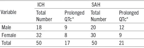
A total of 100 patients with stroke were included in present study; 50 patients had acute hypertensive spon-taneous intracerebral hemorrhages while the other 50 patients developed acute non-traumatic subarachnoid hemorrhage. The mean age of stroke patients was 64±9 years; 40% of them were in the group of 60-69 years. Fe-males outnumbered Fe-males, with a female to male ratio as 1.6:1. Most (82%) of the stroke patients were hyperten-sives, 47% of them were hyperlipidemic, and 42% of them were diabetic (Tables 1-4). There was a significant asso-ciation between the age of the patients and subarachnoid hemorrhage (p<0.001). No significant difference was observed among stroke patients with hemorrhage types with respect to gender (p=0.6).
Variable
Table 1. Patients’ total number, gender, and their QTc interval prolongation with respect to their stroke pattern; intracerebral hemorrhage (n=50), subarachnoid hemorrhage (n=50). *Defined as a corrected QT interval of >440 ms in males and 460 ms in females.
Gender Number SMD P-value 95% Confidence Interval
Lower Upper
Male 9 31.178
0.38
451.336 482.470
Female 8 18.7 465.462 501.753
Total 17 29.635
Table 2. Comparison between males and females within the
intracerebral hemorrhage group who had prolonged QTc interval.* -SMD, standardized mean difference. *Defined as a corrected QT interval of >440 ms in males and 460 ms in females.
Gender Number SMD P-value 95% Confidence Interval
Lower Upper
Male 12 30.28
0.18
459.745 491.857
Female 9 28.31 478.782 528.782
Total 21 32.057
Table 3. Comparison between males and females within the subarachnoid hemorrhage group who had prolonged QTc interval.* -SMD, standardized mean difference., *Defined as a corrected QT interval of >440 ms in males and 460 ms in females.
Variable Number SMD P-value 95% Confidence Interval
Lower Upper
hemorrhage 12 18.76 464.748 491.865
Total 21 26.79 461.652 487.578
Table 4. Comparison between males of the intracerebral hemorrhage group versus those of the subarachnoid hemorrhage group who had prolonged QTc interval.* -SMD, standardized mean difference. *Defined as a corrected QT interval of >440 ms in males and 460 ms in females.
Variable Number SMD P-value
95% Confidence
hemorrhage 9 28.32 483.810 532.822
Total 17 25.132 482.718 506.179
Echocardiography findings were unremarkable among 56% of stroke patients; concentric left ventricular hyper-trophy (and diastolic dysfunction) was found in 40 pa-tients with intracerebral hemorrhage while 12 subarach-noid hemorrhage patients demonstrated left ventricular hypertrophy.
The mean RR interval of stroke patients was 603.8±246.9 msec; 87% of stroke patients had long RR interval. The mean QTc interval of stroke patients was 355.3±86.6 msec; 38% of them demonstrated prolonged QTc interval. A significant association was observed between intracerebral hemorrhage and hypertension (p<0.001) and a similar significance was noted between intracerebral hemorrhage and left ventricular hypertro-phy (p<0.001).
Both hemorrhagic groups revealed no statistical-ly significant difference with respect to the RR interval (p=0.1). The QTc interval was prolonged in 17 intrace-rebral hemorrhage patients while 21 patients with sub-arachnoid hemorrhage showed prolongation of that interval. There was a significant association between subarachnoid hemorrhage and prolongation of the QTc interval (p<0.001). However, no statistically significance gender difference was found within the same group and between the 2 groups (Tables 1-5).
4. DISCUSSION
Neurologists and cardiologists are frequently involved in coordinating the care of patients with a variety of con-ditions, the most common being stroke. A multitude of ECG changes have been observed in patients who presented with acute strokes, both ischemic and hem-orrhagic. In particular, repolarization changes, such as prolongation in the QTc interval, have been noticed in as much as 90% of unselected stroke victims.
The mean age of our patients was 64±9 years. This finding is similar to the results of Al-Asadi and Habib study among Iraqi patients in 2014 (11) which reported a mean age of stroke patients as 63.8±12.3 years. This age reported by our study is in agreement with that reported in many Asian and developing countries (12, 13) but it was about one decade lower than that reported in West-ern countries (14). This difference from WestWest-ern coun-tries might be attributed to health lifestyles adopted by these communities in last decades, in addition to highly organized health services. There was a significant associ-ation between older age of the patients and subarachnoid hemorrhage (p<0.001). This is similar to results of Degos et al study in the USA (2012) (15). In our study, females outnumbered males. This study disagrees with many studies in finding a male preponderance (a male: female ratio of 1.27:1) (16). It was reported that stroke incidence rates are 1.25 times greater in men, but because women tend to live longer than men, more women than men die of stroke each year (17).
The main risk factor for stroke among our patients was hypertension. This is consistent with results of Sidhar-tha et al study in India (2015) (18) which concluded Sidhar-that hypertension was a major risk factor for medical com-plications of hemorrhagic stroke. There was a significant
association between Intracerebral hemorrhage and sys-temic hypertension (p<0.001). A study from Italy (19) found that hypertension is more associated with intrace-rebral hemorrhage rather than ischemic stroke.
Echocardiographic findings were unremarkable in more than half of our patients; 40% of the patients had left ventricular hypertrophy. This is similar to results of Goldstein et al study in USA (2011) (20) which reported that strokes resulting from cardiac diseases and cardiac abnormalities-associated with neuromuscular disorders are examples of the many points of contact between neu-rology and cardiology.
In the present study, the mean RR interval of stroke pa-tients was 603.8±246.9 msec; 87% of them had prolonged RR interval but no statistically significant difference was observed between intracerebral and subarachnoid hem-orrhage patients regarding this RR interval prolongation. The mean QT interval was 371.6±61.9 msec; 15% of the patient demonstrated long QT interval. Long QT inter-vals were significantly found among SAH patients. This is consistent with results of van den Bergh et al study in Netherlands (2003) (21) which reported that ECG ab-normalities frequently occur after aneurysmal subarach-noid hemorrhage (SAH). ECG abnormalities usually disappear within a day with no any change in the neu-rological or cardiac condition (22). They are considered markers of the severity of the SAH but not predictors for potentially serious cardiac complications or clinical outcome (23).
The mean QTc interval of stroke patients in our study was 355.3±86.6 msec; 38% of of the patients had long QTc interval. This is close to results of Malik et al study in Pakistan (2013) (24). Sakr et al found that 34% of the subarachnoid hemorrhage patients had a prolonged QTc (25). On the other hand, Bergh et al found that 61% of
their patients had a prolonged QTc within 72 hours of hospital admission because ruptured cerebral aneurysm (26). Maramattom studied 110 patients with supra-ten-torial intracerebral hemorrhage and concluded that changes in the ECG were observed in 64% of patients; 8% of them had prolonged QTc interval (27). Akbar et al analyzed 84 patients who had developed acute hem-orrhagic stroke; he found that 63.4% and 68.29% of the patients had prolongation of the QTc interval in lead III and VI respectively (28).
Disturbances in the autonomic nervous system and a surge in the sympathetic nervous system output are thought to be responsible for these ECG abnormalities and changes. The frontal lobe, insular cortex, amygdala, and the stellate ganglia play a central role in controlling the autonomic nervous system and therefore, influenc-ing the cardiac conduction system and heart rate (7, 28). When the QTc interval prolongs, the myocardium be-comes unstable and ventricular ectopic beats develop frequently. The latter can readily degenerate into poly-morphic ventricular tachycardia or even ventricular fibrillation (30, 31).
Khechinashvili and Asplund (30) concluded that the presence of QTc interval prolongation in acute stroke (ischemic and hemorrhagic) usually represents pre-ex-isting coronary artery disease rather than a direct con-sequence of the stroke itself on the heart. On the other hand, Soliman and colleagues (32) found that QTc terval prolongation is associated with a significantly in-creased risk of incident stroke independent of traditional stroke risk factors. In addition, Maebuchi and coworkers (33) had linked QTc interval prolongation to the future development of cardiovascular disease in the general population.
Limitations of the study:
1. The number of cases was relatively small.
2. This is a single institutional study which does not reflect the practice of stroke in the whole of Iraq.
3. The target population was composed of patients of Kurdish ethnicity only, who might well have different ge-netic/cardiovascular risk factors from Arab patients (the latter group constitutes the majority of the Iraqi popula-tion and was not involved in the study).
4. There was no “healthy” group as well as no locally or nationally published articles which target the same topic, so that we might compare the results with.
5. The size and location of the stroke and their rela-tionship with the QTc interval prolongation were not assessed.
6. Only a single ECG examination was done, at the time of the emergency room admission. No 24-hour car-diac Holter monitoring was done.
Therefore, the findings might well have been different if the aforementioned factors were addressed.
• Acknowledgments: Many thanks go to our patients and their fami-lies; without their kind help, this study would have not been accom-plished. A special gratitude goes to our emergency room nursing staff for their kind help and care of our patients.
• Conflicts of interests, grants, and permission: None.
REFERENCES
1. Popescu D, Laza C, Mergeani A, Bajenaru OA, Antochi FA.
Lead Electrocardiogram Changes after Supratentorial Intra-cerebral Hemorrhage. Mædica. 2012; 7(4): 290-4.
2. Zhang Y, Post WS, Blasco-Colmenares E, Dalal D, Tomaselli
GF, Guallar E. Electrocardiographic QT Interval and Mortal-ity: A Meta-analysis. Epidemiology. 2011; 22: 660-70.
3. Montanez A, Ruskin JN, Hebert PR, Lamas GA, Hennekens
CH. Prolonged QTc interval and risks of total and
cardiovas-cular mortality and sudden death in the general population: a review and qualitative overview of the prospective cohort studies. Arch Intern Med. 2004; 164: 943-8.
4. Straus SM, Kors JA, De Bruin ML, van der Hooft CS, Hofman
A, Heeringa J, et al. Prolonged QTc interval and risk of sud-den cardiac death in a population of older adults. J Am Coll Cardiol. 2006; 47: 362-7.
5. Dekker JM, Crow RS, Hannan PJ, Schouten EG, Folsom AR.
Heart rate-corrected QT interval prolongation predicts risk of coronary heart disease in black and white middle-aged men and women: the ARIC study. J Am Coll Cardiol. 2004; 43: 565-71.
6. Amin OSM, Myckan HA, Hussein EH. QTc Interval
Prolon-gation and Stroke: Any Differences between Ischemic and Hemorrhagic Strokes? Cukurova Med J. 2014; 39(1): 75-82.
7. Amin OSM, Shwani SS, Noori SF, Hasan AM.
Carbamaze-pine and the QTc interval; any association? Neurology Asia. 2010; 15(2): 119-23.
8. Cowan JC, Yusoff K, Moore M, Amos PA, Gold AE, Bourke
JP, et al. Importance of lead selection in QT interval measure-ment. Am J Cardiol. 1988; 61: 83-7.
9. Hnatkova K, Malik M. “Optimum” formulae for heart rate
correction of the QT interval. Pacing Clin Electrophysiol. 1999; 22: 1683-7.
10. Kligfi eld P, Hancock EW, Helfenbein ED, et al. Relation of QT interval measurements to evolving automated algorithms from different manufacturers of electrocardiographs. Am J Cardiol. 2006; 98: 88-92.
11. Al-Asadi JN, Habib HA. Risk factors and 30-day case fatality of first-ever stroke in Basrah, Iraq. Nigerian Medical Journal. 2014; 55(3): 209-13.
12. Cheung CM, Tsoi TH, Hon SF, Au-Yeung M, Shiu KL, Lee CN, et al. Outcomes after first-ever stroke. Honk Kong Med J 2007; 13: 95-9.
13. Khan SN, Vohra EA. Risk factors for stroke: A hospital based study. Pak J Med Sci. 2007; 23: 17-22.
14. Wolfe CD, Giroud M, Kolominsky-Rabas P, Dunadas R, Le-mesle M, Heuschmann P, et al. Variation in stroke incidence and survival in 3 areas of Europe. European Registries of Stroke (EROS) Collaboration. Stroke. 2000; 31: 2074-9. 15. Degos V, Gourraud PA, Tursis VT, Whelan R, Colonne
C, Ko-rinek AM, et al. Elderly age as a prognostic marker of 1-year poor outcome for subarachnoid hemorrhage patients through its interaction with admission hydrocephalus. Anesthesiol-ogy. 2012; 117(6): 1289-99.
16. Dalal PM, Bhattacharjee M, Vairale J, Bhat P. Mumbai Stroke Registry (2005-2006) Surveillance using WHO Steps Stroke Instrument—Challenges and Opportunities. J Assoc Physi-cians India. 2008; 56: 675-80.
17. Wong KS. Risk factors for early death in acute ischaemic stroke and intracerebral haemorrhage: A prospective hos-pital-based study in Asia. Asian Acute Stroke Advisory Panel. Stroke. 1999; 30: 2326-30.
18. Sidhartha JM, Mohan J, Purma AR, Reddy LVPK, Sagar NK, Teja MP, et al. Risk factors for medical complications of acute hemorrhagic stroke. Journal of Acute Disease. 2015; 4(3): 222-5.
neuro-logic clinic of Modena. Rev Neurol. 1999; 59: 1-7.
20. Goldstein LB, El Husseini N. Neurology and Cardiology: Points of Contact: Update; Systemic Diseases and the Car-diovascular System (III). Rev Esp Cardiol. 2011; 64(4): 319-27. 21. van den Bergh WM, Algra A, Rinkel GJ. Electrocardiographic abnormalities and serum magnesium in patients with sub-arachnoid hemorrhage. Stroke. 2004; 35(3): 644-8.
22. Kuroiwa T, Morita H, Tanabe H, Ohta T. Significance of ST segment elevation in electrocardiograms in patients with ruptured cerebral aneurysms. Acta Neurochir (Wien). 1995; 133: 141-6.
23. Zaroff JG, Rordorf GA, Newell JB, Ogilvy CS, Levinson JR. Cardiac outcome in patients with subarachnoid hemorrhage and electrocardiographic abnormalities. Neurosurgery. 1999; 44: 34-9.
24. Malik S, Abdul Sattar R, Shah S, Rehman H, Tahira, Ismail MA. Frequency of QTc prolongation in patients with hemor-rhagic stroke. J Ayub Med Coll Abbottabad. 2013; 25(3-4): 75-7. 25. Sakr YL, Lim N, Amaral ACKB, Ghosn I, Carvalho FB, Re-nard M, et al. Relation of ECG changes to neurological out-come in patients with aneurysmal subarachnoid hemorrhage. Int J Cardiol. 2004; 96: 369-73.
26. Bergh WMVD, Algra A, Rinkel GJ. Electrocardiographic ab-normalities and serum magnesium in patients with subarach-noid hemorrhage. Stroke. 2004; 35: 644-8.
27. Maramattom BY, Manno EM, Fulghsm JR, Jaffe AS, Wijdicks EF. Clinical importance of cardiac troponin release and car-diac abnormalities in patients with supratentorial cerebral hemorrhages. Mayo Clin Proc. 2006; 81(2): 192-6.
28. Akbar MA, Haider SA, Awan MM, Chaudhary GM. Electro-cardiographic changes in acute stroke. Professional Med J. 2008; 15(1): 91-5.
29. Colkesen AY, Sen O, Giray S, Acil T, Ozin B, Muderrisoglu H. Correlation between QTc interval and clinical severity of subarachnoid hemorrhage depends on the QTc formula used. Pacing Clin Electrophysiol. 2007; 30(12): 1482-6.
30. Khechinashvili G, Asplund K. Electrocardiographic changes in patients with acute stroke: a systematic review. Cerebro-vasc Dis. 2002; 14(2): 67-76.
31. Klingelhofer J, Sander D. Cardiovascular consequences of clin-ical stroke. Baillieres Clin Neurol. 1997; 6(2): 309-35. 32. Soliman EZ, Howard G, Cushman M, Kissela B, Kleindorfer
D, Le A, et al. Prolongation of QTc and risk of stroke: The RE-GARDS (REasons for Geographic and Racial Differences in Stroke) study. J Am Coll Cardiol. 2012; 59(16): 1460-7. 33. Maebuchi D, Arima H, Doi Y, Ninomiya T, Yonemoto K,