Definitions
•
Pathology is a dicipline bridging clinical
practice & basic sience
•
To render diagnosis & guide therapy
•
Identity changes in :
–
Gross
The scientific focus of pathology is :
Etiology
• On the cause of disease
Pathogenesis
• Mechanisme of its development & the
pathways by which morphologic changes
occur
Normal Homeostasis
If cell adjusting structure & function
to accommodate changing demands &
Stages in the cellular response to stress & injurious
stimuli
Stresses/pathologic stimuli the cell:
Adaptation
• Atrophy
• Hypertrophy
• Hyperplasia
• Metaplasia
Perubahan sel & jaringan :
Agenesis
Aplasia
Hypoplasia
Atrophy
Hypertrophy
Hyperplasia
Metaplasia
Dysplasia
Anaplasia
Granuloma
•
Complete absent of an
organ
•
E.g. :
– Renal agenesis
– Ovarial agenesis
– Tubal agenesis, etc.
Agenesis
Aplasia
•
Anlarge is present but
never develops
•
E.g. :
!
•
Anlarge develoved incompletly but the tissue
histhologicaly normal
•
Ex. : microcephaly
Hypoplasia
•
Decrease in the:
–
Size
–
Function of a cell
•
But not dead
"
Causes of atrophy :
1. functional demand (immobilitation in fracture, prolonged
bed rest)
2. Inadequate supply O2 (ischemia)
3. Insufficient nutrients (starvation, inadequate nutrition, chronic disease)
4. Interrruption of trophic signals transmitted by chemical mediators (endocrine system/Neuromusculator transmission) e.g. : thyroid, adrenal cortex, ovarium, testis.
5. Persistent cell injury by chronic inflamation e.g. : chronic gastritis, prolonged pressure 6. Aging : brain, heart (Senile Atrophy)
4
The mechanism of atrophy :
•
synthesis
•
catabolism
•
Influenced by a number of hormones
e.g. :
•
Insulin
•
Tyroid stimulating hormon
•
Glucocorticoids
•
in the size of cell accompanied by augmented
functional capacity
•
Hypertrophy is a response to trophic signals
5
… hypertrophy
Physiological (hormonal) hypertrophy
• in puberty
• production of sex hormon
• Hypertrophy breast tissue
• Abnormal hormon production in cancer
Functional demands
• Exercise
• Pathological conditions (myocardial cell) • Kidney hypertrophy on surgical removed
') &# ) %&' * (- % ' ( ' $ % & &
# + (- % ' + ' * %& ,6# & && %76./ ' % % ' % ( )%+ % &
& 3 *'1 ) % %&'8 &')) *'++) ' &# * %&/ 1- 8' 1+ ( )%+ % ++) ' % ) ' )'9 & % (0 ) & * % ++) &# & ' / &#') ) %&' + %+ ' ' '% & &# & &# % ++) # 8 #-0 & 0#-,' % ) ' 8 + ( * % ++)./
& #' # ( '*'% &' 3 &# + ( &
* % ' % ( )%+ % ++) %+ ' ') 00 &/ ' % % ' % ( )%+ % ++) % & '8' 3 &#
:
Hyperplasia
the number of cells in an organ / tissue
1. Physiologic hyperplasia
–
Hormonal hyperplasia
–
Compensatory hyperplasia
2. Pathologic hyperplasia
–
Excessive hormonal / growth factor stimulation
e.g. : Endometrial hyperplasia
;
/
(
+ )&'( + &'
•
)&
( & ' ( ,#-0 0+ )' .
•
-
% ( )&'
/
%
)
*
%&'
+
(
< ) %
- 0 +-%-& ('
< +-(0# %-& #-0 0+ )'
/
)')&
&
++
$
-< %#
'% ' *+ (( &'
' &# )7'
&#
0' =
&# +' ( * 8')%
< #-0 0+ )'
* &# 1+
0'&# +' (
Metaplasia
Reversible change in which 1 adult cell type is replace by another adult cell type (convertion of 1 differentiated cell type of
another)
Metaplasia is usually reversible if the stimulus is
removed
• Squamous metaplasia of the bronchial epithelium to tobacco
• Lower oesophagus by reflux acidic gastric
• Endocervical metaplasia
> )& % (( ') &# 0+ %( & * + + 0'&# +' (
Metaplasia of normal columnar (left) to squamous epithelium (right) in a bronchus, shown (A) schematically and (B)
histologically
++ + +& &' ' &# )'9 3 )# 0 '9 &' * &# % ++ + % (0 & * &'))
1. Variation in the size & shape of cells
2. Enlargment, irregularity & hyperchromatism of the
nuclei
3. Disorderly arrangement of the cells within the
epithelium
Dysplasia
Dysplasia included in the morphological classification of the stage if intraepithelial neoplasia
Dysplasia is a preneoplastic lession in the sense
that it is a necessary stage in the multistep cellular
evolution to cancer.
•
Normal cell
primitive cell
•
E.g. : Malignant cell
–
Carcinoma
–
Sarcoma
–
Adenocarcinoma
–
Lymphoma
–
Etc.
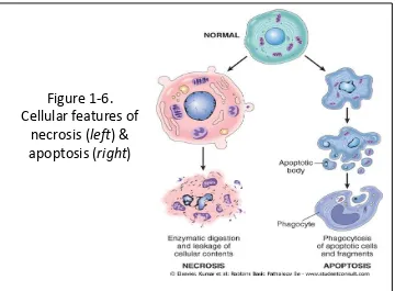
2 principal pattern of cell death :
• Commonly : coagulative necrosis • Cellular swelling
• Protein denaturation • Organellar breakdown • Cell rupture
NECROSIS
• Regulated event • Programmed death
Term
Definition
Necrosis
Antemortem pathologic cell death
Apoptosis
Antemortem programmed cell death
Autolysis
Postmortem cell death
CAUSES OF CELL INJURY
Hypoxia Physical Agent Chemical and drugs Microbiology Agents Immunologic Reaction
Genetic Defects Nutritional Inbalance
!
• Anemia
• Ischemia
• Intoxication CO2
• Aerobic oxidative respiration
• Mechanical trauma
• Extreme temprature : heat, cold
• Radiation: X-ray, sun light
• Electric shock
• Athmosphere pressure
Hypoxia Physical Agent
… CAUSES OF CELL INJURY
• Sufficiently concentrated :
– Glucose – Salt – O2
• Air pollutants
• Insecticides
• Asbestosis
• Ethanol
• Cellular metabolism (i.e. waste products)
• Tape worms
• Rickettsia
• Virus
• Bacteria
• Fungi
… CAUSES OF CELL INJURY
"
… CAUSES OF CELL INJURY
•
Anaphylactic reaction
•
Autoimmune diseases
Immunologic Reaction
Genetic Defects
• Congenital malformation • Sickle cell anemia
• G-6-PD
Nutritional Imbalance
• Protein calori insufficiency • Vitamins defficiency • Diabetes
4
Cellular response to injurious stimuli depends on :
• Injury type • Duration • Severity Current Status : • Nutritional • Hormonal • Adaptibility
of the cell
Intercellular systems : • Cell membrane
integrity • Aerobic
respiration • Protein synthesis • Integrity genetic
apparatus
O2& oxygen derived free radicals :
• Ischemic • Hypoxic
injury
Mechanism of Cell Injury
The ultrastructural features of these stages of cell injury. Normal cell & changes in reversible & irreversible cell injury
5
•
Reduced of :
– Oxidativephosphorylation in mitochondria
– Activity Na Pump
•
Cellular swelling
•
Loss of microvilli
Glycogen depleted ↓ protein synthesis
Formation of cell surface blebs
•
Severe vacuolization of
the mitochondria
•
Damage of :
– Mitochondrial matrix
– Plasma membrane
•
Swelling of lysosomes
•
Accumulation of
amorphous calcium
•
Rich dentities in
mitochondrial matrix
;
Figure 1-6.
Cellular features of
necrosis (
left
) &
apoptosis (
right
)
1. Reversible acute cell injury
2. Necrosis (cell death after irreversible injury)
3. Apoptosis (cell death by suicide)
4. Subcellular alteration as a respond to chronic or persistent injury stimuli
5. Intracellular accumulations of a number of substance
Sublethal Damage
1. Recoverable (but
necrosis is not)
2. Ultrastructural damage mitochondria
3. Swelling of cellular organelles ( hydrophic deg.)
4. Fatty change is impairment of metabolism
Morphologic changes that follow cell death in living tissue
1. Intense eosinophilia of the dead cell is due to
loss of RNA & coagulation of protein
2. Nuclei undergo:
1. Pyknosis 2. Karyorhexis 3. Karyolysis
Leaving a shrunken cell devoid of nucleus
1. Protein may be liberated from the dead cell
# ( 0# +
'% 00
%
*
% )') ')
&#
) +& * &2
))
&' ++- 0 % )) )
A
/
9-( &'% '
)&'
* &# % ++
/
&
&'
* 0 & '
Autolysis
: is a cell death by hydrolitic
enzymes.
Heterolysis
: cell death by the lysosomes of
invading inflammatory cells.
Nuclear Changes: This nucleus is faded -- karyolysis.
#'+ %-& 0+ )('% %# ) )) %' & 2'&# % ++ &# & )0 %'*'%3 %+ %# ) / # + 2 ' '% & ) ( +< 00 ' %+ ) 2#'+ &# )( ++ 2 ' '% & ) %+ ) &# & ') )( ++ 7 << * & ) * 60-7 )')/6 -7 &'% %+ ' ) )& &# & % ++) # 8 ' , ' % )')./
Types of Necrosis
Depends on :
! • (0+' ) 0 ) 8 &' * 1 )'% )& %& + &+' * &#
% + & % ++ C &')) * )0 * -)/
• # )& %& + 0 & ' &# 9-( &'% 0 & ' &# ) 1+ %7' % ++ + 0 & +-)')
• + &' % )') ') % # & ')&'% * #-0 @'% &# * % ++) ' ++ &')) @% 0& &# 1 '
/ / A >- % ' + * %&' , %%+ )' * & ' + ) 00+- .
Coagulative Necrosis
• '? * %&'8 C ++'? &'8 % )')
• &')) &# & 00 ) ) (' +'? ' ) ) +& * ')) + &' * &')) 1- &# %&' * #- +-&'%
9-( )
• / /A % 1 + ' * %&' 3 % )') % ) 1- 1 %& ' + ' */
• ) ) % )')
• % ++ * ( ( 0# ) 0 & ' % ) ( ))3 ' ' + %#'& %& % 1 ) #')& + '% ++-,) *& 2#'& ) (1+' % ( %# ) .
"
• Gumatous Necrosis
• Dead tissue, it is firm & rubbery like caseous necrosis in the spirochetal infection syphilis.
• Hemorrhagic Necrosis
• Dead tissue suffused with extravasated red cell, when cell death is due to blockage
• Fat Necrosis
• Not really necrosis.
• Focal areas of fat destruction tipically occuring following pancreatic injury /after trauma to fat for (ex. in the breast)
• Describes foci of hard yellow material seen in dead adipose tissue
•
Fibrinoid Necrosis
•
Fibrin deposited in damage necrotic vessel
walls in hypertension and vasculitis
•
Gangrene
4
APOPTOSIS
• Responsible for the programmed cell death in several important physiology processes
• Including :
– During embryogenesis (in implantation, organogenesis, & developmental involution)
– Hormon dependent physiologic involution (endometrium, lactating, prostate after castration)
– Cell deletion in proliferating population (intestinal crypt epithelium / cell dead in tumor)
– Deletion of autoreactive T cell in the thymus, cell death of cytokine starved lymphocytes
CLINICAL EFFECTS OF NECROSIS
•
Abnormal function
–
Kidney
: renal failure
–
Cortex in brain : muscle paralysis
–
Heart
: heart failure
–
Lung
: hemoptysis
5
•
Realease of contens of necrotic cells
–
Liver
: elevation SGOT
–
Heart : creatine kinase
•
Systemic effects
–
Fever
–
Inflamatoar Reaction
•
Local effects
–
Hemorrhage
–
Ulceration
Apoptosis of epidermal cells in an
immune-mediated reaction
A. Apoptotic cells are visible in the epidermis with eosinophilic cytoplasm and small, dense nuclei.
B. High power of apoptotic cell in liver in immune-mediated hepatic cell injury.
:
!"" " #
$ $%&
&
'#&($
%
! $% $%&
8'
(
&
0& &'
* &# % ++
/
#-)' +
'%
0& &'
<
(
)
<
) %# ('% + (
' & )
/
&# +
'%
0& &'
<
%&'
*
2 0 & ' )- &# )') 1- &
&
% ++
++
$ -
A
&# * % ++) , 0 (
&
' $ - .
1+ &# + ' $ - ,
0& &'
.
Granuloma
•
Special type of chronic inflamation in tissue
reaction.
•
Cause :
infection :
TBC fungal syphilis,etc
non-infection :
sarcoidosis;
NECROBIOSIS
•
Gradual cell damage
•
Progressive
•
Singly or small group cells.
•
Reversible (+/-)
•
Example : hepar cell
deg.
cell death
healing
fibrosis.
Alterations in structure and function that may
lead to cell death, or at least diminished capacity
of the cell to respond an injury
•
Reduced cell in :
–
Pleomorphic vacuolated mitochondria
Morphologic alteration in :
• Pleomorphic vacuolated mitochondria
•
endoplasmic reticulum
• Disorted Golgi Apparatus
• Accumutaion of lipofuscin pigment
Cellular senescence is multifactorial :
1. The cumulative effects of extrinsic influences:
free radical damage
DEGENERATION
Cloudy
swelling
Fatty
change
Hydropic
Atropy
Hyaline
Mucoid
Amyloid
Calcifica-tion
-
0'% %#
*
)& &'
+
( +
& #' # ( '*'% &' &# ' & %-& 0+ )('% * &
)*