The journal homepage www.jpacr.ub.ac.id
MDA and Histologic Profile of Pancreatic Diabetic-Rats Model
Administered With Extract of
Glycine max (L.) Merr.
Luh Putu Gina*1, Chanif Mahdi1, Aulanni’am1,2
1
Departement of Chemistry, Faculty of Science, Brawijaya University Jl. Veteran Malang 65145, East Java, Indonesia
2
Faculty of Veterinary Medicine, Brawijaya University
*
Corresponding author: [email protected]
Received 14 January 2016; Revised 16 March 2016; Accepted 16 March 2016
ABSTRACT
Diabetes Mellitus is characterized by leveling up glucose in human blood and affects increasing of free radicals in the body as well as leading to cellular oxidative stress. Experimentally, this condition is able to be characterized by increasing malondialdehyde (MDA) level in cell and histological changing in pancreas appearance. Consumption of antioxidant substances was reported able to reduce the MDA quantity as free radicals. Black soybean or Glycine max (L) Merr. was reported contains important antioxidant agents such as anthocyanin and isoflavone. This paper discloses recent investigation on the application of black soybean water extract to reduce the MDA level on diabetes mellitus-rat model induced by STZ (DM) and also reports the pancreas histological changing of the DM rats. Investigation revealed that black soybean water extract significantly affects decreasing of MDA level by 4.9%, 27.1% and 45.7% in three different doses therapy (500, 750, and 1000 mg/kg BW). Histologically, it also clearly indicates repairing of pancreas tissue of the DM rats.
Keywords: black soybean, diabetes mellitus, MLD-STZ, pancreatic beta cells, MDA, histology, antioxidant
INTRODUCTION
Diabetes mellitus (DM) or commonly known as diabetes is a chronic disease caused by body's inability to producing insulin. It is also due to ineffective use of insulin production and affects increasing blood sugar level [1]. DM type 1 is caused by autoimmune destruction of pancreatic beta cells. This is caused by infiltration of mononuclear cells and results in decreasing of insulin production. And this affects leveling up glucose level in the human blood. This condition is called as hyperglycemia. Hyperglycemia dangerously able to promotes free radicals intensity in the cell. And this will cause imbalance between the protective antioxidant and free radicals in the body. Finally, initiate the oxidative damage or commonly known as oxidative stress [1-2]. Oxidative stress itself will increase the glycosylation and oxidation of proteins which is associated to the pathogenesis of diabetic complications. This also contributes to the destruction of islet function and insulin resistance and also worsening the diabetic conditions as an impact of the decreasing catalase activity in the body [3].
radicals in the human body can be measured by appearing of biological substances developed and known as a biomarker or biological marker during oxidative stress in the body. Biological substance known and is often used as a biomarker of lipid peroxidation and oxidative stress is malondialdehyde (MDA) [4-6].
Previous researchers reported application of some plant chemicals including secondary metabolite not only as an antioxidant but also for treatment DM in rats model such as salam (Syzygium polyanthum) [7-8], temu giring (Curcuma heyneana) [3, 8-9], Sorgum spp [10], and jewawut (Setaria italica) [11-12]. Other potency plant contains antioxidant agent is black soybean (Glycine max (L.) Merrit). In Indonesia, black soybean commonly used for soy sauce ingredients, yogurt production [13-14], and biofuels [15]. Young black soybean or un-dried soybean in Lombok region West Nusa Tenggara is also an important ingredient for preparing traditional food Lombok soup or known as Lebui soup. It is also traditionally applied as herbal medicine to treat glucose diseases. It was prepared as water-boiled black soybean or as black solid. Study paper reports the water extract of black soybean from Lombok Island as dekok (boiling water). Some paper reports that black soybean especially water extracts contain secondary metabolite isoflavonoid and anthocyanin [16], [17-19]. This secondary metabolite in some other research isolated from different plants indicated the potency for reducing free radicals in DM rats. However, it was not clear the rule of this secondary metabolite contained in black soybean. Anthocyanin was arisen for their high reactivity due to the activity as hydrogen or electron donor. It has similarity capability as polyphenols which derived radical to stabilize by delocalizing of the unpaired electrons or other radicals [20-21]. This study was conducted to observe the benefits of water extract of black soybean against decreasing in oxidative stress in the rat model of diabetes mellitus. These rats model was prepared by exposing at low-dose of streptozotocin (MLD-STZ) and measuring the levels of malondialdehyde (MDA) and including the histological appearance from groups of rat pancreas.
EXPERIMENT
Chemicals and instrumentation
This research using Ratttus norvegicus Wistar strain male as model rat (200 g weight) and was kept in a clean box as cages with dimension 20x30x40 cm3. Meanwhile some chemicals used including black soybean, streptozotocin (STZ), citrate buffer pH 4.5, alcohol 70%, 80% and 90%, sodium chloride solution 0.9%, phosphate buffer saline (PBS), paraformaldehyde (PFA) 4% formalin, paraffin, dyes hematoxylene-eosin (HE), xylol, distilled water, entellan, 1 N hydrochloric acid and Na-Thio mL 1%, sodium hydrogen phosphate, potassium dihydrogen phosphate, hydrogen peroxide. Meanwhile, the tools used for research including reaction tube, vortex, scissors, tweezers, pasteur pipette, glass objects, spatula, knife, analytical balance, scalpel, surgical instruments, clamp (block holder), incubator, hot plate, water bath, gloves, light microscopes, centrifuges, centrifuge set, cuvette, mortar.
Preparation of black soybean water extracts
Phytochemicals test
The phytochemical test was conducted to determine the presence of anthocyanin compounds in water extracts of black soybeans by reacting 3 ml of water extract of black soybean with 3 mL of 2 M hydrochloric acid solution. It was heated to 100 °C for 5 minutes until raised of red color. Then it was added dropwise of 2 M of sodium hydroxide to form green color. This green color indicates the presence of anthocyanin on the water extract.
LC-MS Analysis
The flavonoid contained in the extract of black soybean (Glycine max (L.) Merr.) was separated and qualitatively identified utilizing LC-MS/MS [16].
Preparation of experiment animals
Several groups of 2-month old rats (200 g weight) and was adapted for a week by giving feed and drinking water. Groups of healthy rats (negative control) was given feed and water every day. Groups of diabetic rat (positive control) beside food and water were also treated by induction of MLD-STZ at 20 mg/kg BW dose for five times 5 days. The blood glucose level of each group rats was measured after induction of MLD-STZ by using glucometer. Group of rats were classified as positive diabetes rats if blood glucose levels over 200 mg/dL.
Groups of DM-rat treated induced with MLD-STZ 20mg/kg BW for 5-times consecutive days were then treated with black soybeans water extract for 14 days. The dosages applied were 500, 750, and 1000 mg/kg of body weight (BW). The blood glucose measurements were undertaken after induction with MLD-STZ and after treatment with water extract of black soybean for 14 days. Measurement was used glucometer. In the end, all groups of rats were sacrificed by neck dislocation way, pancreas dissected and organs were taken for further analysis. All process and procedure using animal as animal model were undergone following Standard Ethic and Procedure using animal as animal model in Brawijaya University.
Embedding of pancreas
Pancreas organ is immersed in a fixative solution, formalin or 4 % PFA for 1-7 days. Then, it was soaked with 70% of ethanol for 24 h and followed with 80% ethanol for 2 h. Finally, it was soaked in 90% of ethanol for 20 minutes and with absolute ethanol 3-times for 20 minutes. The pancreatic organ was moved onto xylol 1 and 2, respectively 20 minutes, and xylol 3 at 60-63oC for 20 minutes. Then, the organ was dipped in liquid paraffin and further poured into the container. The solidified paraffin contain pancreatic organ was in block shape ready for the further process.
Preparations pancreas
Histological and haematoxylin-eosin staining
Preparations included in the multilevel xylol 1-3 each for 5 minutes. Then, it proceeded the dehydration stage. Preparations consist of the fractionation using ethanol starting 1-3 absolute ethanol, 95% ethanol, 80%, and 70% respectively for 5 minutes each. Then, it was soaked in distilled water followed by staining stages. Preparations of haemotoxylin dye to obtain the best color results for about 10 minutes. Then it was washed with flowing water for 30 minutes, rinsed with distilled water. Then, it was dipped into eosin staining for several minutes and soaked with distilled water. The next dehydration stages were undertaken using multilevel preparations in ethanol 80%, 90%, and 95% to absolute ethanol 1-3. The clearing process was carried out by entering preparate in xylol 1, 2, and then the air dried process. Finally was mounting (adhesion) with Entellan [22].
Preparation of standard curve of MDA
Preparation of the MDA standard curve with concentrations of 0, 1, 2 , 3, 4, 5, 6, 7, and 8 µg/mL with total volume maximum 100 μL each, and were put in different tube. Then, each of tubes was added 550 mL of distilled water. Each of these tubes already containing 650 mL of standard solution and was added 100 mL of TCA 100%, 250 mL of 1 N hydrochloric acid solution and 100 mL of 1% Na-Thio. Then, they were homogenized with vortex, and each of tubes were covered with plastic and given a hole. All the tubes were incubated in waterbath at 100°C for 30 minutes and cooled at room temperature. Finally, MDA concentrations were measured at 500-600 nm wave length in order to determine the maximum wavelength of MDA. The standard curve was made by plotting the absorbance resulted in each measurement with their concentration at their maximum wavelength.
Measurements MDA level with Thiobarbituric Acid Test
Pancreas tissue of each group rats was taken respectively (0.5 g) and cut into small pieces. Then, it was crushed with mortar and placed on the cold block of ice. Then, it was added 1 mL of 0.9% sodium chloride. The resulting homogenates of pancreatic tissue was transferred into small test tube and centrifuged at 8000 rpm for 20 minutes. The supernatant resulted was taken and 100 mL of supernatant of pancreatic were added 550 mL of distilled water, 100 mL TCA, and 250 mL 1N hydrochloric acid, and 100 mL of Na-Thio. Each reagent addition, the mixture solution was homogenized with vortex, centrifuged at 500 rpm for 15 minutes. The supernatant was separated and transferred into new reaction tube. Finally, the solution was incubated in a water bath at a room temperature and samples were measured the absorbance at maximum wavelength of TBA test and plotted on a standard curve which has been plotted in order to calculate the concentration of MDA in sample.
RESULT AND DISCUSSION
Phytochemicals and LC-MS analysis of black soybean extract
Moreover, identification the composition of water extract by means of liquid chromatography-mass spectrometry (LC-MS/MS) provided the composition of flavonoid contained on the water extract of black soybean (Table 1) [16].
Figure 1. Schematic anthocyanine structures at different pH [23].
Table 2. Composition of Glycine max (L.) Merr. water extract analyzed by LCMS [16]
Molecule M
(m/z)
Area
% M+1
M+G (m/z)
Area %
M+G+A (m/z)
Area %
Genestein 270.24 14.0 271.24 432.24 1.00 474.24 8.60
Daidzein 254.23 8.20 255.23 416.23 7.30 458.23 4.00
Glycitein 284.26 1.60 289.26 446.26 1.40 488.26 11.1
Delphinidin-3-Gluc 338.80 29.6 339.80 500.80 6.80 542.80 4.60
Cyanidin-3-Gluc 322.80 2.50 323.80 484.80 7.80 526.80 2.30
Petunidin-3-Gluc 317.41 4.00 318.41 479.41 11.3 521.41 13.0
Note: M= Aglycone, M+G =Glycoside, and M+G+A =Acylated Glycoside
Malondialdehyde (MDA) levels on model rats
Measurement of malondialdehyde (MDA) level in 5-different group of rats was undertaken using thiobarbituric acid (TBA) test. Each member of groups was analyzed spectrophotometrically by measuring the absorbance value toward the TBA absorbance at maximum wavelength. The result is presented in Table 2.
Table 2. Malondialdehyde (MDA) pancreatic levels of different groups of model rats
Group MDA levels
(µg/mL)
Decreasing MDA levels (%)
Healthy rats (negative control) 0.227±0.037 - Diabetic rats (positive control) 0.584± 0.058 -
Therapy 500 mg/kgBW* 0.555±0.013 4.90
Therapy 750 mg/kgBW* 0.426±0.032 27.1
Therapy 1000 mg/kgBW* 0.317±0.023 45.7
Note: *therapy using water extract of black soybean at given doses.
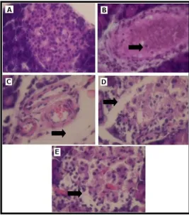
Figure 2. HE staining results with a magnification of 1000x of pancreatic cell preparate. (A) Healthy rats (negative control), (B)
diabetic rats (positive control), (C) therapy rats 500 mg/kgBW, (D)therapy rats 750 mg/kgBW, and therapy rats with dose 1000 mg/kgBW. (BW is body weight of mouse).
black soybean water extract with 750mg/kg BW were relatively giving similar effect. Furthermore, this result requires more investigation to give detailed mechanism.
Histology of pancreatic beta cells on rat model
The decreasing MDA level on cellular level of the DM-model rats after treatment with black soybean water extract theoretically has to in-line with appearance on the pancreatic tissue. Histology observation is presented in figure 2. A haematoxylin and eosin (HE) staining procedure were undertaken for 5 groups of model rats, i.e. negative control (healthy rats), positive control (diabetic rats), and therapy rats treated water extract of black soybean 500, 750, and 1000 mg/kg of rat body weight (BW).
The results of the study are shown in Figure 1 shows the pancreatic beta cells in healthy rats, diabetic rats and therapy rats there is a difference. In healthy rats histology images shown in Figure A there is no cell damage (necrosis) that is characterized by the absence of free space on the islet pancreas and cell nucleus (purple round the islet), in figure B shows necrosis quite severe, visible inflammation of the cells there is cavity because cell nucleus as seen not as dense as in the pancreatic tissue of healthy rats. Rats were treated therapy water extract of black soybean in image C, D and E with dose variation 500 mg/kg of BW, 750 mg/kg of BW and 1000 mg/kg of BW necrosis cavity seen in the results of histopathology but narrower than diabetic rats. Figure C, D and E indicates an improvement in pancreatic tissue after treatment of soybean water extract for 14 days. Figure C shows greater necrosis seen from the amount of free space on pancreatic islet cells as a description of the dissolution junction cell (relatival between cells) due to inflammation. Figure C, D and E pancreatic tissue of therapy rats that received treatment extract of black soybean showed necrosis, but also show improvement when be compared with image B for image diabetic rats pancreatic tissue is characterized by more dense nucleus of cells. This suggests that the pancreatic beta cell damage due to free radicals as induced by MLD-STZ. Pancreatic beta cell damage is what will cause a reduction in insulin production. The results of the study, it is shown in Figure 5.3 shows the black soybean extract with dose variation 500mg/kgBW, 1000mg/kgBW, and 750mg/kgBW can help reduce free radicals in the pancreas that causes damage to the tissues of the pancreas.
CONCLUSION
The conclusion of this study is that the water extract of black soybean (Glycinemax (L.) Merr.) with a dose variation 500mg/kgBW, 750mg/kgBW, and 1000mg/kgBW for 14 days effect on the decreased levels of MDA respectively 4,9%, 27.1%, and 45.7% on therapy with dose of 500mg/kgBW, 750mg/kgBW, and 1000 mg/kgBW. Water extract of black soybean also affects the pancreas tissue repair.
ACKNOWLEDGMENT
Authors declare that no conflicts of interest and all authors have an equal contribution preparing the manuscript.
REFERENCES
[1] American Diabetes Association, Diabetes Care, 2010, 33 (1), S62–S69. [2] K. G. M. M. Alberti and P. ft Zimmet, Diabet. Med., 1998, 15 (7), 539–553.
[4] D. Del Rio, A. J. Stewart, and N. Pellegrini, Nutr. Metab. Cardiovasc. Dis., 2005, 15 (4), 316–328.
[5] R. Mateos, L. Goya, and L. Bravo, J. Chromatogr. B, 2004, 805 (1), 33–39.
[6] V. Peddireddy, B. Siva Prasad, S. D. Gundimeda, P. R. Penagaluru, and H. P. Mundluru, Biomarkers, 2012, 17 (3), 261–268.
[7] T. Widyawati, W. W. Purnawan, I. J. Atangwho, N. A. Yusoff, M. Ahmad, and M. Z. Asmawi, Int. J. Pharm. Sci. Res., 2015, 6 (4), 1698.
[8] A. Saifudin, T. Usia, S. AbLallo, H. Morita, K. Tanaka, and Y. Tezuka, Asian Pac. J. Trop. Biomed., 2016, 6 (1), 38–43.
[9] A. Saifudin, K. Tanaka, S. Kadota, and Y. Tezuka, J. Nat. Prod., 2013, 76 (2), 223– 229.
[10] H. Y. Kil, E. S. Seong, B. K. Ghimire, I.-M. Chung, S. S. Kwon, E. J. Goh, K. Heo, M. J. Kim, J. D. Lim, and D. Lee, Food Chem., 2009, 115 (4), 1234–1239.
[11] V. K. Thirumalairaj, P. A. Anitha, G. Durairaj, S. G. Menon, and L. Shanmugaasokan,
J. Appl. Pharm. Sci. Vol, 2014, 4 (12), 026–029.
[12] Y. Sireesha, R. B. Kasetti, S. A. Nabi, S. Swapna, and C. Apparao, Pathophysiology,
2011, 18 (2), 159–164.
[13] J. Zhang, E. Tatsumi, J. Fan, and L. Li, Int. J. Food Sci. Technol., 2007, 42 (3), 263– 268.
[14] F. Fetriyuna, Int. J. Adv. Sci. Eng. Inf. Technol., 2015, 5 (1), 44–46.
[15] C. U. Zanetta, B. Waluyo, M. Rachmadi, and A. Karuniawan, Energy Procedia, 2015, 65, 29–35.
[16] L. P. Gina, C. Mahdi, and A. Aulanni’am, J. Pure Appl. Chem. Res., 2014, 3 (3), 131– 137.
[17] B. Xu and S. K. Chang, J. Agric. Food Chem., 2008, 56 (16), 7165–7175.
[18] M.-G. Choung, I.-Y. Baek, S.-T. Kang, W.-Y. Han, D.-C. Shin, H.-P. Moon, and K.-H. Kang, J. Agric. Food Chem., 2001, 49 (12), 5848–5851.
[19] J. H. Lee, N. S. Kang, S.-O. Shin, S.-H. Shin, S.-G. Lim, D.-Y. Suh, I.-Y. Baek, K.-Y. Park, and T. J. Ha, Food Chem., 2009, 112 (1), 226–231.
[20] T. Tsuda, Y. Kato, and T. Osawa, FEBS Lett., 2000, 484 (3), 207–210.
[21] J.-M. Kong, L.-S. Chia, N.-K. Goh, T.-F. Chia, and R. Brouillard, Phytochemistry,
2003, 64 (5), 923–933.
[22] M. Masruri, M. Lutfillah, A. Sumaryanto, R. Retnowati, and A. ’am Aulanni’am, J. Trop. Life Sci., 2014, 4 (3), 161–165.
![Figure 1. Schematic anthocyanine structures at different pH [23].](https://thumb-ap.123doks.com/thumbv2/123dok/2861331.1694407/5.595.67.530.430.553/figure-schematic-anthocyanine-structures-different-ph.webp)