PROGRESSION OF DIABETIC RETINOPATHY FOLLOWING CATARACT SURGERY
Yayan Heryanto, Iwan Sovani, Arief Kartasasmita, Erwin Iskandar, Djonggi Panggabean. Dept. of Ophthalmology Medical Faculty Unpad, Cicendo Eye Hospital Bandung
ABSTRACT
Introduction: Diabetes is the most common risk factor for cataract development in underdeveloped countries. Cataract was the principal cause of legal blindness in adult-onset diabetics and the second most common cause of legal blindness after proliferative diabetic retinopathy in those with juvenile-onset diabetes. Cataract surgery in diabetic patients has been associated with progression of retinopathy.
Objctive:to report the progression of diabetic retinopathy following cataract surgery.
Case Report: CaseI, A 55 years old woman was diagnosed as diabetic cataract ODS. She had undergone phacoemulsification+IOL on left eye. Postoperatively the visual acuity in left eye was improved. Afer four months the visual acuity in left eye was decreased, she was diagnosed as immature cataract OD + mild-moderate NPDR OD + macular edema OD + pseudofakia OS + severe NPDR OS + macular edema OS. Intravitreal injection of Avastin® was done in her left eye. One week after injection the visual acuity in left eye was improved and than she had undergone panretinal laser photocoagulation (PRP) in her left eye. Case II, A 59 years old woman was diagnosed as diabetic cataract ODS. phacoemulsification+IOL on right eye and than left eye was done. One week postoperative the visual acuity in both eyes was decreased, she was diagnosed as pseudofakia ODS + PDR ODS. and had undergone intravitreal injection Avastin®, panretinal laser photocoagulation (PRP) in her both eyes. The visual acuity in the left eye was improved
Conclusion: Cataract surgery in diabetic patients has been associated progression of retinopathy and that progression is associated with poor visual prognosis.
Introduction
Diabetes has become the epidemic of the 21stcentury and diabetic retinopathy will in the coming years account for an increasing proportion of the workload of retinal specialists.1
The incidence of diabetes is increasing because of factors that include the ageing of the population, the increase of life expectancy and large population movements from rural areas to urban areas, with dietary and lifestyle changes that result in increase in the incidence of obesity and decrease in physical activity.1
The global prevalence of diabetes is estimated to exceed 220 million people by the year 2010 and 300 milion by the year 2025. Diabetes is the most common risk factor for cataract development in underdeveloped countries. Furthermore, diabetic patients suffer lens opacities at an earlier age than individuals without
diabetes.The Wisconsin Epidemiological Study of Diabetic Retinopathy reported that cataract was the principal cause of legal blindness in adult-onset diabetics and the second most common cause of legal blindness after proliferative diabetic retinopathy in those with juvenile-onset diabetes.2,3
Cataract surgery is indicated when a patient’s visual function is significantly reduced as a result of lenticular opacity. Surgical intervention may also be indicated if a cataract reduces the ophthalmologist’s view of the retina, thus impending the diagnosis and treatment of diabetic retinopathy.2
and macular edema. Some study have reported that cataract surgery causes progression of retinopathy and that progression is associated with poor visual prognosis. Thus it is important for the surgeon to address the preoperative management of diabetic retinopathy, the impact of surgery on the disease’s progression, the appropriate timing of surgery, and the optimal management of postoperative macular edema.2,4
The effect of cataract surgery on the progression of diabetic retinopathy is a somewhat controversial topic. The rate of pathologic condition’s postoperative progression may be influenced by a number variables, including (1) the severity of the preoperative diabetic retinopathy, (2) how long the patient has had diabetes, (3) the adequacy of the patient’s glycemic control, and (4) the cataract surgeon’s operative techniques.2
This case report will discuss about the progression of diabetic retinopathy following cataract surgery.
Case Report Case I :
A 55 years old woman came to the Hospital on April 17th 2009 with chief complain of gradually blurred vision on both of eyes since 1 year ago. Blood pressure was 140/80 mmHg. Laboratory finding showed blood glucose level of 388 mg% . From the ophthalmology examination, visual acuities of both eyes were 0.15 ph 0.3 uncorrected.. No data about posterior segment examination of both eyes. She was diagnosed as diabetic cataract ODS. Phacoemulsification + IOL on left eye was performed after one month DM regulation (blood glucose level at any
moment 113 mg%). one week
postoperative , visual acuity was 0.7 ph 1.0.
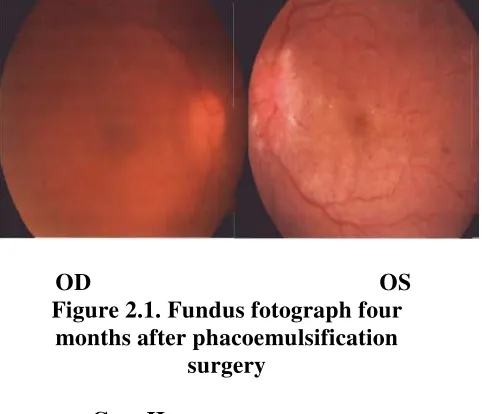
Four months after operation the visual acuity in left eye was 0.1 ph (-),blood glucose level at any moment 216 mg%. The patient consulted to vitreoretinal specialist. She was diagnosed as immature
Cataract OD + mild-moderate NPDR OD + pseudofakic OS + severe NPDR OS. She had undergone intravitreal injection Bevacizumab (Avastin®) in her left eye and panretinal laser photocoagulation (PRP) in her left eye.
OD OS
Figure 2.1. Fundus fotograph four months after phacoemulsification
surgery Case II :
A 59 years old woman came to the hospital on December 28th 2005 with chief complain of gradually blurred vision on both of eyes. She had history of hypertension and diabetes mellitus. blood pressure 170/100 mmHg. Laboratory finding showed blood glucose level at any moment 257 mg% . Opthalmology examination, visual acuity in right eye 1/60 and left eye 0.25 uncorrected. Intraocular pressures were normal for both eyes. No data about posterior segment examination of both eyes. She was diagnosed as diabetic cataract ODS. phacoemulsification surgery + IOL on right eye was performed. One day postoperative the visual acuity in right eye was 0.7 and one week postoperatively was 0.8.
and one week postoperatively visual acuity in right eye was 0.8 and left eye 0.05 ph 0.1. The patient consulted to vitreoretinal specialist and was diagnosed as Pseudofakic ODS + Proliferative Diabetic Retinopathy ODS. She had undergone intravitreal injection Avastin®in both eyes and laser PRP three sessions in both eyes. on December 26th 2007, the visual acuity in right eye was 0.8 and left eye 0.5.
OD OS
Figure 2.2. Fundus fotograph two years (OD) and 1 week (OS) after phacoemulsification surgery
Discussion
Diabetic retinopathy may be defined as the presence of typical retinal microvascular lesions in an individual with diabetes. Diabetic retinopathy remains the leading cause of new blindnes in the United States for adults under the age of 65 years, accounting for 5800 new cases of legal blindness annualy. It is estimated that approximately 25% of the diabetic population have some degree of retinopathy, and that about 5% are affected by more severe disease (proliferative retinopathy). The prevalence of all types retinopathy increases with the duration of diabetes, probably reflecting yhe
consequences of prolonged
hyperglycemia. Patients are usually spared for 3 to 5 years following the onset of systemic disease, but after 20 years of hyperglycemia, nearly all patients with type I diabetes and more than 60% of patients with type II diabetes have some degree of retinopathy. Young persons with type I diabetes are more likely to suffer severe visual complications from proliferative diabetic retinopathy during
their lifetime, whereas a greater total number of older patients with type II diabetes mellitus experience visual loss from macular edema.5
Diabetic retinopathy is broadly classified as nonproliferative diabetic retinopathy (NPDR), and proliferative diabetic retinopathy (PDR). The microvascular changes that occur in NPDR are limited to the confines of the retina, whereas PDR is characterized by the growth and extension of new vessels beyond the internal limiting membrane. Diabetic macular edema (DME), a common cause of decreased central vision, can occur in the setting of NPDR or PDR.
5,6
The fundus findings observed in diabetic retinopathy are a consequence of two important characteristics at the level of the retinal capillaries. These include (1) a variable degree of intraretinal capillary closure, and (2) increased retinal vascular permeability leading to a breakdown of the inner blood-retinal barrier.5
With progression of NPDR, intraretinal hemorrhages resulting from ruptured microaneurysms and decompensated capillaries become visible, with superficial flame-shaped hemorrhages being located in the nerve fiber layer and dot-shaped hemorrhages or blot-shaped hemorrhages within the deeper retina.5
Acceleration of retinal capillary abnormalities eventually affects adjacent arterioles, resulting in arteriolar closure and discrete areas of capillary dropout or nonperfusion. Clinically, the most obvious manifestation of such arteriolar ischemia is the cotton-wool spot, a localized infarct of the nerve fiber layer that results from occlusion of the terminal arteriole (precapillary arteriole). Once the cotton-wool spot resolves, the inner retina layers may become atropic, with the site becoming ophthalmoscopically visible as a focal, depressed area.5
proliferative disease become evident. Examples include beading and dilation of retinal veins adjacent to areas of arteriolar nonperfusion. Another important fundus sign of retinal ischemia is the development of intraretinal microvascular abnormalities (IRMAs), a descriptive term for irregular, segmental dilatations of the retinal capillary bed. IRMAs are thougth to represant either a compensatory dilatation of pre-existing vascular channels (shunt vessels), or a form of intraretinal neovascularization.5
NPDR, also known as background diabetic retinopathy, is further graded into mild, moderate, severe, or very severe. PDR is described as early, moderate, high-risk, or advanced (Early Treatment Diabetic Retinopathy Study/ ETDRS grading)7,8
In case I, the patient was diagnosed mild-moderate NPDR in right eye by the fundus viewed reveal minimally exudates and hemorrhages and severe NPDR in left eye (hemorrhages in 4 quadrants). In case II, the patients was diagnosed PDR in both eyes by the fundus viewed reveal growth of abnormal new vessels, vein dilatation (vein beading), and fibrous tissue (vitreous fibrous), and preretinal hemorrhage in left eye. Diabetic macular edema (DME) reveal in all cases.
Various studies suggest that diabetic retinopathy may progress following cataract surgery. Cataract surgery in diabetic patients may become necessary, not only to improve vision but also to allow assessment and treatment of diabetic retinopathy. Compared to non-diabetic patients, visual outcome after cataract surgery was reported to be worse in diabetic patients – especially in those with diabetic retinopathy. Some investigators also found an increased progression of retinopathy and a higher incidence of macular edema after cataract surgery. Most of these studies were retrospective, and either different operation techniques – such us ICCE or ECCE, phacoemulsification, or different
patients populations such as diabetic patiens without retinopathy and with retinopathy of various degrees – may explain the different outcomes. If postoperative inflammation and breakdown of the blood-retinal barrier are involved in the pathogenesis of progression of diabetic retinopathy after
cataract extraction, then
phacoemulsification surgery should theoritically result in a decreased rate of retinopathy production. Surgical outcome in patients without diabetic retinopathy is comparable to non-diabetic patients, and the outcome of patients with retinopathy appears to depend on the degree of retinopathy at the time of surgery. In general, patients with mild NPDR without laser indication have been proposed to have a good prognosis.2,3,9
The most important factor to be emphasized in the medical management of diabetic retinopathy is maintenance of good glycemic control. Intensive glycemic control is associated with reductions in progression to severe NPDR and PDR, incidence of macular edema, and need for panretinal and focal photocoagulation. Control of hypertension was also benefical in reducing progression of retinopathy and loss of vision.8
Severe visual loss following cataract surgery in diabetics may be due to worsening macular edema, continuing anterior and posterior segment proliferation, posterior capsule opacification, or unrelated events, such as retinal vein occlusion. Risk factors associated with worsening retinopathy after cataract surgery include pre-existing severe treated or untreated retinopathy, poor glycaemic control, increasing age, and planned or unplanned posterior capsule disruption.10
patients shows visual improvement after phacoemulsification surgery but gradually decreased, there are possibilities consequences of progression of diabetic retinopathy and DME.
Non-proliferative diabetic retinopathy can rapidly progress to severe diffuse macular edema in the months following uncomplicated cataract extraction. This suggest that the blood-barrier is significantly impaired, even in diabetics with no retinopathy and that cataract surgery worsens this impairment.
10
Preoperative and early postoperative photocoagulation for macular edema appears to reduce but not to eliminate the risk of visual loss. 10
Adequate panretinal laser
photocoagulation (PRP) is therefore essential if there is severe peripheral retinal ischaemia or early retinal neovascularisation. This photocoagulation should be applied preoperatively or in the early postoperative period. 10 Intravitreal administration of corticosteroids was shown to modestly improve vision in the short term and reduce macular thickness for up to 2 years of follow up.
In this cases, all patients were undergone intravitreal injection of Avastin® (anti VEGF) and panretinal laser photocoagulation (PRP). Anti-VEGF was associated with rapid regression of persistent neovascularization in diabetic retinopathy refractory to PRP as well as visual acuity improve for at least 12 weeks.1,8,11
All patients in these cases were having a good life prognosis. Functional prognosis of the patients eye were dubia. However, their had good response condition of anti-VEGF and photocoagulation laser PRP treatments.
Refferences
1. Cunha-Vaz Jose et al, Euro Times Update Diabetic Retinopathy, 9th Euretina congress, Pfizer Ophthalmics, Nice France. 2009. P 2-9
2. Sam S & Everett, Cataract surgical considerations in Diabetes. Available at:
http://www.crstoday.com/PDF%20Articles /0805/CRST0805_F4_Yang.html.
3. Krepler K et al. Cataract surgery inpatients with diabetic retinopathy: Visual outcome, progression of diabetic retinopathy, and incidence of diabetic macular edema. Graefe’s Arch Clin Exp Ophthalmol (2002) 240: 735-738
4. Henricsson M et al. Diabetic retinopathy befoe and after cataract surgery. British Journal of Ophhalmology 1996;80:79-793 5. Regillo CD et al. Diabetic Retinopathy.
Chapter 10. In: Vitreoretinal Disease the Essentials. Thieme. New York. 1999. P 133-58
6. Olk R Joseph & Lee M Carol. Classification of Diabetic Retinopathy. Chapter 2. In: Diabetic Retinopathy. JB Lipincott Company. Philadelphia. 1993. P 3-18
7. Joussen M Antonia et al. Grading of Diabetic Retinopathy. Chapter 19. In: Retinal Vascular Disease. Springer. NewYork. 2007. P 294
8. American Academy of Opthalmology. Retina and Vitreous, Section 12. San Fransisco: AAO, 2008-2009
9. Borrillo JL et al. Retinopathy progression
and visual outcomes after
phacoemulsification in patients with diabetes mellitus. Tr. Am. Ophth. Vol XCVII. 1999
10. Flanagan DW. Progression of diabetic retinopathy following cataract surgery: can it be prevented?. Br. J Ophthalmol. 1996 Sept; 80(9): 778-779