TESIS
KARAKTERISTIK MORFOLOGI TIPE CACING
Fasciola gigantica MELALUI KAJIAN
MORFOMETRI PADA SAPI YANG
DIPOTONG DI RPH PEGIRIAN
SURABAYA
PENELITIAN OBSERVASIONAL
Oleh
NIM 061414253009 VIRGI ALCITA RAKA JHONI
PROGRAM STUDI MAGISTER
ILMU PENYAKIT KESEHATAN MASYARAKAT VETERINER FAKULTAS KEDOKTERAN HEWAN
UNIVERSITAS AIRLANGGA SURABAYA
2016
iii
KARAKTERISTIK MORFOLOGI TIPE CACING
Fasciola gigantica MELALUI KAJIAN
MORFOMETRI PADA SAPI YANG
DIPOTONG DI RPH PEGIRIAN
SURABAYA
PENELITIAN OBSERVASIONAL
TESIS
untuk memperoleh gelar Magister
dalam Program Ilmu Penyakit Kesehatan Masyarakat Veteriner
pada Fakultas Kedokteran Hewan Universitas Airlangga Surabaya
NIM 061414253009 VIRGI ALCITA RAKA JHONI
PROGRAM STUDI MAGISTER
ILMU PENYAKIT KESEHATAN MASYARAKAT VETERINER
FAKULTAS KEDOKTERAN HEWAN UNIVERSITAS AIRLANGGA
SURABAYA
v
Lembar Pengesahan
TESIS INI TELAH DISETUJUI Tanggal 12 Februari 2016
Oleh :
Pembimbing Ketua
NIP. 19520928 197803 1 002
Prof. Dr. Setiawan Koesdarto, drh., M.Sc.
Pembimbing
NIP. 19610402 198803 1 003
Dr. Soeharsono, drh., M.Si.
Mengetahui, Ketua Program Studi
Ilmu Penyakit Kesehatan Masyarakat Veteriner Fakultas Kedokteran Hewan Universitas Airlangga
NIP. 19620828 198903 2 001
vii
UCAPAN TERIMA KASIH
Puji syukur Kehadirat Allah SWT atas karunia yang telah dilimpahkan sehingga
penulis dapat melaksanakan penelitian dan menyelesaikan tesis dengan judul
Karakteristik Morfologi Tipe Cacing Fasciola gigantica Melalui Kajian Morfometri pada Sapi yang dipotong di RPH Pegirian Surabaya, sebagai
salah satu syarat menempuh gelar magister pada Fakultas Kedokteran Hewan
Universitas Airlangga.
Pada kesempatan ini penulis ingin menyampaikan terima kasih kepada:
1. Prof. Dr. Pudji Srianto, drh., M.Kes., selaku Dekan Fakultas Kedokteran Hewan Universitas Airlangga atas kesempatan mengikuti pendidikan di
Fakultas Kedokteran Hewan Universitas Airlangga.
2. Prof. Dr. Lucia Tri Suwanti, drh., MP., selaku Ketua Program Studi Ilmu Penyakit dan Kesehatan Masyarakat Veteriner Fakultas Kedokteran Hewan
Universitas Airlangga yang telah bersedia membimbing dan memberikan
saran serta nasihat yang berguna kepada penulis.
3. Prof. Dr. Setiawan Koesdarto, drh., M.Sc., selaku pembimbing utama atas kesempatan untuk bersedia memberikan bimbingan, saran dan nasihat yang
berguna selama penelitian serta dalam penyusunan tesis ini.
4. Dr. Suharsono, drh., M.Si, selaku pembimbing kedua yang bersedia memberikan bimbingan, saran dan nasihat yang berguna selama penelitian
viii
5. Prof. Dr. Bambang Sektiari L., drh., DEA., Dr. Benjamin Chr. Tehupuring, drh., M.Si. dan Dr. Kusnoto, drh., M.Si., selaku penguji atas segala nasihat
dan masukan yang diberikan kepada penulis demi kesempurnaan tesis ini.
6. Kedua orang tua, ayahanda Joni Mulyono Cipto dan ibunda Mudji Rahayu serta saudari Deka Isnatu R.J dan seluruh keluarga besar terima kasih atas
doa dan motivasi yang diberikan.
7. Bodhi Agustono, drh., M.Si. terima kasih atas segala bentuk dukungan, doa, semangat dan waktu yang diberikan kepada penulis.
8. Ririn Rohmawati, Dinda Rahma H., Shafia Khairani, Alfiana Laili A., Mia Zakia, dan teman-teman seperjuangan S2 IPKMV angkatan 2014, terima
kasih atas segala bentuk dukungan, doa dan semangat yang diberikan
kepada penulis.
9. Semua pihak lain yang telah membantu penulis dalam penyusunan tesis ini baik langsung maupun tidak langsung. Semoga segala bantuan dan
bimbingan kepada penulis menjadi sebuah amal ibadah yang akan dibalas
oleh Allah SWT.
Penulis menyadari bahwa tesis ini masih banyak kekurangan. Semoga hasil yang
dituangkan dalam tesis ini bermanfaat bagi pembaca dan semua pihak yang
membutuhkan.
Surabaya, Februari 2016
Penulis
ix
RINGKASAN
Karakteristik Morfologi Tipe Cacing Fasciola gigantica Melalui Kajian Morfometri pada Sapi yang dipotong di RPH Pegirian Surabaya
Fasciolosis merupakan salah satu penyakit helmin yang utama pada ternak yang sampai saat ini masih sulit untuk diberantas dan menyebabkan kerugian secara signifikan dalam produktivitas ternak serta bersifat zoonosis. Di Indonesia, fasciolosis lebih sering terjadi pada sapi dan kerbau daripada domba dan kambing, umumnya disebabkan oleh Fasciola gigantica. Strain variasi Fasciola gigantica di Indonesia sangat luas berdasarkan pemeriksaan morfologi dan molekuler terdapat tiga tipe Fasciola gigantica dengan morfologi dan klaster yang berbeda. Identifikasi morfologi merupakan langkah dasar untuk mengetahui keberagaman spesies dalam mempelajari epidemiologi dan melakukan diagnosis, penegakan diagnosis fasciolosis memerluka n uji dan bahan uji dengan spesifitas tinggi sehingga mengetahui perbedaan spesifik spesies sangat diperlukan.
Pemeriksaan morfologi cacing dewasa Fasciola gigantica menggunakan
Scanning Electron Microscope (SEM) dibeberapa wilayah di Indonesia
menunjukkan bahwa cacing dewasa F.gigantica terdiri dari tiga tipe berdasarkan perbedaan morfologi spina, sucker dan cirrus. Fasciola gigantica tipe 1 dijumpai di Indonesia berasal dari domba, kambing, sapi dan kerbau, Fasciola gigantica tipe 2 ditemukan pada kerbau di Jawa Tengah dan sapi perah di Kota Batu Jawa Timur, sedangkan Fasciola gigantica tipe 3 ditemukan pada sapi Bali dan kerbau di Jawa Tengah. Selain penggunaan SEM sebagai alat identifikasi morfologi tehnik pengukuran dengan komputer bermanfaat untuk mengetahui variasi morfometri antara populasi yang berbeda dari Fasciola, teknik ini dapat dilakukan untuk mengetahui keragaman morfologi spesifik dengan menganalisis karakteristik morfologi tertentu antara spesies.
Penelitian ini bertujuan untuk menganalisis struktur tipe morfologi dan morfometri cacing Fasciola gigantica pada sapi yang dipotong di Rumah Potong Hewan Pegirian Surabaya menggunakan optilab camera microscope dan
Scanning Electron Microscope (SEM). Karakteristik tipe cacing Fasciola gigantica menggunakan SEM berdasarkan morfologi sucker, spina dan cirrus,
sedangkan morfometri tipe cacing Fasciola gigantica berdasarkan 15 parameter meliputi Length of body (BL), width of body (BW), length of cone (CL), width of
cone (CW), diameter oral sucker; maximum (OS max), diameter oral sucker; minimum (OS min), diameter ventral sucker maximum (VS max), diameter ventral sucker minimum (VS min), jarak antara anterior body dan ventral sucker (A-VS),
jarak antara oral sucker dan ventral sucker (OS-VS), jarak antara ventral sucker dan vitelline glands (VS-Vit), jarak antara vitelline glands dan posterior body (Vit -P), jarak antara ventral sucker dan posterior body (VS-P), pharynx length (Ph L) dan pharynx width (Ph W).
Hasil pemeriksaan post mortem pada hepar sapi yang dipotong di RPH Pegirian Surabaya didapatkan 5 sampel positif yang berasal dari daerah berbeda
x
yaitu Probolinggo, Pasuruan, Malang, Bali dan Lumajang. Berdasarkan pengamatan morfologi sucker, spina dan cirrus menggunakan SEM di dapatkan tiga tipe yang berbeda dari ke lima daerah masing-masing Fasciola gigantica tipe 1 diidentifikasi pada hepar sapi Peranakan Ongole yang berasal dari Lumajang dan Probolinggo dengan ciri cirrus invaginasi dan bentuk spina tumpul, landai (15˚) mengarah ke posterior, tipe 2 diidentifikasi dari hepar sapi Frisian Holstein yang berasal dari Malang dan Pasuruan dengan ciri ukuran cirrus besar (200 µm) permukaan berduri, spina meruncing, landai (30˚) mengarah ke posterior dan tipe 3 berasal dari hepar sapi Bali yang berasal dari Bali dengan ciri ukuran cirrus besar (181,8 µm) permukaan halus, spina tumpul, ujung bergerigi dan menonjol kearah atas (65˚).
Hasil analisis data morfometri cacing dewasa Fasciola gigantica dengan
SPSS Statistics Version 23 diperoleh parameter pembeda tipe yang paling
dominan yaitu oral sucker (Os max, Os min), diameter ventral sucker (Vs max, Vs min), jarak oral sucker dan ventral sucker, panjang tubuh dan cone (BL,CL), jarak ventral sucker dan vitteline (VsVit) serta diameter pharyng (Ph L, Ph W).
Berdasarkan hasil penelitian disarankan perlu dilakukan studi lebih lanjut mengenai karakterisasi morfologi tipe cacing Fasciola gigantica melalui identifikasi morfologi untuk melihat kemungkinan adanya tipe lain atau subtype dari peneltian yang telah ada serta dengan metode molekuler untuk mengetahui karakterisasi profil yang lengkap pada setiap tipe cacing Fasciola gigantica.
xi
SUMMARY
Morphological characteristics Type of Worm Fasciola gigantica Based on Morphometrics Studies on Cattle Slaughtered in The Pegirian Abattoir
Surabaya
Fasciolosis is an important disease in cattle that is still difficult to eradicate and cause a significant loss in productivity of livestock and zoonotic. In Indonesia, fasciolosis that is more common in cattle and buffaloes instead of sheep and goats, is generally caused by Fasciola gigantica. Strain variation Fasciola gigantica in Indonesia is very broadly based on morphological and molecular examination there are three types of Fasciola gigantica with different morphology and clusters. Morphological Identification of the basic steps to determine the diversity of species in the study of the epidemiology and diagnosis, diagnosis fasciolosis require testing and test materials with high specificity so knowing the species-specific differences are very necessary.
Morphological examination of Fasciola gigantica adult worms using a Scanning Electron Microscope (SEM) in some areas in Indonesia indicate that the adult worms Fasciola gigantica consists of three types based on morphological differences spines, sucker and cirrus. Fasciola gigantica type 1 found in Indonesia comes from sheep, goats, cows and buffalo, Fasciola gigantica type 2 is found in buffalo in Central Java and dairy cattle in Batu East Java, while Fasciola
gigantica type 3 is found in Bali cattle and buffaloes in Central Java. In addition
to use as a means of identification SEM morphology measurements with computer techniques helpful to know the morphometric variation among different populations of Fasciola, this technique can be performed to determine the specific morphological diversity by analyzing certain morphological characteristics between species.
The aim of this research is to analyze the types of morphology and morphometry Fasciola gigantica from liver cattle slaughtered at Pegirian Abbatoir Surabaya using optilab camera microscope and Scanning Electron Microscope (SEM). The characteristics of the types of worm Fasciola gigantica using SEM based on morphology sucker, spine and cirrus, while morphometry types of worm Fasciola gigantica based on 15 parameters including Length of body (BL), width of the body (BW), length of cone (CL), width of the cone (CW), the diameter of the oral sucker; maximum (max OS), the diameter of the oral sucker; The minimum (OS min), the diameter of the ventral sucker maximum (VS max), the diameter of the ventral sucker minimum (VS min), the distance between the anterior body and the ventral sucker (A-VS), the distance between the oral sucker and ventral sucker (OS-VS) the distance between ventral sucker and vitelline glands (VS-Vit), the distance between the vitelline glands and posterior body (Vit -P), the distance between ventral sucker and posterior body (VS-P), pharynx length (Ph L) and pharynx width (Ph W).
xii
The results of post-mortem examination of the liver of cattle slaughtered at the abattoir Pegirian Surabaya obtained 5 positive samples originating from different regions, namely Probolinggo, Pasuruan, Malang, Bali and Lumajang. Based on morphological observation sucker, spina and cirrus using SEM in get three different types of the five regions each Fasciola gigantica type 1 was identified in the liver of Ongole Cross derived from Lumajang and Probolinggo with the characteristics of cirrus invaginasi and form of spina blunt, ramps ( 15˚) directed posteriorly, type 2 identified from liver Holstein Frisian cows from Malang and Pasuruan with the characteristics of cirrus large size (200 m) spiked surface, tapering spines, ramps (30˚) directed posteriorly and type 3 is derived from the liver Bali cattle originating from Bali with a characteristic size of large cirrus (181.8 m) surface is smooth, blunt spines, serrated tip and protruding upward (65˚).
The results of morphometry analysis of adult worm Fasciola gigantica with SPSS Statistics Version 23 obtained parameter differentiating the type most dominant namely oral sucker (Os max, Os min), the diameter of the ventral sucker (Vs max, Vs min), distance oral sucker and ventral sucker, long body and cone (BL, CL), distance ventral sucker and vitteline (VsVit) and pharyngeal diameter (Ph L, Ph W).
Based on the results of the study suggested further studies need to be done on the morphological characterization of the type of worm Fasciola gigantica through morphological identification to see the possibility of another type or subtype of the research that's been there as well as the method to determine the molecular characterization of a complete profile on each type of worm Fasciola gigantica.
xiii
MORPHOLOGICAL CHARACTERISTICS TYPE OF WORM Fasciola Gigantica BASED ON MORPHOMETRICS STUDIES ON CATTLE
SLAUGHTERED IN THE PEGIRIAN ABATTOIR SURABAYA
Virgi Alcita Raka Jhoni
ABSTRACT
The aim of this research is to analyze the types of morphology and morphometry Fasciola gigantica from liver cattle slaughtered at Pegirian Abbatoir Surabaya using optilab camera microscope and Scanning Electron Microscope (SEM). A sample of the adult worm Fasciola gigantica observed morphological differences based sucker, spina and cirrus and measured based on body size, pharyngeal, sucker, and vitteline. The results of morphological observation using SEM, type 1 is identified in the liver of Ongole Cross from Lumajang and Probolinggo with the characteristics of cirrus invaginasi and form of spina blunt, ramps leading to the posterior, type 2 were identified from the liver of cattle Frisian Holstein from Malang and Pasuruan characterized cirrus large size (200 m) spiked surface, tapering spines, ramps leading to the posterior and type 3 is derived from cow liver Bali from Bali with a characteristic size of large cirrus (181.8 m) surface is smooth, blunt spines, serrated tip and stand towards the top. The results of morphometric analysis using factor analysis with SPSS Statistics version 23 obtained a dominant factor to distinguish three kinds of worms, the diameter and disctance oral and ventral suckers, body length and cone, ventral sucker and vitteline distance and diameter of the pharynx.
xiv
DAFTAR ISI
Halaman
HALAMAN SAMPUL DALAM ... ii
PRASYARAT GELAR ... iii
PERNYATAAN ... iv
PERSETUJUAN ... v
PENETAPAN PANITIA PENGUJI ... vi
UCAPAN TERIMA KASIH ... vii
RINGKASAN ... ix
SUMMARY ... xi
ABSTRACT ... xiii
DAFTAR ISI ... xiv
DAFTAR GAMBAR ... xvi
DAFTAR TABEL ... xvii
DAFTAR LAMPIRAN ... xviii
SINGKATAN DAN ARTI LAMBANG ... xix
BAB 1 PENDAHULUAN ... 1
1.1 Latar Belakang ... 1
1.2 Rumusan Permasalahan ... 4
1.3 Tujuan Penelitian ... 4
1.4 Manfaat Hasil Penelitian ... 4
BAB 2 TINJAUAN PUSTAKA ... 5
2.1 Cacing Fasciola gigantica ... 5
2.1.1 Klasifikasi dan Morfologi ... 6
2.1.2 Siklus Hidup ... 8
2.1.3 Epidemiologi ... 9
2.1.4 Pencegahan dan Pengendalian ... 10
2.2 Gambaran Umum RPH Pegirian ... 11
2.3 Gambaran Umum Jenis Sapi ... 12
2.4 Morfometri ... 15
2.4.1 Optilab Camera Microscope ... 16
2.4.2 Scanning Electron Microscope (SEM) ... 16
BAB 3 KERANGKA KONSEPTUAL ... 18
BAB 4 MATERI DAN METODE ... 21
4.1 Jenis Penelitian ... 21
4.2 Lokasi dan Waktu Penelitian... 21
4.3 Populasi dan Besar Sampel ... 21
4.3.1 Populasi ... 21
4.3.2 Besar Sampel ... 22
xv
4.4 Bahan Penelitian ... 22
4.5 Peralatan Penelitian ... 22
4.6 Metode Penelitian ... 23
4.6.1 Cara Pengambilan Sampel ... 23
4.6.2 Tehnik Pewarnaan Semichen-Acetic Carmine ... 23
4.6.3 Tehnik Pengukuran Morfometri ... 24
4.6.4 Metode Scanning Electron Microscope (SEM) ... 24
4.7 Bagan Kerangka Operasional ... 26
4.8 Analisis Data ... 27
BAB 5 HASIL PENELITIAN ... 28
5.1 Identifikasi Morfologi Cacing Fasciola gigantica dengan Scanning Electron Microscope (SEM) ... 28
5.2 Morfometri Cacing Fasciola gigantica dengan Optilab Camera Microscope ... 34
BAB 6 PEMBAHASAN ... 37
6.1 Identifikasi Morfologi Cacing Fasciola gigantica dengan Scanning Electron Microscope (SEM) ... 37
6.2 Morfometri Cacing Fasciola gigantica dengan Optilab Camera Microscope ... 39
BAB 7 KESIMPULAN DAN SARAN ... 42
7.1 Kesimpulan ... 42
7.2 Saran ... 43
DAFTAR PUSTAKA ... 44
xvi
DAFTAR GAMBAR
Gambar Halaman
2.1 Bagian anterior sukcer dan cirrus Fasciola gigantica dengan SEM (A) Tipe 1 (B) Tipe 2 (C) Tipe 3. Sumber : Kurniasih (1995) ... 6
2.2 Siklus hidup Fasciola spp yang dimodifikasi ... 8
2.3 Standar pengukuran cacing dewasa Fasciola gigantica yang di-
modifikasi ... 16
3.1 Kerangka konseptual ... 19
4.1 Kerangka operasional ... 27
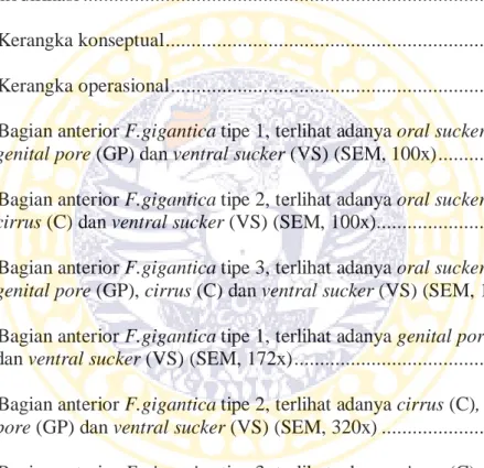
5.1 Bagian anterior F.gigantica tipe 1, terlihat adanya oral sucker (OS),
genital pore (GP) dan ventral sucker (VS) (SEM, 100x) ... 29
5.2 Bagian anterior F.gigantica tipe 2, terlihat adanya oral sucker (OS),
cirrus (C) dan ventral sucker (VS) (SEM, 100x)... 29
5.3 Bagian anterior F.gigantica tipe 3, terlihat adanya oral sucker (OS),
genital pore (GP), cirrus (C) dan ventral sucker (VS) (SEM, 100x) 30
5.4 Bagian anterior F.gigantica tipe 1, terlihat adanya genital pore (GP) dan ventral sucker (VS) (SEM, 172x) ... 31
5.5 Bagian anterior F.gigantica tipe 2, terlihat adanya cirrus (C), genital
pore (GP) dan ventral sucker (VS) (SEM, 320x) ... 31
5.6 Bagian anterior F.gigantica tipe 3, terlihat adanya cirrus (C), genital
pore (GP) dan ventral sucker (VS) (SEM, 320x) ... 32
5.7 Spina (S) F.gigantica tipe 1 a). anterior (SEM, 750x) b). mid-body (SEM, 500x) ... 33
5.8 Spina (S) F.gigantica tipe 2 a). anterior (SEM, 750x) b). mid-body (SEM, 500x) ... 33
5.9 Spina (S) F.gigantica tipe 3 a). anterior (SEM, 750x) b). mid-body (SEM, 1400x) ... 33
xvii
DAFTAR TABEL
Tabel Halaman
5.1 Klasifikasi Morfologi Tipe Cacing Fasciola gigantica Menggunakan
Scanning Electron Microscope (SEM) ... 28
5.2 Nilai Rata-Rata Hasil Pengukuran Cacing Fasciola gigantica dengan
Optilab Camera Microscope dalam millimeter (mm) ... 35
xviii
DAFTAR LAMPIRAN
Lampiran Halaman
1 Analisis data morfometri tipe cacing dewasa Fasciola gigantica .... 50
2 Proses Scanning Electron Microscope (SEM) ... 51
3 Dokumentasi Kegiatan ... 52
xix
SINGKATAN DAN ARTI LAMBANG
CDC = Centers for Disease Control
cm = centimeter
kg = kilogram
mm = milimeter
pH = Power of Hydrogen
RPH = Rumah Potong Pegirian
spp. = spesies
WHO = World Health Organization
µm = mikrometer