www.elsevier.com/locate/jinsphys
Salivary proteins of aphids, a pilot study on identification,
separation and immunolocalisation
Anas Cherqui
1, W. Fred Tjallingii
*Wageningen University, Laboratory of Entomology, PO Box 8031, 6700 EH, Wageningen, The Netherlands
Received 26 July 1999; accepted 13 December 1999
Abstract
Salivary proteins (SPs) of Schizaphis graminum, Acyrthosiphon pisum and Myzus persicae were studied after probing and feeding on different artificial diets. Salivary sheaths as well as apical lumps of saliva were found, presumably representing subsequently excreted saliva of different types. Phenoloxidase, pectinase and peroxidase activities were detected by staining the enzyme-converted products, thus confirming these enzyme activities found earlier by others. Proteinase and cellulase were not found. SPs in three major SDS–PAGE bands, at 154 and 66/69 kDa, were collected in fluid diets (soluble fraction) and as sheath material (solid fraction) attached to the membranes covering these diets. Proteins of both fractions presumably represented the enzymatic activities found, although this could not be proven. The lack of electrophoretic mobility of the undenaturated (isoelectrofocusing and PAGE) active proteins meant that they could not be separated, whereas the mobile denaturated (SDS–PAGE) proteins had lost their enzyme activity. Polyclonal antibodies, anti-SP154 and anti-SP66/69, both cross-reacted to most salivary proteins in Western blots. They also reacted to sheath material and to the principal salivary glands. For further studies of saliva some monoclonal antibodies were developed. The complexity of salivation and the relation of the results obtained to the behaviourally known secretion periods is discussed. 2000 Elsevier Science Ltd. All rights reserved.
Keywords: Salivary glands; Proteins; Immunolocalisation
1. Introduction
The aphid Schizaphis graminum (greenbug) is known as a serious pest for a variety of cereals, causing major economic damage. Stylet penetration, or probing, by S.
graminum often causes chlorosis and sometimes necrosis
in leaves of cereals (Ryan et al., 1990). This damage by the plant’s reactions to aphid probing develops in a time span of days. Presumably, the aphid saliva plays an important role in the plant’s chlorosis as the direct cell damage by the penetrating stylets through the plant tissue is only minute, at least initially, in spite of many intracellular punctures (Tjallingii and Hogen-Esch, 1993). At the level of sieve elements in the phloem, there
* Corresponding author. Fax:+31-317-484821.
E-mail address: [email protected] (W.F. Tjallingii).
1 Present address: Universite´ de Picardie Jules Verne, Faculte´ des Sciences, Laboratoire de Biologie des Entomophages, 33, rue Saint Leu, 80039 Amiens, France.
0022-1910/00/$ - see front matter2000 Elsevier Science Ltd. All rights reserved. PII: S 0 0 2 2 - 1 9 1 0 ( 0 0 ) 0 0 0 3 7 - 8
is also no indication of serious damage. Though careful puncturing of the sieve element by a glass microelec-trode evokes plugging of its sieve plate within minutes (Knoblauch and van Bel, 1998), an aphid is apparently able to puncture and tap sap from a sieve element for hours and days, continuously (Tjallingii, 1995). Possibly here also, the saliva injected before phloem ingestion may play an important role.
from detected enzyme activity (Urbanska et al., 1998). It remains unclear how these observations, all done with aphids probing into artificial diets covered by mem-branes, are related to probing into plants.
Diet-collected saliva, i.e. saliva obtained after excretion by aphids in Parafilm-covered fluid diets, has been analysed and proteins have been separated by electrophoresis (Miles, 1999). Baumann and Baumann (1995) showed that saliva of S. graminum contained three main proteins with homologue sequences in their N terminus, suggesting a structural and synthetic relationship. Miles and Harrewijn (1991), on the other hand, found proteins of different molecular weights in diet-collected saliva from the aphid Myzus persicae. The aphid species as well as the diet compositions used in these studies differed so that it remains unclear what caused these differences. Also, these studies provided no indication where and when these proteins are secreted in plants. Phenoloxidases were found in aphid saliva (Miles, 1985) and there were speculations about their detoxifying function in plant tissues (Leszczynski and Dixon, 1990; Miles and Oertli, 1993). Urbanska et al. (1998) confirmed the phenoloxidase activity in the aphid’s saliva excretions. Pectinase activity has been detected elegantly by Ma et al. (1990) in salivary excretions of S. graminum. The need of pectinase for stylet penetration between cells has been postulated (McAllan and Adams, 1961; Campbell and Dreyer, 1990). However, its interference with the physiology of plants by the release of pectin fragments, known as potent elicitors, may be more important (Ma et al., 1998). Other enzyme activities in aphid saliva have been detected as well (Peng and Miles, 1988; Miles and Peng, 1989; Urbanska et al., 1998).
The injected saliva seems to play a crucial role in the prevention of the plant’s wound responses and damage but, on the other hand, it may act as an elicitor of plants reactions, resulting in damage during a later stage of the infestation. Whether the compatibility between plant and aphid species, the result of the evolutionary develop-ments, really has these conflicting aspects forms the underlying question of this study. First, however, the role of the saliva should be shown and its composition during the various excretion periods must be established. The direct objective of this pilot study, therefore, was to try different diets for the collection of saliva, using dif-ferent aphid species. For saliva analysis, assays for enzyme activity and techniques for protein separation were used to compare results with those obtained by others and to explore their technical prospects. We also wanted to initiate antibody experiments and to test them as a tool for future studies, such as characterisation of salivary components, tracing their origin from different salivary glands and their secretion in different plant tissues and cells. The use of some existing polyclonal
and newly developed monoclonal antibodies can be seen as first attempts that should be extended further.
2. Materials and methods
2.1. Aphids and saliva collection
The three species of aphids were reared in the green-house at 20°C and a 16L:8D photoperiod. Myzus
per-sicae (M1 clone, WAU, Wageningen) was reared on
Brussels sprouts, Acyrthosiphon pisum (green clone from INSA, Villeurbane, France; Dr Y. Rahbe´) on broad beans, and Schizaphis graminum (clone from University of California, Davis, USA; Dr P. Baumann) on wheat. Up to 100 adults corresponding to 50 mg, were trans-ferred from plants to a feeding chamber, a standing cyl-inder (PVC tube), 27 mm in diameter and 20 mm high, covered on top by a double layer of Parafilmwith one of the diets in between (a “sachet”). Each sachet con-tained about 100 µl of diet. Aphids remained in the chamber overnight in an incubator at 20°C, illuminated from above with a constant yellow light (yellow foil filtered). Four types of diets were used (Table 1); (1) distilled water, (2) 15% sucrose, (3) 15% sucrose con-taining 100 mM serine, 100 mM methionine and 150 mM aspartic acid, and (4) an anti-clotting diet. The latter diet contained 50 mM citrate buffer, pH 6.2, and 10 mM EDTA and 120 mM NaCl to reduce clotting or gelling of salivary sheath material. Diets were microbial filtered and experiments were done in sterile conditions to avoid bacterial or fungal contamination.
The saliva collected after incubation of aphids on the diets can be distinguished as two fractions: (1) soluble saliva, which was in the fluid diets, collected after removal of the top membrane of the sachets, in addition to what was washed from the lower membrane with sam-ple buffer (see below; an extra volume of about 20 µl per sachet), and (2) the sheath saliva, i.e. what remained attached to the lower membrane and was not suspended or dissolved in diets 1, 2 and 3. The collected saliva of about five sachets was pooled.
2.2. Sheath observations
Table 1
Composition of diets used for the collection of salivary excretions
Diet 1: water Diet 2: water sucrose Diet 3: amino acidsa Diet 4: anticlotting
Distilled and filtered water Distilled and filtered water+15% Distilled and filtered water+15% Distilled and filtered water+15% sucrose sucrose, 100 mM serine, 100 sucrose, 50 mM citrate buffer, 10 mM
mM methionine, 150 mM EDTA, 120 mM NaCl pH 5.5 aspartic acid pH 6.8
aThe only diet that resulted in honeydew excretion.
2.3. Enzyme activities
Various enzyme substrates were added to the diets in the feeding chambers to detect the presence of enzymes in the excreted saliva. Dihydroxyphenylalanine (dopa), 0.1%, was used to identify phenoloxidase activity (catechol oxidase, EC 1.10.3.1; Miles, 1999) in diets with or without agarose. The black enzyme product, mel-anin, will stain salivary sheaths and halos around the sheaths. For pectinase identification 0.1% pectin was used in a 0.5% agarose diet. The jelly diets were removed from the sachets after aphid exposure and trans-ferred to a Petri dish for staining in 0.01% Ruthenium red (Sterling, 1970). This was done at two pH values, 5.0 and 6.4, to indicate the activity of pectin(methyl)esterase (EC 3.1.1.11) and polygalactu-ronase (EC 3.1.1.15), respectively (Ma et al., 1990). At pH 6.4 incubation was in 50 mM phosphate citrate buffer at 37°C for 4 h. At pH 5, the jelly diets were washed twice in 1% solution of zwitterionic detergent CHAPS, incubated overnight in 50 mM phosphate citrate buffer (pH 5), then kept for 2 h at 37°C. The gels were then stained with Ruthenium red for 1 h and de-stained by several washes with distilled water. At pH 6.4, red halos around the stylet insertion sites in the gel indicated activity of pectinesterase. Non-staining halos in the pink pectin of the background were caused by polygalactu-ronic acid activity at pH 5. Peroxidase (EC 1.11.1.7) activity was demonstrated when the stylet penetrated jelly diets showed a reddish staining after immersion for some minutes in 0.1% diaminobenzidine (DAB, Sigma), 50 mM Tris–HCl (pH 7.5) and 0.1% H2O2 (Sigma).
Other assays for enzyme activity were tried to detect pro-teinase (EC 3.4.99) and cellulase (EC 3.2.1.4) activity. Gelatine, 0.5% in diet 3, was used as a proteinase sub-strate. After 24 h feeding, gels were incubated for 3 h at room temperature at pH 8 in 50 mM Tris–HCl, 100 mM NaCl and 10 mM CaCl2, followed by Coomassie
blue staining overnight. A white spot in a blue back-ground would be a positive proteinase indication. For cellulase, 0.1% carboxymethyl cellulose in 2% agar in diet 3 was the substrate. After feeding exposure, the jelly diets were then incubated for 3 h at 60°C in 50 mM disodium phosphate and 12.5 mM citric acid (pH 6.3) and then stained in 0.1% Congo red and de-stained in
1 M NaCl. Cellulase activity would appear as a red spot on a white background.
2.4. SDS–PAGE and Western blotting
Analytical mini-SDS–PAGE was performed as described by Laemmli (1970). The soluble saliva frac-tion was centrifuged, and then the supernatant and the pellet were treated with a modified sample buffer. The sample buffer (pH 6.8) normally contained 100 mM Tris–HCl, 2% SDS, 20% glycerol and 0.01% bromo-phenol blue. In order to inhibit bromo-phenoloxidase activity and to dissolve the protein for better separation, the sam-ple buffer was modified by adding PTU (phenyl thiourea) and DTT (dithiothreitol), both 10 mM. The solid fraction of the saliva was collected by washing the sheath proteins from the Parafilm membrane with modi-fied sample buffer. Iodoacetamide was added to 10 mM and the samples were boiled for 3 min in a water bath. The small amount of soluble proteins released by the aphids allowed no protein estimation (Bradford, 1976) without TCA precipitation. Proteins were applied to 4% stacking gel, and run in 10% electrophoretic gel at a current of 30 mA and silver stained. For calibration, standard proteins (Sigma kit) were used: myosin (205 kDa), β-galactosidase (114 kDa), phosphorylase b (97 kDa), bovine serum albumin (BSA; 67 kDa), ovalbumin (43 kDa) and bovine carbonic anhydrase (31 kDa) (as shown left of the lanes in Figs. 2 and 3).
Western blot testing by antisera followed the minigel electrophoresis. The salivary protein (SP) was trans-ferred to a polyvinylidene difluoride membrane (Immobilon-P, Millipore) using a liquid transfer appar-atus in a continuous buffer system at pH 11, containing 2.3 g/l of CAPS and 20% methanol, applying 0.8 mA/cm2 for 1 h. Membranes were incubated overnight
at 4°C in PBS pH 7.4 containing 0.1% Tween-20 and 1% BSA. After washing in PBS, the blots were incubated for 2 h with antisera, and diluted 1:2000 in PBS/0.1%BSA. Polyclonal anti-SP154 or anti-SP66/69 was used (against salivary proteins of 154 and 66/69 kDa of the S.
grami-num donated by Dr P. Baumann, University of
per-oxidase-conjugated swine-anti-rabbit diluted to 1:2000 in PBS/0.1% BSA. After another step of washing in PBS, the blots were incubated in PBS containing 0.05% DAB and 0.01% H2O2 until a reddish brown colour
appeared.
2.5. Isoelectrofocusing of saliva
Isoelectrofocusing (IEF) was performed under non-denaturating conditions. Proteins were electrofocused in a pH range 3.5 to 9.5 in precasted gel (Pharmacia) using 10µl samples containing 10 mM of CHAPS. Electrode solutions for cathode and anode were 1 N NaOH and 20 mM H3PO4, respectively. A voltage of 1500 V was
applied for 90 min. For pI calibration a mixture of 12 marker proteins was used (Pharmacia kit, containing: 1, amylo-glycosidase (3.5); 2, methyl red (3.75); 3, soya bean trypsin inhibitor (4.55); 4,β-lactoglobulin A (5.2); 5, bovine carbonic anhydrase (5.85); 6, human carbonic anhydrase B (6.55); 7, horse myoglobulin acidic band (6.85); 8, horse myoglobin basic band (7.35); 9, lentil lectin-acidic band (8.15); 10, lentil lectin-middle band (8.45); 11, lentil lectin-basic band (8.65); 12, trypsino-gen (9.35)). Gels were silver stained.
2.6. Histological procedures and immunodetection
Adult aphids with severed abdomens were submersed in Bouin–Hollande sublimate, a fixative solution con-taining 12% formaldehyde (Vieillemaringe et al., 1984). Then specimens were dehydrated through a graded series of ethanol and amyl acetate and vacuum-embedded in Paraplast-Plus. Serial sections (10 µm) were cut and mounted on poly-l-lysine coated slides, de-paraffinised, blocked with 10% normal swine serum in PBS, and incu-bated for 2 h with the antisera SP154 or SP66/69 diluted 1:2000. To detect these antibodies with a swine-anti-rab-bit peroxidase conjugate, sections were incubated in 0.05% (w/v) DAB in PBS containing 0.01% (v/v) H2O2
until the reddish brown colour appeared. The reaction was stopped by incubation in water.
As the aphid’s own peroxidase activity might cause false positive reactions, fluorescein isothiocyanate (FITC)-conjugated swine-anti-rabbit immunoglobulin (Ig) was used as a stain in a control series, diluted 1:200 with PBS. Sections were observed by using a fluor-escence microscope. For a second control, normal rabbit IgG was used instead of anti-SP antibodies. None of the controls showed any stained tissue.
2.7. Monoclonal antibodies of A. pisum
Principal and accessory salivary glands of A. pisum were isolated in 50 mM citrate buffer, pH 5.5, with 10 mM EDTA to prevent proteolytic degradation. About 1000 glands were homogenised in 0.2 M Tris–HCl, pH
6.8, containing 5 mM 2-mercaptoethanol and 2% SDS and centrifuged at 10,000g at 4°C for 5 min. The super-natant was heated for 5 min in boiling water, and a 2-ml sample was loaded on a preparative SDS–PAGE gel (491 Prep-cell apparatus; BIO-RAD). Fifteen fractions were collected in a molecular weight range of 30–70 kDa and subsequently freeze-dried. After dissolving the frac-tions in PBS they were ultra-filtrated (filtered with 5 kDa cut-off), washed several times with PBS and concen-trated to 200 µl. After 1:1 mixing with Freund’s incom-plete adjuvant, the fractions were injected intraperitone-ally into 15 mice, 1 mouse for each fraction. A second immunisation was given 4 weeks later, and 2 weeks later antiserum samples were collected and immunohistolog-ically tested. Three mice were selected for hybridoma cell fusion experiments. They received a last booster injection 13 weeks after the first immunisation. After 3 days half of the spleen cells of these mice were separ-ately fused with SP2/0 myeloma cells to result in hybridoma cell lines.
Antibody production was assessed by indirect immun-olabelling, similar to the immunolocalisation for the polyclonal antibodies of S. graminum. This screening used salivary gland sections instead. Collected glands from A. pisum adults in 10-µm sections were mounted in poly-l-lysine coated wells of ELISA plates. After incubations with primary antiserum and secondary rab-bit-anti-mouse peroxidase conjugate and staining, the sections could easily be screened.
3. Results
3.1. Salivary sheaths
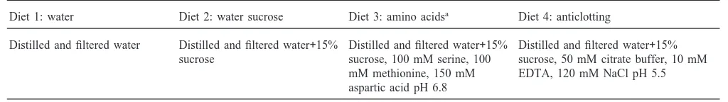
Excreted salivary sheaths of three different aphid species in fluid diets (1, 2 and 3, Table 2) were clearly visible by light microscope after staining with Coomas-sie blue, indicating the protein nature of the sheath material. Sheaths remained attached to the penetrated lower membrane of the sachet. In addition to sheaths, distinct secretions at the end of the salivary sheaths were sometimes observed as stained swellings, referred to as “lumps” (Table 2, Fig. 1). In diet with anti-clotting buffer (diet 4, Table 2) fewer and smaller sheaths were found. Polyclonal antibodies SP154 and anti-SP66/69 both stained the sheath material. The apical lumps of protein material, as observed in Coomassie blue stained sheaths (Fig. 1a), also reacted positively to the antisera when excreted by S. graminum (Fig. 1d).
3.2. Enzyme identification
Table 2
Detection of proteins and enzymatic activities of salivary excretions in different diets for three aphid speciesa
Diet Substrate or stain Aphids Observations (LM) Indications
Diets 1, 2 and 3 Coomassie blue S A M sheaths, halos proteins
Diets 1, 2 and 3 Dopa S A M sheaths, halos phenoloxidase
Diet 4 Dopa S A M sheaths none
Diet 3+agarose Dopa S A M sheaths, halos phenoloxidase
Diet 3+agarose Pectin S A M sheaths, halos pectin methylesterase
Diet 3+agarose Pectin S A M sheaths, halos polygalacturonase
Diet 3 DAB A M sheaths, halos peroxidase
aAbbreviations: Dopa, dihydroxyphenylalanine; DAB, diaminobenzidine; LM, light microscopy; S, Schizaphis graminum; A, Acyrthosiphon
pisum; M, Myzus persicae.
Fig. 1. Salivary sheaths excreted in diets by Schizaphis graminum. Sheath and lumps stained and observed after 24 h probing in a fluid or agarose diet: (a) sheath with lump in fluid diet 3 (without agarose) stained with Coomassie blue; (b) sheath and a concentration of dark material, which later will become a halo, in agarose diet 3 containing 0.1% dopa (dihydroxyphenylalanine); (c) agarose diet 3 containing 0.1% pectin, the Ruthenium red stain has disappeared around the sheath (light circular area) whereas it remained in the surrounding (pink, but grey in the photograph); (d) fluid diet 3 stained with anti-SP154 and horseradish peroxidase as secondary antibody. Bars 40µm.
due to phenoloxidase activity after a few hours of aphid exposure. Also, halos were seen around some sheaths in fluid diets with dopa and they seemed to originate from the sheath’s apex. In agarose diets with dopa we observed halos as well as concentrations of dark material (Fig. 1b) around some (but not all) sheaths, which later became distinct halos. These halos and the dark material in the agarose diets are similar to the halos shown in fluid diets. All the visible staining indicated melanin due to phenoloxidase activity.
Pectinesterase and polygalacturonase activities were
detected in diet 3 with agarose but not, or less obvious, in diets 1 and 2. Clear halos were found (Fig. 1c), which also seemed to originate from the sheath ends. With anti-clotting buffer, diet 4 (Table 1), no phenoloxidase or pectinase activity was observed.
A weak peroxidase activity was found in salivary sheaths of A. pisum and M. persicae, but not in salivary sheaths of S. graminum (Table 2). Thus, some detected enzyme activities seemed to depend on the diet compo-sition and the aphid species. No activity could be detected in assays for cellulase and proteinase.
3.3. Salivary protein separations
SDS–PAGE of the soluble saliva fraction from S.
gra-minum showed two main bands, SP154 and SP66/69
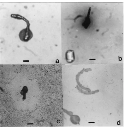
(Table 3). Depending on the diet, some differences were shown (Table 3, Fig. 2). When the time of silver staining was extended in diet 3 (containing sucrose and amino acids), some more bands appeared near the 43 kDa marker band (Fig. 2a; 40 and 30 kDa in Table 3), which were not present in diet 2 (without amino acids). In diet 4 (anti-clotting) we observed a major band at 154 kDa and a sharp band at 240 kDa, but neither at 66/69 kDa nor near 43 kDa were any proteins detected (Fig. 2b; Table 3). In the solid, salivary sheath fractions from diets 2 and 3, the SDS–PAGE showed only an SP66/69 band, no SP154. In the solid fraction of diet 3, as in the soluble fraction, additional bands were shown near 43 kDa, but not in diet 2 sheaths. Sheaths from the anti-clotting diet 4 showed a major band at 66/69 and a weak band at 154 kDa (Table 3).
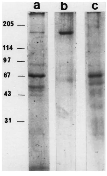
In Western blots of the soluble fractions of all S.
gra-minum probed diets, the polyclonal anti-SP154 reacted
Table 3
Electrophoretic pattern of salivary fractions collected after exposure of S. graminum to different diets
Diet Saliva fraction Electrophoretic pattern Polyclonal anti-SP154 Polyclonal anti-SP66-69 (kDa)
Diet 2 soluble 154, 66/69 all all, except 154
sheath 66/69 all all
Diet 3 soluble 154, 66/69, 40, 30 all all, except 154
sheath 66/69, 40, 30 all all
Diet 4 soluble 240, 154 all none
sheath weak 154, 66/69 all all, except 154
Fig. 2. Electrophoretic separation of salivary proteins. Proteins col-lected in two diets after 24 h exposure to S. graminum and submitted to SDS–PAGE under reducing conditions and silver stained. Pattern of soluble protein fractions in diet 3 (0.2µg, lane a) and diet 4 (0.1
µg, lane b), and solid fraction (sheath material) in diet 3 (0.1µg, lane c). Left, kDa values of calibration kit.
M. persicae reacted to the S. graminum anti-SP154 and
anti-SP66/69.
Repeated attempts to use IEF for separation of compo-nents in soluble saliva, released during diet feeding, showed that the material applied in the samples did not migrate. Addition of zwitterionic detergent, 10 mM CHAPS, 6 M urea or 0.1% triton could not mobilise it. All loaded material remained in the deposit area as shown by silver staining. Immunoblots of the IEF gel showed that the same single spot was recognised by the anti-SP154 and the anti-SP66/69. Thus, classical separ-ation procedures and subsequent biochemical techniques (Madhusudhan et al., 1994) could not be applied to the
Fig. 3. Western blots of Greenbugs’ salivary proteins. Soluble frac-tion, lanes a and b; solid fracfrac-tion, lane c. Antibody staining after appli-cation of anti-SP154, lanes a and c; anti-SP66/69, lane b. Left, kDa values of calibration kit. Antibody binding visualised by horseradish peroxidase conjugated to the secondary antibodies.
proteins without denaturation. Similarly, native PAGE electrophoresis was tried, without SDS pretreatment, at different pH values, with the same persistent lack of electrophoretic movement.
3.4. Immunolocalisation in aphids
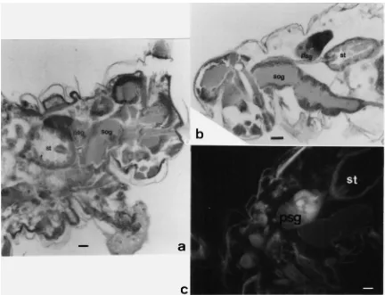
Fig. 4. Immunodetection of salivary proteins in salivary glands. Sections of whole aphids treated with anti-SP154 and horse radish peroxidase as secondary antibody (a and b) or fluorescine (c). sog, sub-oesophageal ganglion; st, stomach; psg, principle salivary gland; asg, accessory salivary gland; bars, 50µm in a and b, 25µm in c.
glands might give a false positive immunodetection (antibody detection was based on peroxidase conjugation) we incubated whole S. graminum sections without antibody treatment in DAB as controls. Staining anti-SP154 and anti-SP66/69 with fluorescein-anti-rabbit confirmed their specific binding to the posterior part of the principal salivary glands in S. graminum sections (Fig. 4d). No fluorescence was observed in accessory glands or other tissues. Salivary glands of A. pisum and
M. persicae showed no reaction to the two S. grami-num antibodies.
The six best monoclonal antibodies reacted to the accessory and principal salivary glands or only to the latter. Some cross reactivity to certain areas in the cer-ebral ganglia was observed as well. Tested on salivary sheaths, some showed positive reactions, others did not.
4. Discussion and conclusions
4.1. Saliva excretion and collection
The salivary sheaths and the apical lumps, excreted in diets, reacted similarly to protein staining by Coomassie
blue and polyclonal antibodies. Phenoloxidase substrate (dopa), however, reacted to salivary sheaths but not to the clearly bordered lumps, resulting in more indistinct halos, which differed in appearance from the lumps. Therefore, the relation between lumps and halos is still unclear. However, we can conclude that although the sheath saliva and the lumps both contain salivary pro-teins, their enzyme activity is different. Phenoloxidase might be diffused from the lumps where it was not bound, as it was in the sheath material, or it might be secreted as “watery saliva”, independent of the lumps. Since we did not combine dopa diets with Coomassie blue, this remains unanswered at present.
The halos seem to be the result of activity by diffused enzymes, proteins that were not bound (polymerised or not embedded in the gelling saliva) to the sheath, rather than the diffused enzyme products themselves. The pec-tinase halos, for example, reflect the absence of pectin (the substrate) converted by the enzymes, not the pres-ence of the reaction product. Hpres-ence, the clear halos indi-cate the position of the enzymes themselves. Whether or not halos are caused by separate watery salivation or must be considered as the consequence of the protein lumps remains unclear. Both halos and lumps were not shown with all sheaths but halo absence could also be explained by a time lapse between sheath secretion and halo development. After 24 h exposure, only the sheaths produced early might have already developed halos, whereas near the later ones sufficient enzyme activity has not yet taken place. The intermediates, the observed dark concentrations (Fig. 1b), made it clear that there is a certain time needed for the halos to develop. Our find-ings do not seem to disagree with watery salivation occurring intermittently with or after sheath salivation, a suggestion made by McLean and Kinsey (1965) and Urbanska et al. (1998). Possibly this salivation is ident-ical to one of the watery salivation periods identified in EPG studies (Tjallingii and Cherqui, 1999). Watery vation occurs in plants, intermittently with sheath sali-vation, during short intracellular punctures along the stylet pathway and their corresponding waveforms have been observed in EPGs from diets as well (waveform pd; Tjallingii, 1985; Powell et al., 1995). However, these excretion periods are very short, only about 1 s, so that their contribution to the relatively large halo deposits seems questionable. Also, they would presumably not result in apical halos. Another watery salivation occurs in sieve elements after reaching the phloem, which is also responsible for the inoculation of persistent plant viruses (Gildow and Gray, 1993; Prado and Tjallingii, 1994). This waveform has also been shown by aphids on diets (waveform E1; Tjallingii, 1988). However, this saliva is thought to be secreted by accessory glands, at least partly, which may not be in agreement with the principal gland indications we found. Hence, further studies combining enzyme detection in diets with EPG recording are needed.
4.2. Identification of enzyme activity
Phenoloxidase activities in aphid saliva have also been detected by Miles and Peng (1989), Ma et al. (1990) and Urbanska et al. (1998). Catalytic reactions transform dopa to melanin in two steps. First, a hydroxylation of monophenol to o-diphenol and, subsequently, an oxi-dation of o-diphenols to o-quinones and their polymer, melanin (Cherqui et al., 1998). In insects, phenoloxi-dases are the key enzymes. They are involved in the tan-ning process of the cuticle and in the haemolymph for
the defence reactions. In Homoptera (Miles, 1972) and the Heteroptera (Laurema et al., 1985) phenoloxidase seems to be present in the saliva and related glands. The phenoloxidase activity was observed in the salivary sheaths of aphids (Miles, 1972) and also in less structural deposits (halos) around the sheath material when excreted in agarose gels (Urbanska et al., 1998). Phenol-oxidases probably oxidise the plant polyphenols to o-quinones. Plant penetration by aphids causes accumu-lation of polyphenols, especially catechin, presumably as a plant defence reaction (Peng and Miles, 1988; Miles, 1999). It remains unclear, however, what the benefit of this phenoloxidase excretion is for an aphid. Some sug-gest that the main function for the aphids is an in situ detoxification of phenols (Miles and Oertli, 1993; Lesz-czynski and Dixon, 1990). However, since there seems to be no ingestion of plant fluids during salivary sheath formation (pathway)—at least no substantial ingestion (Tjallingii, 1988)—this explanation is disputable.
Pectinases, pectinesterase and polygalacturonase have been suggested to be of major importance for intercellu-lar stylet penetration, dissolving the pectin middle lamella (McAllan and Adams, 1961; Dreyer and Campbell, 1987; Campbell and Dreyer, 1990). However, transmission electron microscopy has shown that stylets predominantly follow the secondary cell walls, penetrat-ing between the layers of cellulose fibres and not via the middle lamella (Tjallingii and Hogen-Esch, 1993). Pectinases may break down the pectin between the cellu-lose and hemicellucellu-lose fibres, but stylet penetration seems to go faster than the enzyme activity would allow. On the other hand, functional for stylet penetration or not, the pectinases will also cause pectin fragments, some of which might act as strong elicitors causing plant reactions (Ma et al., 1998).
4.3. Protein separation
presum-ably due to anti-clotting, although this is not com-pletely understood.
The specificity of the polyclonal antibodies anti-SP154 and anti-SP66/69 to the saliva proteins appeared rather low, which may support the common origin. On the other hand, there were no cross-reactions observed to salivary proteins of other aphid species. Therefore, the low specificity within S. graminum proteins may indicate a common epitope for this aphid species as well.
4.4. Immunolocalisation
Aphids have two pairs of salivary glands, the principal and the accessory glands (Ponsen, 1972). Principal glands are divided into two parts, an anterior membra-nous part and a posterior glandular part. Polyclonal anti-bodies stained only the posterior cells of the principal salivary glands; neither the accessory glands nor the anterior cells of the principal glands were stained. Both peroxidase and fluorescence labels linked to the anti-bodies were unequivocal. This indicates that the princi-pal glands secrete SP154, SP66/69 and other positively reacting proteins in our blots. SP69 and SP66 probably form the bulk proteins of the sheath material and are secreted by the posterior principal glands. The accessory glands might secrete the smaller proteins (near 43 kDa), as we suggested above. However, no antibody binding was found to the accessory glands: either there was no binding or it was too weak.
The results obtained demonstrated once more the complexity of saliva secretion by aphids. They also showed that understanding the functions of salivary secretions in the separate stages of plant penetration by the mouthparts requires a better knowledge of the secretion phases and how the two glands are involved. The role of the accessory glands, in particular, remained unknown in this study. With the diet collection method we presumably did not obtain all secretions of aphid sali-vation into plants. Diet assays, on the other hand, will remain important and should be done together with plant studies as circumstances can be controlled. This study has also shown that further studies need new tools and new combinations, for example combining behavioural and chemical techniques. The development of additional (and more specific) antibodies than we could realise in this study is another important challenge, which we think may provide important knowledge in the field of aphid– plant interactions and its impact on higher trophic levels as well.
Acknowledgements
We thank Dr Paul Baumann (Davis, UCA) for using his polyclonal antibodies, Dr Hans Smid for his help and advice, and Dr Arjen Schots and his group for
co-oper-ation and help in developing the MABs. This work was supported by the EC HCM contract ERBCHRXCT940660.
References
Baumann, L., Baumann, P., 1995. Soluble salivary proteins secreted by Schizaphis graminum. Entomologia Experimentalis et Applicata 77, 56–60.
Bradford, M., 1976. A rapid and sensitive method for the quantification of microgram quantities of protein utilizing the principle of pro-tein–dye binding. Analytical Biochemistry 72, 248–254.
Campbell, B.C., Dreyer, D.L., 1990. The role of plant matrix polysac-charides in aphid–plant interactions. In: Campbell, R.K., Eiken-bary, R.D. (Eds.) Aphid–Plant Genotype Interactions. Elsevier, Amsterdam, pp. 149–170.
Cherqui, A., Duvic, B., Reibel, C., Brehelin, M., 1998. Cooperation of dopachrome conversion factor with phenoloxidase in the eumelanin pathway in haemolymph of Locusta migratoria (Insecta). Insect Biochemistry and Molecular Biology 28, 839–848.
Dreyer, D.L., Campbell, B.C., 1987. Chemical basis of host–plant resistance to aphids. Plant Cell and Environment 10, 353–361. Gildow, F.E., Gray, S.M., 1993. The aphid salivary gland basal lamina
as a selective barrier associated with vector-specific transmission of barley yellow dwarf luteoviruses. Phytopathologia 83, 1293–1302. Knoblauch, M., van Bel, A.J.E., 1998. Sieve tubes in action. The Plant
Cell 10, 35–50.
Laemmli, U.K., 1970. Cleavage of structural proteins during the assembly of the head of bacteriophage T4. Nature 277, 680–685. Laurema, S., Varis, A.L., Miettinen, H., 1985. Studies of enzymes in
the salivary glands of Lygus rugulipennis (Hemiptera Miridae). Insect Biochemistry 15, 211–224.
Leszczynski, B., Dixon, A.F.G., 1990. Resistance of cereals to aphids: interaction between hydroxamic acids and the aphid Sitobion
avenae (Homoptera Aphididae). Annals of Applied Biology 117,
21–30.
Ma, R., Reese, J., Black, W.C., Bramel-Cox, P., 1990. Detection of pectinesterase and polygalacturonase from salivary secretions of living greenbug, Schizaphis graminum (Homoptera: Aphididae). Journal Insect Physiology 36, 507–512.
Ma, R., Reese, J., Black, W.C., Bramel-Cox, P., 1998. Chlorophyll loss in a greenbug-susceptible sorghum due to pectnases and pectin fragments. Journal of the Kansas Entomological Society 71, 51–60. Madhusudhan, V.V., Taylor, G.S., Miles, P.W., 1994. The detection of salivary enzymes of phytophagous hemiptera: a compilation of methods. Annals of Applied Biology 124, 405–412.
Martin, B., Collar, J.L., Tjallingii, W.F., Fereres, A., 1997. Intracellular ingestion and salivation by aphids may cause acquisition and inocu-lation of non-persistently transmitted plant viruses. Journal of Gen-eral Virology 78, 2701–2705.
McAllan, J.W., Adams, J.B., 1961. The significance of pectinase in plant penetration by aphids. Canadian Journal of Zoology 39, 305–310.
McLean, D.L., Kinsey, M.G., 1965. Identification of electrically recorded curve patterns associated with aphid salivation and inges-tion. Nature, London 205, 1130–1131.
Miles, P.W., 1959. The secretion of two types of saliva by an aphid. Nature 183, 756.
Miles, P.W., 1972. The saliva of Hemiptera. Advances in Insect Physi-ology 9, 183–255.
Miles, P.W., 1985. Dynamic aspects of the chemical relation between the rose aphid and rose buds. Entomologia Experimentalis et Applicata 37, 129–135.
Miles, P.W., Harrewijn, P., 1991. Discharge by aphids as soluble secretions into dietary sources. Entomologia Experimentalis et Applicata 59, 123–134.
Miles, P.W., Oertli, J.J., 1993. The significance of antioxidants in aphid–plant interaction: the redox hypothesis. Entomologia Experi-mentalis et Applicata 67, 273–285.
Miles, P.W., Peng, Z., 1989. Studies of the salivary physiology of plant bugs: detoxification of phytochemicals by the salivary peroxidase of aphids. Journal Insect Physiology 35, 865–872.
Peng, Z., Miles, P.W., 1988. Acceptability of catechin and its oxidative condensation products to the rose aphid, Macrosiphum rosae. Ento-mologia Experimentalis et Applicata 47, 255–265.
Ponsen, M.B., 1972. The site of potato leafroll virus multiplication in its vector, Myzus persicae (Thesis). Mededelingen Landbouwhoge-school Wageningen 16, 1–147.
Powell, G., Pirone, P., Hardie, J., 1995. Aphid stylet activities during polyvirus acquisition from plants and an in vitro system that corre-late with subsequent transmission. European Journal of Plant Path-ology 101, 411–420.
Prado, E., Tjallingii, W.F., 1994. Aphid activities during sieve element punctures. Entomologia Experimentalis et Applicata 42, 157–165. Ryan, J.D., Morgan, A.T., Richardson, P.E., Johnson, R.C., Mort, A.J., Eikenbary, R.D., 1990. Greenbugs and wheat: a model system for the study of phytotoxic Homoptera. In: Campbell, R.K., Eikenbary, R.D. (Eds.) Aphid–Plant Genotype Interactions. Elsevier, Amster-dam, pp. 171–186.
Sterling, E., 1970. Crystal-structure of ruthenium red and stereochemi-stry of its pectin stain. American Journal of Botany 57, 172–175. Tjallingii, W.F., 1985. Membrane potentials as an indication for plant cell penetration by aphid stylets. Entomologia Experimentalis et Applicata 38, 187–193.
Tjallingii, W.F., 1988. Electrical recording of stylet penetration activi-ties. In: Minks, A.K., Harrewijn, P. (Eds.). Aphids, their Biology, Natural Enemies and Control, vol. 2B. Elsevier, Amsterdam, pp. 95–108.
Tjallingii, W.F., 1995. Regulation of phloem sap feeding by aphids. In: Chapman, R.F., de Boer, G. (Eds.) Regulatory Mechanisms in Insect Feeding. Chapman and Hall, New York, pp. 190–209. Tjallingii, W.F., Cherqui, A., 1999. Aphid saliva and aphid–plant
inter-actions. Experimental and Applied Entomology 10, 169–174. Tjallingii, W.F., Hogen-Esch, T., 1993. Fine structure of aphid stylet
routes in plant tissues in correlation with EPG signals. Physiologi-cal Entomology 18, 189–200.
Urbanska, A., Tjallingii, W.F., Dixon, A.F.G., Leszczynski, B., 1998. Phenol oxidizing enzymes in the grain aphid’s saliva. Entomologia Experimentalis et Applicata 686, 197–203.