Structure and Function: Clinical Implications
Joan Kaufman, Paul M. Plotsky, Charles B. Nemeroff, and Dennis S. Charney
Child abuse is associated with markedly elevated rates of major depression and other psychiatric disorders in adult-hood. This article reviews preclinical studies examining the effects of early stress, factors that modify the impact of these experiences, and neurobiological changes associated with major depression. Preclinical studies demonstrate that early stress can alter the development of the hypothalamic-pitu-itary-adrenal axis, hypothalamic and extrahypothalamic cor-ticotropin releasing hormone, monoaminergic, andg -ami-nobutyric acid/benzodiazepine systems. Stress has also been shown to promote structural and functional alterations in brain regions similar to those seen in adults with depression. Emerging data suggest, however, that the long-term effects of early stress can be moderated by genetic factors and the quality of the subsequent caregiving environment. These effects also can be prevented or reversed with various pharmacologic interventions. Preclinical studies of early stress can provide valuable insights in understanding the pathophysiology and treatment of major depression. They also can provide an important tool to use to investigate interactions between genes and environments in determining an individual’s sensitivity to stress. More research is needed to understand how inherent factors interact with experiences of abuse and other psychosocial factors to confer vulnera-bility to develop depression. Biol Psychiatry 2000;48: 778 –790 © 2000 Society of Biological Psychiatry
Key Words: Depression, corticotropin-releasing hormone (CRH), child abuse, animal models
Introduction
C
hild abuse is a pervasive social problem that is fre-quently associated with long-term significant psychiatric sequelae. Each year there are more than one million substan-tiated reports of child maltreatment (U.S. Department of Health and Human Services, National Center on Child Abuse and Neglect 1997), and numerous studies indicate thatcountless other cases never come to the attention of author-ities (e.g. Wolfner and Gelles 1993). In clinic (Brown and Anderson 1991; Windle and Houston 1995), community (Bifulco et al 1991), and representative epidemiologic sam-ples (Holmes and Robins 1987), experiences of early child maltreatment have been associated with elevated rates of major depression (MDD), anxiety, and other psychiatric disorders.
Preclinical studies suggest that stress early in life can promote long-term changes in multiple neurotransmitter systems and brain structures implicated in the etiology of MDD (Arborelius et al 1999; Heim et al 1997). It is hypothesized that neurobiological changes associated with adverse early experiences can confer a vulnerability for the development of MDD and other psychiatric disorders. It is also suggested that preclinical studies of early stress can provide a valuable model for understanding the patho-physiology of MDD, even when it occurs independent of early childhood trauma (Charney and Deutch 1996).
This article is organized into five sections. The first section reviews functional connections among key structures and neurotransmitter systems involved in the initiation and con-trol of the stress response. The second section reviews preclinical studies examining the long-term effects of early stress on hypothalamic-pituitary-adrenal (HPA) axis function and central corticotropin releasing hormone (CRH), mono-aminergic (norepinergic, dopmono-aminergic, serotonergic), and
g-aminobutyric acid/benzodiazepine (GABA/BZ) systems. Structural and functional brain changes associated with early or severe stress are also reviewed. The next section reviews factors that modify the impact of early stress, and pharma-cologic interventions that can prevent or reverse the neuro-biological alterations are delineated. Lastly similarities in the neurobiological correlates of stress and depression in adults are highlighted, and the clinical implications of the research are discussed.
Key Structures and Neurotransmitter
Systems Involved in the Stress Response:
Overview
The brain appears to respond to stress in a complex and orchestrated manner, with both general and
stimuli-spe-From the Department of Psychiatry, Yale University School of Medicine, New Haven, Connecticut (JK), the Department of Psychiatry and Behavioral Sciences, Emory University School of Medicine, Atlanta, Georgia (PMP, CBN), and National Institute of Mental Health, Bethesda, Maryland (DSC). Address reprint requests to Joan Kaufman, Ph.D., Yale University School of
Medicine, University Towers, Suite 2H, 100 York Street, New Haven CT 06511.
Received May 17, 2000; revised July 12, 2000; accepted July 25, 2000.
cific components to the stress response (Lopez et al 1999). Knowledge about the structural and functional compo-nents of the stress system is still evolving. The review of the stress response included in this section is not exhaus-tive. It focuses on key components of the stress system and emphasizes the structures and neurotransmitter systems most extensively studied in preclinical studies examining the long-term effects of early stress. The reader is referred to Chrousos (1998) and Lopez et al (1999) for a more detailed discussion of the central and peripheral compo-nents of the stress system.
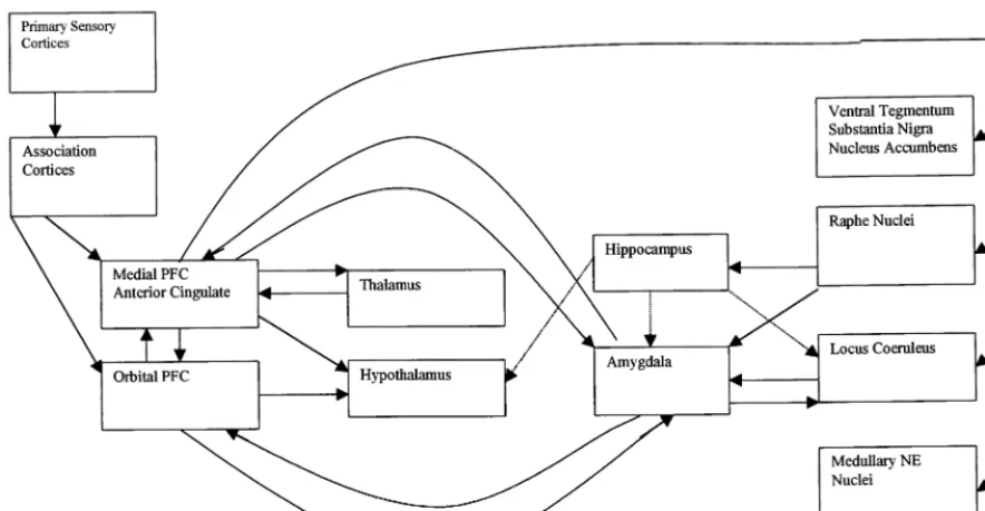
Figure 1 depicts the functional connections among the different cortical and subcortical brain regions involved in the stress response. There is growing appreciation of the role of cortical inputs, with medial prefrontal cortex (PFC), anterior cingulate, and orbital PFC currently un-derstood to play an important role in relaying information from primary sensory and association cortices to subcor-tical structures involved in the stress response (Lopez et al 1999). Medial and orbital PFC are reciprocally intercon-nected, and each has indirect connections with the hypo-thalamus and amygdala via inputs to the periaqueductal gray and parabrachial nucleus (An et al 1998; Bernard and
Bandler 1998; Krout et al 1998). The medial and orbital prefrontal cortices also provide direct inputs to the hypo-thalamus and are reciprocally connected with the amyg-dala (Ongur et al 1998). These prefrontal regions appear to be critical in restraining the acute stress response and facilitating negative feedback inhibition of the system (Herman and Cullinan 1997). The medial prefrontal cortex (mPFC) also is reciprocally connected with the mediodor-sal thalamic nucleus (Groenewegen 1988) and has exten-sive connections with the ventral tegmental area, substan-tia nigra, nucleus accumbens, raphe, locus coeruleus, and brainstem autonomic nuclei (Drevets et al 1998).
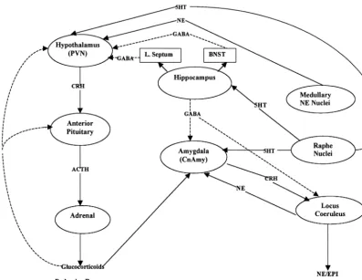
Figure 2 depicts in more detail the relationships among subcortical structures involved in the stress response and the neurotransmitter systems involved in the transmission of information between the different brain regions. Corti-cotropin releasing hormone is the neurohormone that initiates the endocrine response to stress. It is secreted from the paraventricular nucleus (PVN) of the hypothala-mus. Among the numerous inputs to the PVN, noradren-ergic inputs are primary in promoting the synthesis and release of CRH into the median eminence and hypophysial portal system (Plotsky et al 1989). The main noradrenergic
inputs into the PVN appear to be derived from medullary sources, the nucleus of the solitary tract (NTS) and the ventrolateral medullary oblongata (Pacak et al 1995). Corticotropin releasing hormone then binds to receptors at the anterior pituitary gland and, through a cascade of intracellular events, increases pro-opiomelanocortin (POMC) gene expression and the release of POMC-derived peptides such as adrenocorticotropin andb -endor-phin. Adrenocorticotropin (ACTH) then promotes the synthesis and release of glucocorticoids (cortisol in pri-mates, corticosterone in rats) from the adrenal cortex (Arborelius et al 1999). Glucocorticoids regulate energy substrate availability and utilization and provide negative
feedback to the stress system at the pituitary, hypothala-mus, and other central sites involved in the stress response. In addition, CRH appears to act centrally as a neurotrans-mitter to initiate the autonomic and behavioral changes observed in response to stress (Valentino et al 1998), as central nervous system administration of CRH has been found to produce physiologic and behavioral changes similar to those reported in animals subjected to stress (Owens and Nemeroff 1991). Centrally administered CRH antagonists also have been found to reduce stress induced increases in plasma catecholamines, tyrosine hydroxylase mRNA in the locus coeruleus (LC), and CRH mRNA and type 1 CRH receptor mRNA in the PVN (Jezova et al 1999).
The LC appears to be the critical site in initiating the catecholamine response to stress because local application of exogenous CRH in the LC has been found to increase norepinephrine (NE) release in the PVN, hippocampus, and prefrontal cortex (Page and Abercrombie 1999; Val-entino et al 1998). The LC receives endogenous CRH inputs from the central nucleus of the amgydala (CnAmy). The amygdala is activated during stress by ascending catecholamine neurons originating in the brainstem and by cortical association neurons involved in processing stress-ful stimuli via direct and indirect medial and orbital prefrontal cortical connections (Lopez et al 1999). Corti-cotropin releasing hormone neurons of the CnAmy re-spond positively to glucocorticoids and activate the LC/NE component of the stress system (Lopez et al 1999). The hippocampus, in contrast, serves to inhibit the stress response via multiple direct and indirect links with several of the brain structures activated during stress (Lopez et al 1999). For example, CRH synthesis in the CnAmy is inhibited by GABA inputs from the hippocam-pus (Owens and Nemeroff 1991). The hippocamhippocam-pus also inhibits the LC via direct connections and inhibits the PVN via indirect inputs through the lateral septum and bed nucleus of the stria terminalis.
The stress response is further modified by serotonin inputs from the dorsal and median raphe to the basolateral and central nucleus of the amygdala, serotonin inputs from the dorsal raphe to the PVN, and serotonin inputs form the median raphe to the hippocampus (Lopez et al 1999). These latter serotonin (5-HT) neurons terminate on inhib-itory GABA neurons.
Both the PVN CRH and LC/NE systems also innervate the mesocortical and mesolimbic components of the do-paminergic (DA) system (Chrousos 1998). The mesocor-tical system includes DA neurons of the ventral tegmen-tum that send projections to the medial prefrontal cortex, and the mesolimbic system consists of DA neurons from the same region that innervate the nucleus accumbens. The mesocortical system is involved in anticipatory and cog-nitive functions and exerts a suppressive effect on the stress system, and the mesolimbic system is involved in processing motivation and reward aspects of experience.
Neurobiological Effects of Early Stress:
Preclinical Studies
Building on the seminal work of Levine and colleagues (Coe et al 1978; Levine et al 1993; Wiener et al 1987), numerous investigators have demonstrated long-term neu-robiological changes in animals subjected to multiple prenatal and postnatal stress paradigms (Graham et al 1999; Takahashi and Kalin 1991). Similar neurobiological alterations have been reported across experimental
condi-tions, with multiple systems implicated in the pathophys-iology of depression affected by these investigational manipulations.
Extensive research has been conducted examining the neurobiological effects of early maternal separation, with these experiences associated with increased CRH and NE drive in adulthood (Francis et al 1999a; Ladd et al 1996; Liu et al 2000). Rat pups separated from their mothers 6 hours per day during the first 3 weeks of life have been found to have increased basal and stress induced ACTH concentrations and decreased CRH binding in the anterior pituitary (Ladd et al 1996). Maternal deprivation has also been associated with increased CRH mRNA expression in the hypothalamic paraventricular nucleus (PVN) and in-creased CRH concentration in the median eminence (Plotsky and Meaney 1993). It also has been associated with increased CRH mRNA expression in the CnAmy, increased CRH content in the parabrachial nucleus (a region that adjoins the LC, increased CRH binding in the LC, and increased NE concentration in the PVN (Menza-ghi et al 1993). Nonhuman primates subjected to maternal separation early in life have also been found to have elevated cerebral spinal fluid NE in response to an acute stressor (Kraemer et al 1989). For reviews, see Francis et al (1999) and Ladd et al (2000).
The increase in CRH and NE drive in maternally deprived rats also is associated with a decrease in tone of the inhibitory GABA/BZ system (Caldji et al 2000; Francis et al 1999b). Specifically, adult rats subjected to repeat separations from their mothers during the first 3 weeks of life have been found in adulthood to have reduced GABAAreceptor binding in the CnAmy,
basolat-eral nuclei of the amygdala (BnAmy), and the frontal cortex. They also have been found to have reduced central benzodiazapine binding in the CnAmy, BnAmy, LC, and NTS. These effects were associated with decreased ex-pression of mRNA for theg2subunit that encodes for the benzodiazapine site of the GABAA receptor. In addition,
adult rats separated from their mothers during the first 3 weeks of life also had increased mRNA expression for the
a2 and a3 subunits and decreased expression of the a1
subunit mRNA (Caldji et al 2000). This profile is associ-ated with decreased GABA binding (Wilson 1996). It is likely that the dampened GABAergic tone in rats exposed to maternal separation animals contributes to the enhanced CRH expression in the amygdala and the increased stress-induced activation of the noradrenergic systems (Francis et al 1999b).
Specifically, Octodon Degus rodents were separated from their mothers three times a day for 1 hour from postnatal day 1 to postnatal day 21. After weaning, the deprived animals were reared in isolation in single cages until postnatal day 45. They were then sacrificed, and mono-amine fiber innervation to the frontal areas was exmono-amined using immunocytochemical detection of tyrosine hydrox-ylase (TH) and 5-HT.
When compared with rodents reared under undisturbed conditions, maternally deprived and socially isolated rats had significantly reduced TH-positive fiber innervation in the precentral medial, anterior cingulate, and prelimbic cortex subdivisions of the mPFC and increased 5-HT-positive fiber densities in the infralimbic cortex (nomen-clature according to Groenewegen 1988). The number of TH-positive somata in the ventral tegmental area and in the substantia nigra did not differ between the groups, suggesting that the reduced fiber densities were likely due to suppressed axonal sprouting and aborization. Mater-nally deprived and socially isolated animals, in addition to having altered DA/5-HT balance in the mPFC, were also found to have a significant decrease of nicotinamide adenine dinucleotide phosphate (NADPH)-diphorase-reactive neurons in the anterior cingulate cortex and the core region of the nucleus accumbens, as well as a trend for reduced NADPH-diphorase-reactive neurons in the infralimbic, precentral medial, and prelimbic prefrontal areas (Poeggel et al 1999). Because some NADPH-containing neurons have been found to be GABAergic, the reduced NADPH-diphorase neurons may represent a loss of inhibitory interneurons in this cortical region, which plays a critical role in integrating affective and cognitive information processing.
Rhesus monkeys reared under isolated nursery condi-tions from 8 weeks of age also have been found to exhibit enhanced anxiety, increased self-directed behaviors, de-creased social interaction, and impaired cognitive perfor-mance when tested at 18 to 24 months of age (Sanchez et al 1998). These animals also have shown reductions in medial and caudal midbody corpus callosum volume, probably reflecting a reduction in the number of cross-hemispheric fibers (Sanchez et al 1998). The nursery-reared animals also exhibited increased CRH1 receptor binding sites in the dentate gyrus of the hippocampus and the orbital prefrontal cortex, with increased CRH2 recep-tor binding in amygdala subregions (Sanchez et al 1999). These findings do not contradict the hypothesis concern-ing the role of central CRH oversecretion in the patho-physiology of depression because peptide oversecretion does not always result in receptor down regulation and has been found to cause upregulation in certain brain regions (Imaki et al 1996; Mansi et al 1996).
In an attempt to more closely parallel the experience of
neglectful parenting and exposure to stressful environments in young human infants, Coplan et al (1996) subjected macaque infant–mother dyads to variable foraging demands. Primates in the low foraging demand condition had easy access to food; primates in the high foraging demand condi-tion had to work hard to find food, but foraging demands and food supply were predictable; and primates in the variable foraging demand condition experienced changing and unpre-dictable access to food. In adulthood, consistent with the maternal deprivation rodent studies discussed above, mon-keys reared in the variable foraging condition had higher cerebral spinal fluid CRH concentration than did monkeys reared under the two other more predictable and less stressful experimental conditions (Coplan et al 1996). The variable foraging condition was also associated with over activity of the NE system, with these animals as adults showing en-hanced behavioral response to yohimbine, ana2adrenergic antagonist (Rosenblum et al 1994).
In contrast, to the negative effects of early stress, rats provided positive stimulation via 15 minutes of han-dling per day during the first 3 weeks of life have been found to have reduced stress reactivity in adulthood compared with nonhandled or maternally separated rats (Plotsky and Meaney 1993). Specifically, in adulthood, rats handled in the first 3 weeks of life showed decreased fearfulness in novel environments. The neu-robiological alterations associated with early handling are essentially the opposite of those reported in mater-nally separated rats. Handled rats showed reduced ACTH and corticosterone response to exogenous stres-sors, with quicker return of corticosterone to baseline levels. They showed enhanced negative feedback of circulating glucocorticoids and increased glucocorticoid receptor mRNA expression and glucocorticoid receptor number in the hippocampus and the frontal cortex, sites involved in the inhibitory control of CRH synthesis in PVN neurons. Accordingly, handled rats had reduced CRH mRNA levels in the PVN and reduced basal CRH concentration in the median eminence. Handled rats also had reduced CRH mRNA concentrations in the CnAmy and lower CRH content in the LC (Francis et al 1999b; Ladd et al 2000). They also had attenuated CRH induced activation of the LC and smaller resulting increases in extracellular NE levels in the PVN after acute restraint stress (Liu et al 2000). Handled rats had increased GABAA receptor levels in noradrenergic cell
The maternal deprivation and postnatal handling studies clearly highlight the importance of early experience on the development of the brain and multiple neurotransmitter systems. The stress effects on hippocampus development are likely mediated by a minimum of three forms of structural plasticity: neuronal atrophy, neurotoxicity, and neurogenesis. Neuronal atrophy in the CA3 region of the hippocampus can be caused by 3 weeks of exposure to stress or stress levels of glucocorticoids (Sapolsky 1996; Woolley et al 1990). At this level, glucocorticoids produce a reversible decrease in number of apical dendritic branch points and length of apical dendrites of sufficient magni-tude to impair hippocampal dependent cognitive processes (Watanabe et al 1992). More sustained stress or glucocor-ticoid exposure can lead to neurotoxicity—actual perma-nent loss of hippocampal neurons. Rats exposed to high concentrations of glucocorticoids for approximately 12 hours per day for 3 months experience a 20% loss of neurons specific to the CA3 region of the hippocampus (Sapolsky et al 1985). Evidence of stress-induced neuro-toxicity of cells in this region has been reported in nonhuman primates as well (Sapolsky 1996; Uno et al 1994). Reductions in hippocampal volume may also be affected by decreases in neurogenesis (Gould and Cam-eron 1996). The granule cells in the dentate gyrus of the hippocampus continue to proliferate into adulthood, and neurogenesis in this region is markedly reduced by stress. See articles in this volume by Sapolsky, Gould, and McEwen for further discussion of these topics.
Factors Moderating the Impact of Early
Stress
The studies reviewed in the prior section demonstrate that early life experiences can have profound effects on brain structure and function; however, there is emerging data to suggest that the subsequent caregiving environ-ment can moderate the adverse effects of early stress. In conducting the handling experiments, Meaney and col-leagues noted that there were marked differences in the maternal behavior of the mothers of handled and non-handled pups, with the former group spending signifi-cantly more time licking and grooming their offspring than the latter group (B. C. Woodside, M. J. Meaney, J. Jans, unpublished observations).
To determine if the differences in maternal behavior were related to differences in stress reactivity of handled and nonhandled rats, Meaney and colleagues examined multiple indices of stress reactivity in adult rats reared by mothers with similar naturally occurring differences in maternal behaviors (Caldji et al 1998; Francis et al, unpublished data; Liu et al 1997) 1999). They found that the adult offspring of high licking and grooming mothers
reared without any experimental manipulations showed greater exploration in novel environments and had reduced plasma ACTH and corticosterone response to acute stress. The animals also showed increased hippocampal cocorticoid receptor mRNA expression, enhanced glu-cocorticoid negative feedback sensitivity, and decreased hypothalamic CRH mRNA levels. They also had de-creased CRH mRNA expression in the CnAmy, inde-creased central benzodiazepine receptor number in the CnAmy and LC, decreased CRH receptor density in the LC, and decreased stress induced NE secretion from the PVN. These results parallel the findings observed in handled rats and suggest that maternal licking and grooming behaviors may “program” the development of the neural systems that mediate reactivity to stress (Caldji et al 1998). These studies raised questions as to whether the neurobiological changes associated with handling were due to the early experimental manipulation or to subsequent differences in maternal behavior.
To determine if the neurobiological changes associated with early handling could be altered by subsequent care-giving experiences, rat pups exposed to early handling or maternal separation experiences were cross-fostered with dams whose pups were assigned the same or opposite condition (Gonzalez et al 1999). In the initial set of experiments, handled pups were either cross-fostered to other dams assigned to the handled condition or to dams assigned to the maternal separation condition. Similar cross-fostering was performed on pups exposed to the maternal separation condition. When tested as adults, the handled pups cross-fostered to dams assigned to the maternal separation condition reacted to novel stressors like rats subjected to maternal separation during the neonatal period. Conversely, maternally separated pups reared by dams assigned to the handling condition were more similar to handled animals.
stimulation) in regulating physiologic systems involved in the stress response (Caldji et al 1998; Kuhn and Schanberg 1998).
The cross-fostering experiments clearly demonstrate that the effects of early experiences can be moderated by subsequent rearing experiences. Because the influence of genetic factors or strain effects has been well established in preclinical studies of stress reactivity (Dhabhar et al 1997), the cross-fostering studies raise questions as to whether manipulations in parenting can overcome genetic and breed differences in stress reactivity. To address this question, Meaney and colleagues subjected BALB/cByJ and C57BL/6ByJ mice to early handling experiences and randomly assigned them to BALB/cByJ or C57BL/6ByJ mothers for subsequent rearing (Anisman et al 1998; Zaharia et al 1996). The BALB/cByJ mice are inherently high reactors and have elevated corticosterone and brain catecholamine responses to acute stressors. In addition, mice of this strain exhibit impaired performance on a Morris water-maze that is exacerbated by foot-shock application. Early handling of BALB/cByJ mice reduced the learning impairments seen when mice were tested in the Morris water-maze as adults and prevented stress-induced elevations of corticosterone and disturbances with task performance. Likewise, cross-fostering BALB/cByJ mice with C57BL/6ByJ dams prevented corticosterone hyperactivity and performance deficits; however, cross-fostering and handling did not alter stress-induced changes in NE concentration in the hypothalamus, LC, hippocam-pus, or prefrontal cortex. Early handling and cross-foster-ing of the more resilient C57BL/6ByJ mice had no impact on maze performance, corticosterone stress reactivity, or brain NE. A similar set of findings was reported by investigators studying two different high- and low-reactive rat species (Steimer et al 1998). Effects of handling and cross-fostering were only observed in the high-reactive rats, and these experimental manipulations only affected stress induced corticosterone levels, not central NE measures.
These studies highlight the need for a better understand-ing of the interactions between genes and environmental interactions in determining an individual’s stress reactivity and vulnerability to depression. They suggest that species with more intrinsic reactivity are more responsive to the effects of environmental manipulations than are less in-trinsically reactive species and that environmental manip-ulations have greater impact on some neurobiological systems (e.g., HPA axis) than on others (e.g., central NE). The clinical and research implications of these findings are far reaching. They imply that there are multiple pathways to the development of depression and that phenotypes with similar neurobiology may have distinct etiologies. The Human Genome Project and evolving methodologies such
as gene chips that permit the simultaneous analysis of thousands of genes will help to identify the relevant genes that promote hyperstress reactivity and will facilitate the identification of homogenous subgroups of patients with depression for future neurobiological and genetic studies (Watson and Akil 1999).
Neurobiological Effects of Early Stress:
Pharmacologic Prevention and Treatment
The long-term, stress-related changes in brain structure can be altered by multiple pharmacologic interventions. Emerging data suggest that in addition to glucocorticoids, serotonin and excitatory amino acids (EAA) are involved in the mechanisms that promote neuronal atrophy, neuro-toxicity, and neurogenesis (Gould 1999; McEwen et al 1997; Sapolsky 1996). Consistent with these findings, several classes of medications have been found to prevent dendritic atrophy caused by stress, including serotonin reuptake enhancers (e.g., tianeptine), benzodiazepine ago-nists (e.g., adinazolam), the antiseizure drug phenytoin (which reduces EAA release), and adrenal steroid inhibi-tors (Magarinos et al 1999; McEwen et al 1997; Sapolsky 1996). Tianeptine, in addition to preventing reduction in number of apical dendritic branch points and length of apical dendrites, also reverses already established hip-pocampal atrophy (Magarinos et al 1999). Surprisingly, given the clinical effectiveness of selective serotonin reuptake inhibitors (SSRI) in treating depression, fluox-etine and fluvoxamine do not block dendritic atrophy caused by repeated restraint stress (Magarinos et al 1999). Paroxetine, an SSRI with less serotonergic-specific prop-erties can reverse the HPA axis alterations observed in adult rodents subject to repeat maternal separation during the neonatal period (Plotsky, unpublished data). In addi-tion, in adult primates subject to maternal deprivation in infancy, electroconvulsive therapy and chronic tricyclic antidepressant (e.g., imipramine) treatments have also been found to reverse HPA axis overactivation following acute and chronic stress (Lopez et al 1999; Suomi 1991).
Similarities in the Neurobiological
Correlates of Stress and Depression:
Clinical Implications
1987; Carroll 1982); and blunted ACTH secretion in response to administration of endogenous CRH (Gold et al 1986; Holsboer et al 1987; Plotsky et al 1998). They also have been found to have increased central CRH drive, as evidenced by reports of elevated concentrations of cere-bral spinal fluid CRH (Nemeroff et al 1992), and reduced CRH receptor binding site number in the frontal cortex of suicide victims (Nemeroff et al 1988). Depressed adults also have been found to have higher cerebral spinal fluid NE concentration (Wong et al 2000) and decreased corti-cal GABA measured in vivo using proton magnetic resonance spectroscopy (Sanacora et al 1999). Altered DA/5-HT balance also has been reported in adults with depression (Reddy et al 1992), with fluoxetine treatment associated with increases in cerebral spinal fluid DA/5-HT metabolite concentration ratios (De Bellis et al 1993; for a discussion of the similarities in the neurobiological
corre-lates of stress and clinical findings in studies of patients with posttraumatic stress disorder, see Arborelius et al 1999; Charney et al 1998).
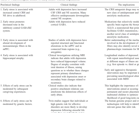
Studies of adults with depression have also reported structural and functional alterations in the mPFC. Specif-ically, adults with depression have been found to have reduced gray matter volume in the subgenual PFC (Dre-vets et al 1997), as well as decreased blood flow and glucose metabolism in this region (Drevets et al 1992). In addition, preliminary histopathologic data from the sub-genual PFC suggest that depressed patients have a de-crease in glia number in this region without a correspond-ing loss of neurons (Drevets et al 1998). Reduced glucose metabolism in the ventromedial PFC (Buchsbaum et al 1997) and decreased blood flow in the dorsal anterior cingulate also has been reported in depressed patients (Mayberg et al 1997). These two regions are contained in Table 1. Neurobiological Effects of Early Stress: Clinical Implications
Preclinical findings Clinical findings The implications 1. Early stress is associated with
increased central CRH and NE drive in adulthood.
Adults with depression have increased CSF CRH and NE secretion. Many effective antidepressants downregulate central NE receptors.
The CRH antagonist drugs may represent a new class of antidepressant and anxiolytic medications. 2. Early stress promotes
decreased tone in the inhibitory central GABA/BZ system.
Adults with depression have reduced cortical GABA.
Medications that selectively modulate in specific brain regions the biosynthesis of ALLO, a neurosteroid that potently facilitates GABA transmission, may be another novel class of antidepressant and anxiolytic medication.
3. Early stress is associated with altered development of monoaminergic fibers in the mPFC.
Studies of adults with depression have reported structural and functional alterations in the mPFC and in connected brain regions (e.g., amygdala).
Better understanding of the mechanisms involved in the development of mPFC fibers may also identify noval targets for pharmacologic treatments for MDD. 4. Early stress is associated with
hippocampal atrophy.
Clinical investigations utilizing MRI indicate that adults with depression have reduced hippocampal volume. Degree of atrophy correlates with total duration of illness, raising questions as to whether these changes represent primary disturbances associated with depression onset or secondary brain changes related to recurrence.
Longitudinal studies of depressed patients with repeat neurobiological assessments at different stages of illness are needed (e.g. first episode vs. third or greater). Early and aggressive therapeutic
intervention may be important in preventing neurobiological alterations associated with MDD.
5. Effects of early stress can be moderated by subsequent caregiving experiences.
Clinical studies also indicate that positive attachment relations can ameliorate the deleterious effects of early abuse.
This highlights the importance of clinical interventions aimed at securing permanent and secure placements for maltreated children to optimize the likelihood of good long-term outcomes. 6. Effects of early stress can be
moderated by genetic factors.
Twin studies suggest that individuals at high genetic risk for affective disorders are more likely to develop depression following stressful life events than are individuals at low genetic risk.
The human genome project and evolving technologies will help to identify the relevant genes that infer risk.
the mPFC and overlap with the subgenual PFC. Altered glutamatergic transmission also has been reported in the anterior cingulate of depressed patients (Auer et al 2000). Structural changes in the hippocampus have also been reported in several (Bremner et al 2000; Mervaala et al 2000; Shah et al 1998; Sheline et al 1996), but not all (Axelson et al 1993; Hauser et al 1989; Vakili et al 2000), studies of adults with depression. In two of the positive studies, degree of hippocampal atrophy was found to correlate with total duration of illness (Bremner et al 2000; Sheline et al 1996). This raises questions as to whether these changes represent primary disturbances associated with the onset of disorder or secondary brain changes related to recurrence and extended glucocorticoid expo-sure. Hippocampal volume reductions also have been reported in adults with posttraumatic stress disorder (Bremner et al 1995, 1997; Gurvits et al 1996; Stein et al 1997). Adults with depression also have been found to have a reduced volume of core amygdala nuclei (Sheline et al 1998), with abnormalities in resting blood flow and glucose metabolism reported in this area as well (Drevets et al 1999).
The section on factors that modify the impact of early stress discussed the importance of subsequent positive rearing experiences in ameliorating the deleterious effects of postnatal stress. As in the preclinical studies, the availability of a supportive parent or alternate guardian has been demonstrated to be one of the most important factors that distinguishes abused children with good developmen-tal outcomes from those with more deleterious outcomes (Kaufman and Henrich 2000; Pynoos et al 1995). The importance of positive attachment relationships in modi-fying the adverse effects of early abuse has been demon-strated in studies examining the development of depres-sive disorders in maltreated children (Kaufman 1991), the intergenerational transmission of abuse (Egeland et al 1988; Kaufman and Zigler 1989), the persistence of antisocial behavior from adolescence to adulthood in maltreated youth involved with protective services (Wi-dom 1991), and the severity of posttraumatic stress reac-tions in response to a wide array of stressors (Pynoos et al 1995). Facilitating the formation of permanent and secure positive relations is essential in promoting adaptive out-comes for children with a history of early child abuse and is an important focus for intervention with this clinical population (Kaufman and Henrich 2000; Larrieu and Zeanah 1998). Unfortunately, the development of positive and secure attachments is compromised for many mal-treated children by failures in the child protection system (for further discussion, see Kaufman and Zigler 1996).
The section on factors that modify the impact of early stress also discussed the importance of genetic factors in modifying the impact of postnatal stress. Family and twin
studies of adults with depression have highlighted the importance of genetic factors in understanding individual differences in stress reactivity and vulnerability to develop MDD. For example, in a recent, large, population-based twin study, individuals at high genetic risk for affective disorders were found to be more vulnerable to develop depression following stressful life events than were indi-viduals at low genetic risk (Kendler et al 1995). Genetic and environmental effects are not easily separated, how-ever, as both parent and child genetic factors influence the quality of the parent– child relationship and subsequent environmental experiences (Kendler 1996). More research is needed to understand the manner in which inherent factors interact with experiences of abuse and other psychosocial stressors to confer a vulnerability to develop depression (Kaufman et al 1998). Because child abuse is associated with an increased risk for a range of disorders (e.g., posttraumatic stress disorder, alcohol abuse, antiso-cial personality), a better understanding of the relevant genetic and environmental risk factors will help to explain differences in the clinical outcome of adults with a history of early child abuse.
Many of the medications found to prevent or reverse the behavioral and neurobiological effects of early stress discussed in the section on pharmacological interventions are effective agents in the treatment of adult depression. The finding of central CRH overdrive in preclinical studies of early stress and clinical studies of depressed adults suggest that CRH antagonists may represent a novel and effective antidepressant and anxiolytic medication for the treatment and prevention of stress-related mood and anxiety disorders (Arborelius et al 1999; Holsboer 1999). CRH1antagonist drugs are currently being developed, and
randomized controlled trials in clinical populations will be forthcoming. Drugs that selectively modulate the biosyn-thesis of allopregnalone, a neurosteroid that potently facilitates central GABA transmission, may represent another novel class of antidepressant and anxiolytic med-ication (Guidotti and Costa 1998). No such medmed-ications currently exist.
addition, because many of the medications that are effica-cious in adults with depression are no better than placebo in the treatment of children and adolescents with depres-sion (Keller et al 1998), the utilization of a developmental framework in future preclinical and clinical studies will help to enhance our understanding of maturational changes in the long-term effects of early stress and help to explain developmental differences in the neurobiological correlates and treatment response of depressed patients of various ages.
Conclusion
The problem of child maltreatment is enormous in terms of both its costs to the individual and to society. Despite decades of preclinical research documenting the effects of early stress on the HPA axis and more recent research demonstrating the impact of stress on brain development, there has been surprisingly little research on the neurobi-ological sequelae of child abuse. Given the pervasiveness of this social problem, there is an urgent need for more research in this area.
The preclinical studies reviewed in this manuscript have important implications for understanding the pathophysi-ology and treatment of MDD. The studies provide a valuable heuristic for generating hypotheses regarding neurotransmitter systems, cortical structures, and neuronal circuits involved in the etiology of depression and suggest novel pharmacologic interventions that warrant future experimental investigation. They also highlight the impor-tance of increasing our understanding of the genetic and environmental factors that confer vulnerability for the development of depression and other stress-related disorders.
Aspects of this work were presented at the conference “Depression in the Twenty-First Century: New Insights into Drug Development and Neu-robiology,” February 21–22, 2000, Dana Point, California. The confer-ence was sponsored by the Society of Biological Psychiatry through an unrestricted educational grant provided jointly by Pharmacia & Upjohn and Janssen Pharmaceutica.
References
American Psychiatric Association (1987): The dexamethasone suppression test: An overview of its current status in psychi-atry. The APA Task Force on Laboratory Tests in Psychipsychi-atry.
Am J Psychiatry 144:1253–1262.
An X, Bandler R, Ongur D, Price JL (1998): Prefrontal cortical projections to longitudinal columns in the midbrain peri-aqueductal gray in macaque monkeys. J Comp Neurol 401: 455– 479.
Anisman H, Zaharia MD, Meaney MJ, Merali Z (1998): Do early-life events permanently alter behavioral and hormonal responses to stressors? Int J Dev Neurosci 16:149 –164.
Arborelius L, Owens MJ, Plotsky PM, Nemeroff CB (1999): The role of corticotropin-releasing factor in depression and anxi-ety disorders. J Endocrinol 160:1–12.
Auer DP, Putz B, Kraft E, et al (2000): Reduced glutamate in the anterior cingulate cortex in depression: An in vivo proton magnetic resonance spectroscopy study. Biol Psychiatry 47: 305–313.
Axelson DA, Doraiswamy PM, McDonald WM, et al (1993): Hypercortisolemia and hippocampal changes in depression.
Psychiatry Res 47:163–173.
Bagdy G (1998): Serotonin, anxiety, and stress hormones. Focus on 5-HT receptor subtypes, species and gender differences.
Ann N Y Acad Sci 851:357–363.
Barbazanges A, Vallee M, Mayo W, et al (1996): Early and later adoptions have different long-term effects on male rat off-spring. J Neurosci 16:7783–7790.
Bernard JF, Bandler R (1998): Parallel circuits for emotional coping behaviour: New pieces in the puzzle. J Comp Neurol 401:429 – 436.
Bifulco A, Brown GW, Adler Z (1991): Early sexual abuse and clinical depression in adult life. Br J Psychiatry 159:115–122. Braun K, Lange E, Metzger M, Poeggel G (2000): Maternal separation followed by early social deprivation affects the development of monoaminergic fiber systems in the medial prefrontal cortex of Octodon degus. Neuroscience 95:309 – 318.
Bremner JD, Narayan M, Anderson ER, et al (2000): Hippocam-pal volume reduction in major depression. Am J Psychiatry 157:115–118.
Bremner JD, Randall P, Scott TM, et al (1995): MRI-based measurement of hippocampal volume in patients with com-bat-related posttraumatic stress disorder. Am J Psychiatry 152:973–981.
Bremner JD, Randall P, Vermetten E, et al (1997): Magnetic resonance imaging-based measurement of hippocampal vol-ume in posttraumatic stress disorder related to childhood physical and sexual abuse—a preliminary report. Biol
Psy-chiatry 41:23–32.
Brown GR, Anderson B (1991): Psychiatric morbidity in adult inpatients with childhood histories of sexual and physical abuse. Am J Psychiatry 148:55– 61.
Buchsbaum MS, Wu J, Siegel BV, et al (1997): Effect of sertraline on regional metabolic rate in patients with affective disorder. Biol Psychiatry 41:15–22.
Caldji C, Francis D, Sharma S, et al (2000): The effects of early rearing environment on the development of GABAA and central benzodiazepine receptor levels and novelty-induced fearfulness in the rat. Neuropsychopharmacology 22:219 – 229.
Caldji C, Tannenbaum B, Sharma S, et al (1998): Maternal care during infancy regulates the development of neural systems mediating the expression of fearfulness in the rat. Proc Natl
Acad Sci U S A 95:5335–5340.
Carroll BJ (1982): The dexamethasone suppression test for melancholia. Br J Psychiatry 140:292–304.
Charney DS, Grillon CG, Bremner JD (1998): The neurobiolog-ical basis of anxiety and fear: Circuits, mechanisms, and neurochemical interactions (Part II). Neuroscientist 4:122– 132.
Chrousos GP (1998): Stressors, stress, and neuroendocrine inte-gration of the adaptive response. The 1997 Hans Selye Memorial Lecture. Ann N Y Acad Sci 851:311–335. Coe CL, Mendoza SP, Smotherman WP, Levine S (1978):
Mother-infant attachment in the squirrel monkey: Adrenal response to separation. Behav Biol 22:256 –263.
Coplan JD, Andrews MW, Rosenblum LA, et al (1996): Persis-tent elevations of cerebrospinal fluid concentrations of corti-cotropin-releasing factor in adult nonhuman primates exposed to early-life stressors: Implications for the pathophysiology of mood and anxiety disorders. Proc Natl Acad Sci U S A 93:1619 –1623.
De Bellis MD, Gold PW, Geracioti TD Jr, et al (1993): Association of fluoxetine treatment with reductions in cere-bral spinal fluid concentrations of corticotropin-releasing hormone and arginine vasopressin in patients with major depression. Am J Psychiatry 150:656 – 657.
Dhabhar FS, McEwen BS, Spencer RL (1997): Adaptation to prolonged or repeated stress— comparison between rat strains showing intrinsic differences in reactivity to acute stress.
Neuroendocrinology 65:360 –368.
Drevets W, Gadde K, Krishnan K (1999): Neuroimaging studies of mood. In: Charney D, Nestler E, Bunney BS, editors.
Neurobiology of Mental Illness. New York: Oxford Press,
394 – 418.
Drevets WC, Ongur D, Price JL (1998): Neuroimaging abnor-malities in the subgenual prefrontal cortex: Implications for the pathophysiology of familial mood disorders. Mol
Psychi-atry 3:220 –226, 190 –191.
Drevets WC, Price JL, Simpson JR Jr, et al (1997): Subgenual prefrontal cortex abnormalities in mood disorders. Nature 386:824 – 827.
Drevets WC, Videen TO, Price JL, et al (1992): A functional anatomical study of unipolar depression. J Neurosci 12: 3628 –3641.
Egeland B, Jacobvitz D, Sroufe LA (1988): Breaking the cycle of abuse. Child Dev 59:1080 –1088.
Francis D, Diorio J, Liu D, Meaney MJ (1999a): Nongenomic transmission across generations of maternal behavior and stress responses in the rat. Science 286:1155–1158. Francis DD, Caldji C, Champagne F, et al (1999b): The role of
corticotropin-releasing factor–norepinephrine systems in me-diating the effects of early experience on the development of behavioral and endocrine responses to stress. Biol Psychiatry 46:1153–1166.
Gold PW, Calabrese JR, Kling MA, et al (1986): Abnormal ACTH and cortisol responses to ovine corticotropin releasing factor in patients with primary affective disorder. Prog
Neuropsychopharmacol Biol Psychiatry 10:57– 65.
Gonzalez M, Ladd C, Huot R, et al (1999): Prevention of HPA axis changes in response to neonatal maternal separation by providing dams with foster pups during the absence of her litter. Soc Neurosci Abstr 25:365.
Gould E (1999): Serotonin and hippocampal neurogenesis.
Neu-ropsychopharmacology 21:46S–51S.
Gould E, Cameron HA (1996): Regulation of neuronal birth, migration and death in the rat dentate gyrus. Dev Neurosci 18:22–35.
Graham YP, Heim C, Goodman SH, et al (1999): The effects of neonatal stress on brain development: Implications for psy-chopathology. Dev Psychopathol 11:545–565.
Groenewegen HJ (1988): Organization of the afferent connec-tions of the mediodorsal thalamic nucleus in the rat, related to the mediodorsal-prefrontal topography. Neuroscience 24: 379 – 431.
Guidotti A, Costa E (1998): Can the antidysphoric and anxiolytic profiles of selective serotonin reuptake inhibitors be related to their ability to increase brain 3 alpha, 5 alpha-tetrahydropro-gesterone (allopregnanolone) availability? Biol Psychiatry 44:865– 873.
Gurvits TV, Shenton ME, Hokama H, et al (1996): Magnetic resonance imaging study of hippocampal volume in chronic, combat-related posttraumatic stress disorder. Biol Psychiatry 40:1091–1099.
Hauser P, Altshuler LL, Berrettini W, et al (1989): Temporal lobe measurement in primary affective disorder by magnetic reso-nance imaging. J Neuropsychiatry Clin Neurosci 1:128 –134. Heim C, Owens MJ, Plotsky PM, Nemeroff CB (1997): The role
of early adverse life events in the etiology of depression and posttraumatic stress disorder. Focus on corticotropin-releas-ing factor. Ann N Y Acad Sci 821:194 –207.
Herman JP, Cullinan WE (1997): Neurocircuitry of stress: Central control of the hypothalamo-pituitary-adrenocortical axis. Trends Neurosci 20:78 – 84.
Holmes S, Robins L (1987): The influence of childhood disci-plinary experiences on the development of alcoholism and depression. J Child Psychol Psychiatry Allied Professions 28:399 – 415.
Holsboer F (1999): The rationale for corticotropin-releasing hormone receptor (CRH-R) antagonists to treat depression and anxiety. J Psychiatr Res 33:181–214.
Holsboer F, Gerken A, Stalla GK, Muller OA (1987): Blunted aldosterone and ACTH release after human CRH administra-tion in depressed patients. Am J Psychiatry 144:229 –231. Imaki T, Naruse M, Harada S, et al (1996):
Corticotropin-releasing factor up-regulates its own receptor mRNA in the paraventricular nucleus of the hypothalamus. Brain Res Mol
Brain Res 38:166 –170.
Jezova D, Ochedalski T, Glickman M, et al (1999): Central corticotropin-releasing hormone receptors modulate hypotha-lamic-pituitary-adrenocortical and sympathoadrenal activity during stress. Neuroscience 94:797– 802.
Kaufman J (1991): Depressive disorders in maltreated children.
J Am Acad Child Adolesc Psychiatry 30:257–265.
Kaufman J, Birmaher B, Brent D, et al (1998): Psychopathology in the relatives of depressed-abused children. Child Abuse
Negl 22:171–181.
Kaufman J, Henrich C (2000): Exposure to violence and early childhood trauma. In: Zeanah C Jr, editor. Handbook of Infant
Mental Health. New York: Guilford, 195–207.
Kaufman J, Zigler E (1989): The intergenerational transmission of child abuse. In: Cicchetti D, Carlson V, editors. Child
Maltreatment: Theory and Research of the Causes and Consequences of Child Abuse and Neglect. Cambridge, UK:
Kaufman J, Zigler E (1996): Child abuse and social policy. In: Zigler E, Kagan S, Hall N, editors. Children, Families and
Government: Preparing for the Twenty-First Century. New
York: Cambridge University Press, 233–255.
Keller MB, Ryan N, Birmaher B, et al (1998, May): Multi-center trial of paroxetine and imipramine in the treatment of adoles-cent depression. Paper presented at the meeting of the American Psychiatric Association, New York.
Kendler KS (1996): Parenting: A genetic-epidemiologic perspec-tive. Am J Psychiatry 153:11–20.
Kendler KS, Kessler RC, Walters EE, MacLean C, Neale MC, Heath AC, Eaves LJ (1995): Stressful life events, genetic liability, and onset of an episode of major depression in women. Am J Psychiatry 152:833– 842.
Kraemer GW, Ebert MH, Schmidt DE, McKinney WT (1989): A longitudinal study of the effect of different social rearing conditions on cerebrospinal fluid norepinephrine and bio-genic amine metabolites in rhesus monkeys.
Neuropsycho-pharmacology 2:175–189.
Krout KE, Jansen AS, Loewy AD (1998): Periaqueductal gray matter projection to the parabrachial nucleus in rat. J Comp
Neurol 401:437– 454.
Kuhn CM, Schanberg SM (1998): Responses to maternal sepa-ration: Mechanisms and mediators. Int J Dev Neurosci 16:261–270.
Ladd CO, Huot RL, Thrivikraman KV, et al (2000): Long-term behavioral and neuroendocrine adaptations to adverse early experience. In: Mayer E, Saper C, editors. Progress in Brain
Research: The Biological Basis for Mind Body Interactions,
Vol 122. Amsterdam: Elsevier, 79 –101.
Ladd CO, Owens MJ, Nemeroff CB (1996): Persistent changes in corticotropin-releasing factor neuronal systems induced by maternal deprivation. Endocrinology 137:1212–1218. Larrieu J, Zeanah C (1998): Intensive intervention for maltreated
infants and toddlers in foster care. Child Adolesc Psychiatr
Clin North Am 7:357–371.
Levine S, Wiener SG, Coe CL (1993): Temporal and social factors influencing behavioral and hormonal responses to separation in mother and infant squirrel monkeys.
Psycho-neuroendocrinology 18:297–306.
Liu D, Caldji C, Sharma S, et al (2000): Influence of neonatal rearing conditions on stress-induced adrenocorticotropin re-sponses and norepinepherine release in the hypothalamic paraventricular nucleus. J Neuroendocrinol 12:5–12. Liu D, Diorio J, Tannenbaum B, et al (1997): Maternal care,
hippocampal glucocorticoid receptors, and hypothalamic-pituitary-adrenal responses to stress. Science 277:1659 –1662. Lopez JF, Akil H, Watson SJ (1999): Neural circuits mediating
stress. Biol Psychiatry 46:1461–1471.
Maccari S, Piazza PV, Kabbaj M, et al (1995): Adoption reverses the long-term impairment in glucocorticoid feedback induced by prenatal stress. J Neurosci 15:110 –116.
Magarinos AM, Deslandes A, McEwen BS (1999): Effects of antidepressants and benzodiazepine treatments on the den-dritic structure of CA3 pyramidal neurons after chronic stress.
Eur J Pharmacol 371:113–122.
Mansi JA, Rivest S, Drolet G (1996): Regulation of corticotrop-in-releasing factor type 1 (CRF1) receptor messenger ribonu-cleic acid in the paraventricular nucleus of rat hypothalamus by exogenous CRF. Endocrinology 137:4619 – 4629.
Mayberg HS, Brannan SK, Mahurin RK, et al (1997): Cingulate function in depression: A potential predictor of treatment response. Neuroreport 8:1057–1061.
McEwen BS, Conrad CD, Kuroda Y, et al (1997): Prevention of stress-induced morphological and cognitive consequences.
Eur Neuropsychopharmacol 7(suppl 3): S323–S328.
Meaney MJ, Aitken DH, Bhatnagar S, Sapolsky RM (1991): Postnatal handling attenuates certain neuroendocrine, ana-tomical, and cognitive dysfunctions associated with aging in female rats. Neurobiol Aging 12:31–38.
Meaney MJ, Bhatnagar S, Diorio J, et al (1993): Molecular basis for the development of individual differences in the hypotha-lamic-pituitary-adrenal stress response. Cell Mol Neurobiol 13:321–347.
Menzaghi F, Heinrichs SC, Pich EM, et al (1993): The role of limbic and hypothalamic corticotropin-releasing factor in behavioral responses to stress. Ann N Y Acad Sci 697:142– 154.
Mervaala E, Fohr J, Kononen M, et al (2000): Quantitative MRI of the hippocampus and amygdala in severe depression.
Psychol Med 30:117–125.
Nemeroff CB, Bissette G, Akil H, Fink M (1991): Neuropeptide concentrations in the cerebrospinal fluid of depressed patients treated with electroconvulsive therapy. Corticotrophin-releas-ing factor, beta-endorphin and somatostatin. Br J Psychiatry 158:59 – 63.
Nemeroff CB, Owens MJ, Bissette G, et al (1988): Reduced corticotropin releasing factor binding sites in the frontal cortex of suicide victims. Arch Gen Psychiatry 45:577–579. Ongur D, An X, Price JL (1998): Prefrontal cortical projections to the hypothalamus in macaque monkeys. J Comp Neurol 401:480 –505.
Owens MJ, Nemeroff CB (1991): Physiology and pharmacology of corticotropin-releasing factor. Pharmacol Rev 43:425– 473.
Pacak K, Palkovits M, Kopin IJ, Goldstein DS (1995): Stress-induced norepinephrine release in the hypothalamic paraven-tricular nucleus and pituitary-adrenocortical and sympatho-adrenal activity: In vivo microdialysis studies. Front
Neuroendocrinol 16:89 –150.
Page ME, Abercrombie ED (1999): Discrete local application of corticotropin-releasing factor increases locus coeruleus dis-charge and extracellular norepinephrine in rat hippocampus.
Synapse 33:304 –313.
Patchev VK, Almeida OF (1998): Gender specificity in the neural regulation of the response to stress: New leads from classical paradigms. Mol Neurobiol 16:63–77.
Pihoker C, Owens MJ, Kuhn CM, Schanberg SM, Nemeroff CB (1993): Maternal separation in neonatal rats elicits activation of the hypothalamic-pituitary-adrenocortical axis: A putative role for corticotropin-releasing factor.
Psychoneuroendocri-nology 18:485– 493.
Plotsky PM, Cunningham ET Jr, Widmaier EP (1989): Cat-echolaminergic modulation of corticotropin-releasing factor and adrenocorticotropin secretion. Endocr Rev 10:437– 458. Plotsky PM, Meaney MJ (1993): Early, postnatal experience
Plotsky PM, Owens MJ, Nemeroff CB (1998): Psychoneuroen-docrinology of depression. Hypothalamic-pituitary-adrenal axis. Psychiatr Clin North Am 21:293–307.
Poeggel G, Lange E, Hase C, et al (1999): Maternal separation and early social deprivation in Octodon degus: Quantitative changes of nicotinamide adenine dinucleotide phosphate-diaphorase-reactive neurons in the prefrontal cortex and nucleus accumbens. Neuroscience 94:497–504.
Pynoos R, Steinberg A, Wraith R (1995): A developmental model of childhood traumatic stress. In: Cicchetti D, Cohen D, editors. Developmental Psychopathology: Risk, Disorder,
and Adaptation, Vol 2. New York: Wiley, 72–95.
Reddy PL, Khanna S, Subhash MN, et al (1992): CSF amine metabolites in depression. Biol Psychiatry 31:112–118. Rosenblum LA, Coplan JD, Friedman S, et al (1994): Adverse
early experiences affect noradrenergic and serotonergic func-tioning in adult primates. Biol Psychiatry 35:221–227. Sanacora G, Mason GF, Rothman DL, et al (1999): Reduced
cortical gamma-aminobutyric acid levels in depressed pa-tients determined by proton magnetic resonance spectros-copy. Arch Gen Psychiatry 56:1043–1047.
Sanchez MM, Hearn EF, Do D, et al (1998): Differential rearing affects corpus callosum size and cognitive function of rhesus monkeys. Brain Res 812:38 – 49.
Sanchez MM, Young LJ, Plotsky PM, Insel TR (1999): Different rearing conditions affect the development of corticotropin releasing factor (CRF) and arginine vasopressin (AVP) sys-tems in non-human primates. Soc Neurosci Abstr 25:422. Sapolsky RM (1996): Stress, glucocorticoids, and damage to the
nervous system: The current state of confusion. Stress 1:1–19. Sapolsky RM, Krey LC, McEwen BS (1985): Prolonged glu-cocorticoid exposure reduces hippocampal neuron number: Implications for aging. J Neurosci 5:1222–1227.
Schildkraut J, Green A, Mooney J (1989): Mood disorders: Biochemical aspects. In: Kaplan HI, Sadock B, editors.
Kaplan and Saddock’s Comprehensive Textbook of Psychia-try, Vol I. Baltimore: Williams & Wilkins, 868 – 879.
Shah PJ, Ebmeier KP, Glabus MF, Goodwin GM (1998): Cortical grey matter reductions associated with treatment-resistant chronic unipolar depression. Controlled magnetic resonance imaging study. Br J Psychiatry 172:527–532. Sheline YI, Gado MH, Price JL (1998): Amygdala core nuclei
volumes are decreased in recurrent major depression.
Neuro-report 9:2023–2028.
Sheline YI, Wang PW, Gado MH, et al (1996): Hippocampal atrophy in recurrent major depression. Proc Natl Acad Sci
U S A 93:3908 –3913.
Steimer T, Escorihuela RM, Fernandez-Teruel A, Driscoll P (1998): Long-term behavioural and neuroendocrine changes in Roman high-(RHA/Verh) and low-(RLA-Verh) avoidance rats following neonatal handling. Int J Dev Neurosci 16:165– 174.
Stein MB, Koverola C, Hanna C, et al (1997): Hippocampal volume in women victimized by childhood sexual abuse.
Psychol Med 27:951–959.
Suomi SJ (1991): Early stress and adult emotional reactivity in rhesus monkeys. Ciba Found Symp 156:171–183.
Takahashi LK, Kalin NH (1991): Early developmental and temporal characteristics of stress-induced secretion of pitu-itary-adrenal hormones in prenatally stressed rat pups. Brain
Res 558:75–78.
Uno H, Eisele S, Sakai A, et al (1994): Neurotoxicity of glucocor-ticoids in the primate brain. Horm Behav 28:336 –348. U.S. Department of Health and Human Services, National Center
on Child Abuse and Neglect (1997): Child Maltreatment
1995: Reports from the States to the National Center on Child Abuse and Neglect Data System. Washington, DC: U.S.
Government Printing Office.
Vakili K, Pillay SS, Lafer B, et al (2000): Hippocampal volume in primary unipolar major depression: A magnetic resonance imaging study. Biol Psychiatry 47:1087–1090.
Valentino RJ, Curtis AL, Page ME, et al (1998): Activation of the locus ceruleus brain noradrenergic system during stress: Circuitry, consequences, and regulation. Adv Pharmacol 42: 781–784.
Vazquez DM (1998): Stress and the developing limbic-hypotha-lamic-pituitary-adrenal axis. Psychoneuroendocrinology 23: 663–700.
Watanabe Y, Gould E, McEwen BS (1992): Stress induces atrophy of apical dendrites of hippocampal CA3 pyramidal neurons. Brain Res 588:341–345.
Watson SJ, Akil H (1999): Gene chips and arrays revealed: A primer on their power and their uses. Biol Psychiatry 45:533– 543.
Widom CS (1991): The role of placement experiences in medi-ating the criminal consequences of early childhood victim-ization. Am J Orthopsychiatry 61:195–209.
Wiener SG, Johnson DF, Levine S (1987): Influence of postnatal rearing conditions on the response of squirrel monkey infants to brief perturbations in mother-infant relationships. Physiol
Behav 39:21–26.
Wilson MA (1996): GABA physiology: Modulation by benzo-diazepines and hormones. Crit Rev Neurobiol 10:1–37. Windle PE, Houston S (1995): COMIT: Improving patient
outcomes. Nurs Manage 26:64AA, 64DD, 64FF– 64II. Wolfner GD, Gelles RJ (1993): A profile of violence toward
children: A national study. Child Abuse Negl 17:197–212. Wong ML, Kling MA, Munson PJ, et al (2000): Pronounced and
sustained central hypernoradrenergic function in major de-pression with melancholic features: Relation to hypercorti-solism and corticotropin-releasing hormone. Proc Natl Acad
Sci U S A 97:325–330.
Woolley CS, Gould E, McEwen BS (1990): Exposure to excess glucocorticoids alters dendritic morphology of adult hip-pocampal pyramidal neurons. Brain Res 531: 225–231. Zaharia MD, Kulczycki J, Shanks N, et al (1996): The effects of
early postnatal stimulation on Morris water-maze acquisition in adult mice: Genetic and maternal factors.