Left ventriCuLAr SyStOLiC funCtiOn in SeLeCted
tyPe 1 diAbetiC PAtientS with Or withOut
diAbetiC retinOPAthy And miCrOALbuminuriA
Kajo bućan1, Lovro bojić1, damir fabijanić2, davor Galetović1, vesna Čapkun3, dobrila Karlica utrobičić1 and ivona bućan4
1Clinical department of Ophthalmology, 2Clinical department of Cardiology, 3Clinical department of nuclear medicine, Split university hospital Center; 4School of medicine, university of Split, Split, Croatia
SummAry – vascular endothelial dysfunction is a basic etiologic factor for the development of late clinical complications in patients with diabetes mellitus type 1, such as diabetic retinopathy, diabetic nephropathy (which is characterized at the very beginning by microalbuminuria), and left ventricular cardiac dysfunction. The aims of this study were to determine the prevalence of asymp-tomatic left ventricular systolic dysfunction in patients with diabetes mellitus type 1 and with or wi-thout diabetic retinopathy and microalbuminuria, and to correlate the duration of diabetes with the dynamics of diabetic retinopathy, microalbuminuria and asymptomatic left ventricular dysfunction development in these patients. One-hundred and twenty selected patients with diabetes mellitus type 1 were examined by ophthalmologist and cardiologist. All patients underwent ergometric te-sting and two-dimensional (2-d) echocardiography with pulsed doppler. Patients were divided into three groups according to their fundus findings and microalbuminuria: (1) patients without diabetic retinopathy and without microalbuminuria (n=40); (2) patients with diabetic retinopathy without microalbuminuria (n=40); and (3) patients with diabetic retinopathy and microalbuminuria (n=40). All three groups of patients with diabetes mellitus type 1 (with low cardiovascular risk, regulated blood sugar, and without diabetic neuropathy) had echocardiographic values in the normal range. we found no statistically significant correlation between the duration of diabetes mellitus type 1 and echocardiographic values.
Key words: Diabetes mellitus, type 1; Retinopathy; Albuminuria; Ventricular function, left
Correspondence to: Prof. Kajo Bućan, MD, PhD, Clinical depar-tment of Ophthalmology, Split university hospital Center, Spin-čićeva 1, hr-21000 Split, Croatia
e-mail: kajo.bucan@st.t-com.hr
received June 30, 2014, accepted August 19, 2014 Introduction
diabetes mellitus type 1 (dm1) is a complex, polygenetic, chronic, autoimmune disease character-ized by selective destruction of ß-cells in pancreatic islets1,2. diabetes mellitus (dm) causes generalized microangiopathy in late stages and is the most im-portant cause of cardiovascular diseases3. endothelial dysfunction is the most important etiologic factor for
microangiopathy, and affects mostly small vessels in the retina, renal glomeruli, peripheral nerves, and all cardiovascular system components including small blood vessels, great arteries and the heart4-6.
microangiopathic heart changes are the same as in other organs, so these patients often, in the be-ginning, have asymptomatic dysfunction of the left ventricle7,8. regarding diffuse microangiopathy in pa-tients with dm, we can suppose that papa-tients with diabetic retinopathy (which appears frequently and before microalbuminuria), as a sign of systemic mi-croangiopathy, also have myocardial microangiopa-thy, which may cause systolic dysfunction of the left ventricle. Systolic heart dysfunction implies more
dif-ficult left ventricular discharge, which manifests with lower ejection fraction (ef)9.
The aim of this study was to determine left ven-tricular systolic dysfunction in patients with dm1 and clinically (un)evident diabetic retinopathy (dr) with or without associated microalbuminuria, and to evaluate correlation between the duration of dm1 and the first appearance of dr, microalbuminuria, and asymptomatic left ventricular dysfunction. Material and Methods
Statistics
data analysis was performed with the help of the SPSS17.0 statistical package. distribution of categor-ical and metric variables was performed and the dis-tribution of metric variables tested (normal or gauss-ian, Kolmogorov Smirnov test). if the distribution of metric variables proved normal, parametric methods (AnOvA and duncan test; AnCOvA) were used to test differences among the three test groups. if the dis-tribution of metric variables was not normal, nonpara-metric methods were used (Kruskal-wallis analysis of variance and mann-whitney test). we used χ2-test to determine the association of categorical variables with the three groups of patients. Logistic regression analysis was used for each variable (cardiac function) separately to assess correlation of the test variables with dr and dr together with microalbuminuria. results were interpreted at the level of p<0.05. Methods
we examined 154 patients with dm1. inclusion criteria were the onset of the disease at the age be-tween 12 and 30 years; duration of the disease ≥5 years; regulation of glycemia expressed as hbA1c value <9%; systolic blood pressure ≤130 mm hg, diastolic blood pressure ≤80 mm hg; absence of diabetic auto-nomic neuropathy, absence of complete opacification of the optic media and of other vascular ophthalmic diseases; absence of other heart, kidney and hemato-logic anomalies and diseases.
we excluded 34 patients because of hypertension (n=14), presence of autonomic diabetic neuropathy (n=7), poor metabolic control of the disease (n=6), positive coronary reserve test (n=4), lens opacification (n=2), and urinary tract infection (n=1). All patients
voluntarily participated in the study (n=120) and signed the informed consent form.
All patients underwent direct and indirect oph-thalmoscopy in mydriasis, stereo fundus photography, and fluorescein angiography. fundus photography and fluorescein angiography were done with a fundus camera (Zeiss ff 450 plus ir). Apart from urinary sediment analysis, turbidimetric evaluation of mi-croalbuminuria in 24-h urine was also done twice in all patients, 15 days apart, using the muLtiGent immunoassay on the ArChiteCt cSyStemS (Abbott diagnostics). if the mean value of two mea-sures of microalbuminuria was ≥30 mg/24 h, it was considered as positive finding. Patients with dm1 (n=120) were divided into three groups according to funduscopy and microalbuminuria findings: group 1, patients without dr and without microalbuminuria (n=40); group 2, patients with dr without microal-buminuria (n=40); and group 3, patients with dr and microalbuminuria (n=40).
Ophthalmologist evaluated the autonomic nerve system by measuring pupil cycle time (PCt)10. The mean PCt value was obtained by calculating it from values measured on each eye. we considered the time to 954 milliseconds (msec) and the difference between the two eyes of 70 msec as reference val-ues11-13. in order to exclude the presence of coronary disease or to evaluate its severity, coronary reserve was tested by cardiologist (bruce’s protocol) using ergo-metric testing (marquette electronics inc., CASe 12, uSA). echocardiography was performed by the same cardiologist using standardized two-dimen-sional (2-d) technique on the ultrasound machine vivid 3-s/w version 1.2 e, with 1.7 mhz probe. we measured left ventricular diameter at the end of diastole (Lvidd); left ventricular diameter at the end of systole (Lvids); diastolic septal thickness (ivSd); left ventricular posterior wall thickness in diastole (LvPwd); and left atrial diameter (LA) at the level of aortal valve. All values were expressed in centimeters (cm). from Lvidd and Lvids values, we calculated systolic function of the left ventricle expressed as ef. ef of the left ventricle (ratio between stroke volume and end diastolic volume) was expressed as percent-age (%) and calculated according to the formula: ef = (Lvidd3 - Lvids3) : Lvidd3 x 100. The ef value >50% was considered normal.
Results
The study included 120 patients with dm1, 64 (53.3%) men and 56 (47.7%) women.
Out of 120 study patients, 40 had dm1 without microangiopathic complications (group 1); 40 patients
total
(n=120) Group 1(n=40) Group 2(n=40) Group 3(n=40) without diabetic retinopathy (n) 40 40 0 0 diabetic retinopathy (n): nonproliferative (n) proliferative (n) 80 68 12 0 0 0 40 37 3 40 31 9
Table 1. Number of patients with diabetic retinopathy according to groups
Table 2. Mean (median) age and duration of diabetes mellitus type 1 (DM1)
total
(n=120) Group(n=40)1 Group(n=40)2 Group(n=40)3 p† Age (years) (17-59)33 (17-45)20.5 (18-59)41.5 (17-58)43.5 <0.001 duration of dm1
(years) (5-34)11 (5-22)6.5 (5-31)16 (5-34)17.1 <0.001 dm1 = diabetes mellitus type 1; †Kruskal-wallis test (post hoc: mann-whitney test)
total (n=120) Group 1 (n=40) Group 2 (n=40) Group 3 (n=40) p Lvidd (cm) 4.7±0.49 4.7±0.50 4.7±0.43 4.7±0.56 0.580* Lvids (cm) (1.7-3.9)2.8 (1.9-3.9)2.7 (1.9-3.6)2.8 (1.7-3.94)2.9 0.245† ivSd (cm) 0.916±0.13 0.873±0.13 0.944±0.11 0.93±0.14 0.883* LvPwdd (cm) 0.96±0.12 0.934±0.12 0.99±0.12 0.96±0.13 0.379* LA (cm) 3.18±0.43 3.05±0.42 3.22±0.41 3.26±0.44 0.635* ef (%) 71.7±6.60 73.8±5.80 70.3±5.90 70.2±7.60 0.264*
Lvidd = left ventricular diameter at the end of diastole; Lvids = left ventricular diameter at the end of systole; ivSd = diastolic septal thickness; LvPwdd = left ventricular posterior wall thickness in diastole; LA = left atrial diameter; ef = ejection fraction; *AnCOvA (covariables: age, duration of disease; post hoc: fisher LSd test); †Kruskal-wallis test (post hoc:
mann-whitney test)
Table 3. Descriptive values (X¯¯¯ ±SD) or medians (min-max) of echocardiographic variables in all groups
had dr along with dm1 (group 2); and 40 patients had dm1, dr, and microalbuminuria (group 3) (ta-ble 1).
The mean age (median) of all patients (n=120) was 33 years. The lowest age median was recorded in group
1. The age of group 2 (Z=5.4; p<0.001) and group 3 (Z=5.2; p<0.001) patients was significantly higher than in group 1. The median age of group 2 and group 3 patients was twofold that in group 1 (table 2).
The mean (median) duration of dm1 was 11 years (lower quartile: 7 years, upper quartile: 18.5 years). The duration of the disease in all patients was ≥5 years. The duration of dm1 was significantly higher in group 2 (Z=5.7; p<0.001) and group 3 (Z=5.8; p<0.001) than in group 1. in groups 2 and 3, the median of dm1 duration was twofold that in group 1.
table 3 shows echocardiographic measures. in groups 2 and 3, echocardiographic values differed sig-nificantly from those recorded in group 1, but all were within the reference values. Therefore, we aimed to determine the relationship between the median of ef and dr, microalbuminuria, and duration of dm1. ef values were classified into two categories: ≤median and >median.
we performed χ2-test in order to determine ef distribution according to dm1 duration (≤11 years and >11 years) and groups. This was followed by uni-variate logistic regression. The χ2-test and univariate logistic regression according to groups as predictor provide answer to the ef correlation with diabetic retinopathy and microalbuminuria.
multivariate ‘forward stepwise (wALd)’ analysis was performed to determine the relation of ef with the duration of dm1 (≤11 years and >11 years) and age (≤33 years and >33 years) as predictors.
Left ventricular ef (expressed in %) was not sig-nificantly correlated with the group (dr and mi-croalbuminuria) (χ2=4.49; p=0.106), but group 1 was significantly different from groups 2 and 3 together (χ2=4.3; p=0.038) (table 4). neither univariate logistic regression (p=0.065) nor multivariate logistic regres-sion (predictors: duration of dm1 and age) yielded significant correlation of ef ≤71% with the duration of dm1 (p=0.366) (table 5).
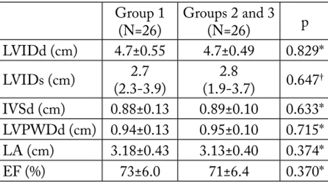
echocardiographic values, age and duration of the disease did not significantly differ between group 2 (dm1 and dr) and group 3 (dm1, dr and microal-buminuria) patients. Since patients in these groups were older than group 1 patients (dm1 without com-plications), we matched the group 2 and 3 patients with group 1 patients according to age and duration of dm1. we obtained two groups of 26 patients each, matched for age and dm1 duration. The mean (me-dian) age in group 1 was 22 years (min-max: 17-45 years), and in the other group of patients it was 21
Table 4. Distribution of patients and odds ratio (univariate logistic regression) according to groups and duration of diabetes mellitus type 1 (DM1) considering median values of ejection fraction (EF) ≤71
ef (%)
Predictor ≤71 >71 p† Odds ratio (95% Ci) p‡
Group 1* 16 24 0.106 (0.92-2.22)1.4 0.119 2 25 15 3 23 17 duration of dm1 (yrs) <11* 28 34 0.064 (0.96-4.1)2.0 0.065 >11 36 22
dm1 = diabetes mellitus type 1; ef = ejection fraction; *values reported as basal values; †χ2-test; ‡logistic
regression
Table 5. Odds ratio (multivariate logistic regression) for incidence of ejection fraction ≤71% according to duration of diabetes mellitus type 1 (DM1) and age
Predictor Odds ratio 95% Ci p
duration of dm1
(years) <11* 1.6 0.57-4.5 0.366
>11
Age (years) <33* 1.3 0.48-3.8 0.576
>33
years (min-max: 18-49 years) (p=0.978). echocardio-graphic results in groups 1, 2 and 3, matched accord-ing to age and dm1 duration, did not show any sig-nificant difference (table 6).
Discussion
in patients with dm1, tests offer better possibili-ties of determining the relation between microangio-pathic complications than in patients with diabetes mellitus type 2 because dm1 develops in younger patients with less adjunctive variables associated with metabolic syndrome and senile atherosclerotic fac-tors.
The results of our study showed that the mean echocardiographic values were within the nor-mal range in all three groups of patients with dm1 (n=120) (group 1 without microangiopathic compli-cations, group 2 with dr, and group 3 with dr and microalbuminuria). data analysis showed significant difference in echocardiographic variables in patients without complications (group 1) as compared with ei-ther patients with dr or those with dr and microal-buminuria (groups 2 and 3). There was no significant difference between the patients with dm1 and dr and those with dm1, dr and microalbuminuria. Left ventricular systolic dysfunction values were within the normal range in all patients. borow et al. did not find
any left ventricular systolic or diastolic dysfunction in patients with dm114.
Some authors found significant correlation be-tween left ventricular systolic dysfunction in patients with dm1 when compared with normal popula-tion15-17. Some other authors found normal left ven-tricular systolic function and their patients showed pathologic values only on physical exercise18. Other authors report on normal echocardiographic values of left ventricular systolic function in patients with dm1 even during physical exercise19.
most authors describe normal left ventricular sys-tolic function in dm1 patients free from microangio-pathic complications and with low cardiovascular risk (normotensive patients without cardiac autonomic neuropathy, dyslipidemia, coagulation dysfunction, and proteinuria), and with regulated glycemia20-24.
Our results showed that there was no significant correlation between dr and ef <71%, and between dr and microalbuminuria on the one hand and ef <71% on the other. it was also confirmed that there was no correlation between ef <71% and duration of the disease. raev also examined the relation between ef and duration of dm1 and reports a rare occur-rence of systolic dysfunction until the age of 1823.
rajan and Gokhale report normal ef (mean value 73%) in the group of patients with dm1 and mi-croangiopathic complications21, while Konduracka et
al. report abnormal systolic function in 185 examined patients with dm1 and mean duration of the disease of 22.8 years24,25. On the contrary, raev reports dete-rioration of systolic function in 39% of patients with dm1 and microangiopathic complications (retinopa-thy, nephropathy and autonomic neuropathy)26. Some authors think that hypertension and cardiac autonom-ic neuropathy (whautonom-ich is present in more than 40% of patients) have great influence on systolic dysfunction development, so with this perspective we can observe the incidence of systolic dysfunction in the previously mentioned study27-30.
in an attempt to correct our data, we matched pa-tients according to age and duration of dm1, but we did not obtain any significant difference among the matched groups in any of the echocardiographic vari-ables or ef.
The most important reasons for different results found in other studies may be different inclusion and Group 1 (n=26) Groups 2 and 3(n=26) p Lvidd (cm) 4.7±0.55 4.7±0.49 0.829* Lvids (cm) (2.3-3.9)2.7 (1.9-3.7)2.8 0.647† ivSd (cm) 0.88±0.13 0.89±0.10 0.633* LvPwdd (cm) 0.94±0.13 0.95±0.10 0.715* LA (cm) 3.18±0.43 3.13±0.40 0.374* ef (%) 73±6.0 71±6.4 0.370*
Lvidd = left ventricular diameter at the end of diastole; Lvids = left ventricular diameter at the end of systole; ivSd = diastolic septal thickness; LvPwdd = left ventricular posterior wall thickness in diastole; LA = left atrial diameter; ef = ejection fraction; *t-test;
†mann-whitney test
Table 6. Descriptive values of echocardiographic variables (X¯¯¯ ±SD) or median (min-max) in group 1 patients, and in groups 2 and 3 patients, adjusted for duration of diabe-tes type 1 and age
exclusion criteria (especially hypertension, neuropa-thy, and poor blood glucose regulation), and differ-ent diagnostic methods employed to reveal complica-tions31.
based on the data gathered on 120 patients with dm1, we have drawn the following conclusions: 1. mean values of all echocardiographic variables
ex-amined in this study were within the normal range in each of the three groups of examined patients with dm1 (with low cardiovascular risk, and without diabetic neuropathy).
2. There were significant differences in echocardio-graphic variables in the group of patients without complications (group 1) when compared with the group of patients with dr and microalbuminuria together (groups 2 and 3).
3. There was no significant difference in echocardio-graphic variables between the group of patients with dm1 and dr and the group of patients with dm1, dr and microalbuminuria.
4. There was significant correlation between the du-ration of dm1, the presence of dr and microal-buminuria.
5. Systolic function, expressed as left ventricular ef, was normal in all patients.
6. There was no significant correlation between dr and ef <71%, or between dr, microalbuminuria and ef <71%.
7. There was no correlation between ef <71% and duration of the disease.
References
1. CheSSLer Sd, LernmArK A. type 1 (insulin-de-pendent) diabetes mellitus. in: dAvidSOn JK, editor. Clinical diabetes mellitus, Third edition. new york: Thieme, 2000:37-57.
2. American diabetes Association. diagnosis and classification of diabetes mellitus. diabetes Care 2010;33(Suppl 1):S62-S69.
3. SChneLL O, StAndL e. diabetes and cardiovascu-lar disease. Current status of trials. Clin Res Cardiol Suppl
2010;5:27-34.
4. CALLeS-eSCAndOn J, CiPOLLA m. diabetes and endothelial dysfunction: a clinical perspective. endocr rev 2001;22(1):36-52.
5. verGOuwe y, SOedAmAh-muthu SS, ZGibOr J, ChAturvedi n, fOrSbLOm C, SneLL-ber-GeOn JK, mAAhS dm, GrOOP Ph, rewerS m,
OrChArd tJ, fuLLer Jh, mOOnS KG. Progression to microalbuminuria in type 1 diabetes: development and val-idation of a prediction rule. diabetologia 2010;53(2):254-62. 6. StehOuwer COen dA. endothelial dysfunction in diabetic nephropathy: state of the art and potential signifi-cance for non-diabetic renal disease. nephrol dial transplant 2004;19(4):778-81.
7. mcmurrAy Jv, mcdOnAGh tA, dAvie AP, CLe-LAnd JGf, frAnCiS Cm, mOrriSOn C. Should we screen for asymptomatic left ventricular dysfunction to pre-vent heart failure? eur heart J 1998;19:842-6.
8. LAm CArOLyn SP, brutSAert dL. endothelial dys-function. A pathophysiologic factor in heart failure with pre-served ejection fraction. J Am Coll Cardiol 2012;60(18):1787-9. 9. ZiLe mr, brutSAert dL. new concepts in diastolic
dysfunction and diastolic heart failure: Part i. Circulation 2002;105:1387-93.
10. mArtyn Cn, ewinG dJ. Pupil cycle time: a simple way of measuring an autonomic reflex. J neurol neurosurg Psy-chiatry 1986;49:771-4.
11. hOyOunG L, yOunGKOOK K, JOnGSeOK P. Pupil cycle time and contrast sensitivity in type ii diabetes mellitus patients: a pilot study. indian J Ophthalmol 2011;59(3):201-5. 12. dAttA S, biSwAS nr, SAxenA r, deePAK KK,
menOn v, GArG SP, tAndOn r. Ocular and car-diovascular autonomic function in diabetic patients with varying severity of retinopathy. Indian J Physiol Pharma-col 2005;49:171-8.
13. bOJić L, ČAGALJ S, rAČić G, KArAmAn-KrALJević K. Cardiovascular reflex testing and pu-pil cycle time in open-angle glaucoma. Ophthalmologica 1993;206:158-61.
14. bOrOw Km, JASPAn Jb, wiLiAmS KA, et al. myocar-dial mechanics in young adult patients with diabetes mellitus: effects of altered load, inotropic state and dynamic exercise. J Am Coll Cardiol 1990;15:1508-17.
15. di COri A, di beLLO v, miCCOLi r, tALini e, PAL-AGi C, delle dOnne mG, PennO G, et al. Left ventricu-lar function in normotensive young adults with well-controlled type 1 diabetes mellitus. Am J Cardiol 2007;99:84-90. 16. fAnG Zy, yudA S, AnderSOn v, ShOrt L, CASe
C, mArwiCK th. echocardiographic detection of early diabetic myocardial disease. J Am Coll Cardiol 2003;41:611-7. 17. SCOGnAmiGLiO r, CASArA d, AvOGArO A.
myocardial dysfunction and adrenergic innervation in pa-tients with type 1 diabetes mellitus. diabetes nutr metab 2000;13:346-9.
18. KArLefOrS t. haemodynamic studies in male diabetics. Acta med Scand Suppl 1996;449:45-80.
19. PALmieri v, CAPAdOLO b, ruSSO C, iACCArinO m, PeZZuLLO S, QuintAvALLe G, di minnO G, riCCArdi G, CeLentAnO A. uncomplicated type 1
diabetes and preclinical left ventricular myocardial dysfunc-tion: insights from echocardiography and exercise cardiac per-formance evaluation. diabetes res Clin Pract 2008;79:262-8. 20. SChAnnweLL Cm, SChnePPenheim SP, Per-inGS S, PLehn G, StrAuer be. Left ventricular dia-stolic dysfunction as an early manifestation of diabetic car-diomyopathy. Cardiology 2002;98:33-9.
21. rAJAn SK, GOKhALe Sm. Cardiovascular function in pa-tients with insulin-dependent diabetes mellitus: a study using noninvasive methods. Ann ny Acad Sci 2002;958:425-30. 22. rOmAnenS m, fAnKhAuSer S, SAner b,
mi-ChAud L, SAner h. no evidence for systolic or dia-stolic left ventricular dysfunction at rest in selected patients with long-term type 1 diabetes mellitus. eur J heart failure 1999;1:169-75.
23. rAev dC. which left ventricular function is impaired ear-lier in the evolution of diabetic cardiomyopathy? An echocar-diographic study of young type 1 diabetic patients. diabetes Care 1994;17(7):633-9.
24. KOndurACKA e, GACKOwSKi A, rOStOff P, GALiCKA-LAtALA d, frASiK w, PiwOwArSKA w. specific cardiomyopathy in type 1 diabetes-mellitus: no evidence for its occurrence in the era of intensive insulin therapy. eur heart J 2007;28:2465-71.
25. KOndurACKA e, GACKOwSKi A, rOStOff P, GALiCKA-LAtALA d, frASiK w, PiwOwArSKA w. diabetic cardiomyopathy: a controversial entity: reply. eur heart J 2008;29:565.
26. rAev dC. Left ventricular function and specific diabetic complications in other target organs in young insulin-depen-dent diabetics: an echocardiographic study. heart vessels 1994;9:121-8.
27. mAiSCh b, ALter P, PAnKuweit S. diabetic cardio-myopathy – fact or fiction? herz 2011;36(2):102-15. 28. mOnteAGudO Pt, mOiSeS vA, KOhLmAn O Jr,
ribeirO Ab, LimA vC, ZAneLLA mt. influence of autonomic neuropathy upon Lv dysfunction in insulin-de-pendent diabetic patients. Clin Cardiol 2000;23:371-5. 29. Lim hS, macfAdyen rJ, LiP Gyh. diabetes mellitus,
the renin-angiotensin-aldosterone system, and the heart. Arch intern med 2004;164:1737-48.
30. macdOnALd mr, Petrie mC, hAwKinS nh, Petrie Jr, fiSher m, mcKeLvie r, AGuiLAr d, Krum h, mcmurrAy J. diabetes, left ventricular systolic dysfunction and chronic heart failure. eur heart J 2008;29:1224-40.
31. viniK Ai, erbAS t. diabetic autonomic neuropathy. handb Clin neurol 2013;117:279-94.
Sažetak
SiStOLiČKA funKCiJA LiJeve KLiJetKe u OdAbrAnih bOLeSniKA SA ŠećernOm bOLeSti tiP 1 S diJAbetiČnOm retinOPAtiJOm i miKrOALbuminuriJOm iLi beZ nJih
K. Bućan, L. Bojić, D. Fabijanić, D. Galetović, V. Čapkun, D. Karlica Utrobičić i I. Bućan
endotelno-vaskularna disfunkcija je temeljni čimbenik u etiologiji razvoja kasnih kliničkih komplikacija u bolesnika sa šećernom bolesti tip 1(Šbt1) uključujući dijabetičnu retinopatiju, dijabetičnu nefropatiju (u samom početku obilježenu mikroalbuminurijom) i disfunkciju lijeve srčane klijetke. Ciljevi rada bili su odrediti asimptomatsku sistoličku disfunkciju lijeve srčane klijetke u bolesnika sa Šbt1 i klinički (ne)evidentnom dijabetičnom retinopatijom i s mikroalbuminurijom ili bez nje, te utvrditi povezanost trajanja Šbt1 s pojavom dijabetične retinopatije, mikroalbuminurije i asimptomatske sistoličke disfunkcije lijeve srčane klijetke. ispitano je 120 odabranih bolesnika sa Šbt1 (normotenzivni, bez dijabetične neuropatije), koji su s obzirom na nalaz fundoskopije (oftalmoskopski, fundus fotografija i fluoresceinska angiografija) i mi-kroalbuminurije (turbidimetrijskom metodom) svrstani u tri skupine: 1) bolesnici bez dijabetične retinopatije i bez mikro-albuminurije (n=40); 2) bolesnici s dijabetičnom retinopatijom bez mikromikro-albuminurije (n=40); 3) bolesnici s dijabetičnom retinopatijom i mikroalbuminurijom (n=40). Kod svih bolesnika izvršeno je ergometrijsko testiranje i ehokardiografsko ispitivanje dvodimenzijskom (2-d) i pulsnom doppler-ehokardiografskom tehnikom. Kod sve tri skupine bolesnika sa Šbt1 (niskog profila kardiovaskularnog rizika, uredne reguliranosti i bez dijabetične neuropatije) srednje vrijednosti ehokardiografskih varijabla bile su unutar referentnih vrijednosti. nismo utvrdili statistički značajnu povezanost između trajanja Šbt1 i ispitivanih ehokardiografskih varijabla.