Hotaru seibutsu hakko o riyoshita shinki imejingu shisutemu no kaihatsu
Bebas
94
0
0
Teks penuh
(2) Contents Chapter 1. Introduction. 1. 1-1. Chemistry of bioluminescence. 2. 1-2. Firefly bioluminescence. 6. 1-3. Applications of firefly bioluminescence. 8. 1-4. Structure activity relationship of firefly luciferin. 12. Chapter 2. Synthesis of firefly luciferin analogues and evaluation of the luminescent properties. 15. 2-1. Introduction. 16. 2-2. Results and discussion 2-2-1. Synthesis of firefly luciferin analogues. 18. 2-2-2. Bioluminescence. 22. 2-2-3. Chemiluminescence. 26. 2-2-4. Inhibitory activity of bioluminescence. 28. 2-2-5. Adenylation of synthetic compound. 30. 2-2-6. Bioluminescence in vivo. 33. 2-3. Summary. 35.
(3) Chapter 3. Development of a luminescence-controllable firefly luciferin analogue using selective enzymatic cyclization. 37. 3-1. Introduction. 38. 3-2. Results and discussion 3-2-1. Synthesis of N-Ac-γ-glutamate luciferin. 40. 3-2-2. Cyclization with aminoacylase. 41. 3-2-3. Reaction rate of cyclization. 43. 3-2-4. Bioluminescence with aminoacylase. 45. 3-3. Summary. 48. Chapter 4. Conclusion. 49. Experimental Section. 53. References. 81. Acknowledgements. 87.
(4) Abbreviations Ac. acetyl. Ala. alanine. AMP. adenosine monophosphate. aq. aqueous. ATP. adenosine triphosphate. BFP. blue fluorescent protein. BRET. bioluminescence resonance energy transfer. Boc. tert-butoxycarbonyl. Bu. butyl. BL. bioluminescence. Co.. cooperation. DAL. diaminophenylpropyl-aminoluciferin. DAST. diethylaminosulfur trifluoride. DCC. dicyclohexylcarbodiimide. DMAP. N,N-dimethyl-4-aminopyridine. DMF. N,N-dimethylformamide. DMSO. dimethyl sulfoxide. ESI. electrospray ionization. Et. ethyl. ffLuc. fluorescent protein fused luciferase.
(5) GFP. green fluorescent protein. Gly. glycine. HPLC. high performance liquid chromatography. isoAm. isoamyl. Imd.. imidazole. LLR. luciferin-luciferase reaction. Me. methyl. MOM. methoxymethyl ether. NMR. nuclear magnetic resonance. NO. nitric oxide. NOC7. N-methyl-3-(1-methyl-2-hydroxy-2nitrosohydrazino)-1-propanamine. PTLC. preparative thin-layer chromatography. quant.. quantitative. SAR. structure activity relationship. Ser. serine. TBAF. tetra-n-butylammonium fluoride. TBS. tert-butyldimethylsilyl. TFA. trifluoroacetic acid. THF. tetrahydrofuran. TMS. trimethylsilyl.
(6) Chapter 1. Introduction. 1.
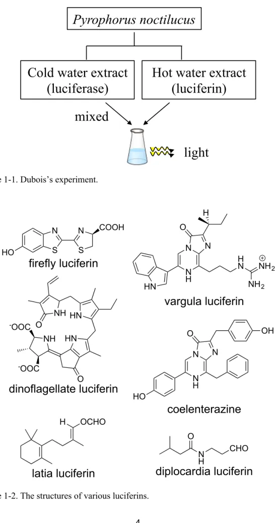
(7) 1-1. Chemistry of bioluminescence. Bioluminescent organisms are found among a wide variety of species, including bacteria, insects, and marine life.[1] Because the mechanisms of bioluminescence (BL) and the diversity of luminescent colors are species-dependent, these luminescence phenomena have long attracted the interest of scientific researchers. In 1667, Sir Boyle demonstrated that luminescent bacteria and mushrooms could not emit light in reduced pressure environments. However, they regained their BL activity when the pressure returned to normal. This result indicated that an aerobic enzyme is required for the BL of luminescent bacteria and mushroom.[2] Spallanzani showed that water extracts of dried sea firefly and jellyfish emitt light. This experiment suggested the existence of a chemical substance that is responsible for light emission.[3] In 1883, Dubois studied the chemistry of BL in more detail. When a cold water extract of the luminescent organ of Pyrophorus noctilucus was added to a hot water extract of the organ, the mixture emitted light strongly. Dubois proposed that the hot water extract contained the compound luciferin, whereas the cold water extract contained the enzyme luciferase. This reaction was named the luciferin-luciferase reaction (LLR) (Figure 1-1).[4] A luciferin and luciferase pair can be found in most of bioluminescent organisms (Figure 1-2).[5–10] BL in the LLR system is a consequence of the luciferase-catalyzed transformation of luciferin in the presence of O2, which generates the excited state of oxyluciferin, followed by light emission (Figure 1-3, route a).[11] In firefly luciferin and sea firefly 2.
(8) luciferin, photons are generated only by the LLR because oxyluciferin contains a fluorescent chromophore. In contrast, in Latia neritoides and Microscolex phosphoreus, the LLR is not produced by a fluorescent chromophore. Therefore, these species induce BL by energy transfer to a different chromophore (Figure 1-3, route b).[12] However, the chromophore and the BL mechanism of these spexies have not been identified. A typical fluorescent chromophore is green fluorescent protein (GFP). GFP was first purified from Aequorea victoria by Shimomura;[13] it is used widely in biological studies.[14–19] In Aequorea victoria, the photoprotein aequorin, which is generated by a combination of coelenterazine, apoaequorin, and O2, is excited by reaction with Ca2+. The excited mixture protein, blue fluorescent protein BFP, normally emits blue light (λmax = 460 nm). BFP then transfers energy to GFP by bioluminescence resonance energy transfer (BRET) (Figure 1-4). As a result, Aequorea victoria emits green light (λmax = 515 nm). [1]. 3.
(9) Pyrophorus noctilucus. Cold water extract (luciferase). Hot water extract (luciferin). mixed light Figure 1-1. Dubois’s experiment.. H. HO. N. N. S. S. O. COOH. N. N. firefly luciferin� HN. H N. N H. NH 2 NH 2. vargula luciferin� -OOC. O. NH HN NH. OH. O. HN. N. N. -OOC. N H. O. dinoflagellate luciferin�. HO. coelenterazine� H. OCHO O N H. CHO. diplocardia luciferin �. latia luciferin� Figure 1-2. The structures of various luciferins. 4.
(10) singlet excited state� (oxyluciferin)*�. energy�. high�. low�. O2. Light. luciferin + luciferase �. singlet excited state� chromophore�. energy transfer�. (a). light. (b). oxyluciferin� ground state�. chromophore� ground state�. Figure 1-3. Energy level diagram of the LLR.. apoaequrin�. O O N. Ca2+ O. OH. Ca2+. NH. OH. HO. Ca2+. N. O. aequrin�. excited BFP�. excited GFP�. OH. O. energy transfer�. N. 460 nm�. N H HO. NH. N. Ca2+. N. *. O. -CO2. N H. O2, EDTA, DDT. Ca2+. 515 nm�. coelenterazine�. apoaequrin�. BFP�. Figure 1-4. The mechanism of BL in Aequorea victoria BL.. 5. GFP�.
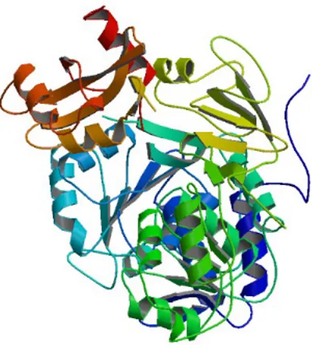
(11) 1-2. Firefly bioluminescence. The most well-known BL is that of the firefly. In 1916, Harvey proved that the mechanism of firefly BL is the LLR.[11] After that, firefly luciferin (1) was isolated (1957) from Photinus pyralis;[20] it was synthesized and its structure was determined (1961) by McElroy.[5] Regarding firefly luciferase, the three-dimensional structure of Photinus pyralis luciferase (1996) was reported by Conti (Figure 1-5).[21] The firefly BL mechanism is proposed as follows (Figure 1-5): the first step is the adenylation of firefly luciferin in the presence of luciferase and adenosine triphosphate (ATP-Mg2+). Next, firefly luciferin carrying adenosine monophosphate (AMP) is oxidized by luciferase. De-adenylation of AMP firefly luciferin generates peroxide. Upon the decarbonylation of this intermediate, excited oxyluciferin occurs and emits yellow-green light (λmax=553 to 559 nm) with a high quantum efficiency (ϕBL = 41%) (Figure 1-6).[22–23]. 6.
(12) Figure 1-5. 3D structure of firefly luciferase (Protein Data Bank number 1LCI).. N HO. COOH + Firefly Luciferase + ATP - Mg 2+. N. S S firefly luciferin (1) N. N. COOAMP. S S firefly luciferin AMP N. HO. N. S S peroxide. N O2. S S peroxide. HO. O O O. N HO. N. S S oxyluciferin. Figure 1-6. The mechanism of firefly BL.. 7. N. N. COOAMP. S S firefly luciferin AMP. HO. +. HO. N. O O O. + AMP. O + CO 2. + Light.
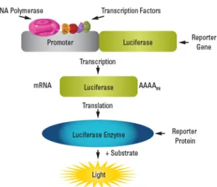
(13) 1-3. Applications of firefly bioluminescence. The firefly LLR system has been widely used in biological studies because of its high efficiency and the ready availability of firefly luciferase and luciferin.[24–27] ATP is necessary for firefly BL; the BL intensity is concentration-dependent. Using this property, Kikkoman Co. Ltd. developed a device for detecting the growth of microorganisms and bacteria in foods (Figure 1-7, a).[28] Firefly luciferase has been applied as a reporter gene to evaluate the transcriptional activation potential of a specific gene (Figure 1-7, b).[29] Further more, if the N-terminal and C-terminal domains of firefly luciferase are separated, the luciferase regains its original function when the interaction is restored. This split-luciferase system enables quantitative evaluation of protein-protein interactions (Figure 1-7, c).[30] Recently, the most high-impacted research in this field has been the development of fluorescent protein-fused luciferase (ffLuc-cp156).[31] This enzyme binds to luciferase and the fluorescent protein Venus; its emits stronger light than natural firefly luciferase by BRET from luciferase to Venus (Figure 1-8).. 8.
(14) b a. c. Lu. c-N. L. Lu. -C uc. c-N. Lu. c-C. splicing light�. Lu protein A. protein B. c-N. -C. c Lu. luciferase. Figure 1-7. a) Lumitester PD-30. b) Schematic of the luciferase reporter assay. c) The Schematic of the split-luciferase assay.. firefly luciferase excited state. luciferin luciferase ground state. cp156Venus. *. cp156Venus. Light. cp156Venus. Figure 1-8. BRET of ffLuc-cp156 and BL images of anesthetized CAG-ffLuc-cp156 transgenic adult mice. 9.
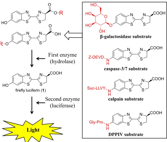
(15) Many imaging techniques using firefly luciferin derivatives have been reported. In 1987, Miska and Geiger prepared BL-inactive luciferin derivatives with recognition sites for a protease enzyme. They demonstrated that the addition of a second enzyme (luciferase) to these derivatives generates BL after the activated protease reacts with the derivatives (Figure 1-9).[32] In 2000, Nishimura reported the detection of galactosidase in vivo using a firefly luciferin derivative with a galactosidase recognition site.[33] Promega Co. Ltd. synthesized various modified luciferin derivatives that react with various. enzymes. (Figure. 1-9).[34]. Urano. developed. a. BL. probe,. diaminophenylpropyl-aminoluciferin (DAL), that can detect NO in vivo (Figure 1-10).[35]. 10.
(16) O. HO. R. O. N. N. S. S. N. N. S. S. HO O. R. HO HO. O. Z-DEVD. S. S. COOH. N H. N. N. S. S. COOH. caspase-3/7 substrate. COOH. N. N. β-galactosidase substrate. S S firefly luciferin (1). HO. O. N. OH. OH. First enzyme (hydrolase)� N. O. Suc-LLVY. Second enzyme (luciferase)�. N H. N. N. S. S. COOH. calpain substrate. Gly-Pro. Light. N H. N. N. S. S. COOH. DPPIV substrate. Figure 1-9.Enhanced BL of luciferin derivatives due to enzyme activity.. N H. H 2N. N. N. S. S. DAL�. NH 2. COOH. NO� O2�. N H. HN N N. almost non-luminescent �. N. N. S. S. COOH. DAL - T� highly luminescent�. Figure 1-10. Molecular design of DAL and detection of NO with DAL in a luc-TAg rat with NOC7.. 11.
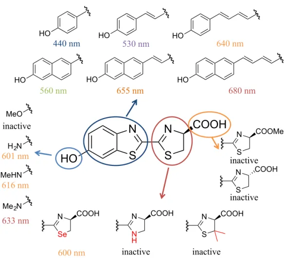
(17) 1-4. Structure activity relationship (SAR) of firefly luciferin. The mechanistic pathway for the reaction catalyzed by firefly luciferase, leading to BL, has been fully elucidated. In addition, much effort has been dedicated to expand the substrate scope of this process.[36–39] The results of these studies showed that generation of BL requires the use of substrates that, similar to firefly luciferin, possess α-amino acid centers with the D-absolute configuration.[40] In particular, aminoluciferin, in which the phenolic hydroxyl group of the parent substrate is replaced with an NH2 group, was found to have BL activity.[41–42] In an earlier study, Maki demonstrated that the emission wavelength maxima for BL by analogues of firefly luciferin were dependent on the nature of the benzothiazole ring. For example, incorporation of a more extensively conjugated chain in this moiety induces a red shift of the luminescence wavelength.[43–44] Importantly, analogues of firefly luciferin that display red-shifted BL are suitable for use in noninvasive, whole-body imaging systems because they generate near-IR light, which can penetrate thick tissues more deeply than the short-wavelength light arising from the original LLR. However, only a few SAR studies of the thiazoline moiety have been published. Results of earlier studies of luciferin analogues, in which the sulfur atom of the thiazoline ring was replaced by a selenium atom and NH and Me2C groups, have been reported (Figure 1-11).[45–47]. In this study, the effects of the thiazoline moiety on BL. and the development of a new imaging tool using the SAR results are described.. 12.
(18) HO. HO. 440 nm. HO. 530 nm. HO. HO. 560 nm. HO. 655 nm. 680 nm. MeO. inactive H 2N. 601 nm. 640 nm. HO. N. N. S. S. COOH. COOMe. S. inactive N. MeHN. 616 nm. S. COOH. inactive. Me 2N. 633 nm. N. N. COOH. Se. 600 nm. N. COOH. N. COOH. N H. S. inactive. inactive. Figure 1-11. Previous SAR study of firefly luciferin.. 13.
(19) 14.
(20) Chapter 2. Synthesis of firefly luciferin analogues and evaluation of the luminescent properties. 15.
(21) 2-1. Introduction. The investigation described below was guided by the goal of expanding the substrate range of the luciferase-promoted reaction and to produce new substances that efficiently produced BL. In these efforts, substances were prepared and explored in which groups of the thiazoline moiety of firefly luciferin (1) were replaced with those derived biosynthetically from D-cysteine.[23, 48–49] Herein, several novel analogues are described in which the thiazoline ring in 1 is replaced by glycine, D-alanine, L-alanine, D-serine, L-serine,. side chains (2–6, Figure 2-1), D-oxazoline, and D-pyrroline rings (7 and 8,. Figure 2-1). All compounds were evaluated to explain the role of the thiazoline moiety in BL.. 16.
(22) HO. N. N. S. S. COOH. 1�. COOH N HO. HN. N. H S. COOH. COOH. HN. S. HO. 2�. O. HO. 3� COOH. COOH N. HN. S. O. OH HO. S. O. OH HO. 5�. HO. O. 4�. HN. N. S. Me. Me. O. N. HN. 6�. N. N. S. O. COOH HO. 7�. N. N. S. C H2. 8�. COOH. Figure 2-1. Luciferin analogues 2–8 derived from amino acids prepared in this study.. 17.
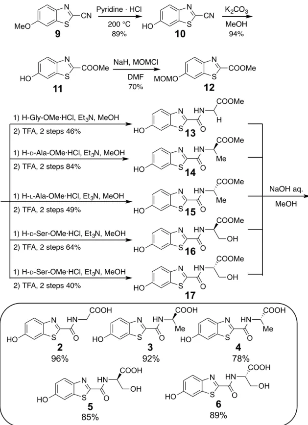
(23) 2-2. Results and discussion. 2-2-1. Synthesis of firefly luciferin analogues. The route used for the synthesis of the benzothiazoylamino acids 2–6 was initiated by the. pyridine. hydrochloride. promoted. thermal. transformation. of. 2-cyano-6-methoxybenzothiazole (9) to form phenol 10.[50] Conversion of the nitrile group in 10 into the corresponding methyl ester in 11,[51] by treatment with MeOH/K2CO3, was followed by MOM protection of the phenolic hydroxyl group to give 12. Coupling reactions of 12 with acid hydrochlorides of glycine, D-alanine, L-alanine, D-serine,. and L-serine followed by removal of the MOM group in each amide. by using TFA, formed the respective ester derivatives 13–17. Finally, treatment of 13– 17 with a solution of NaOH in methanol generated the respective benzothiazoylamino acids 2–6 (Figure 2-2). Preparation of the oxazoline benzothiazole 7 began with selective acetylation of the phenolic hydroxyl group of the serine-derived intermediate 16 (Figure 2-3). This process produced monoacetate 18 in only low yield because of competitive esterification of the primary alcohol group in 16. Importantly, the direct cyclization reaction of unprotected phenol 16 did not take place cleanly. Treatment of 18 with DAST promoted the desired cyclization to generate oxazoline 19, which underwent sequential lipase-promoted deacetylation and methyl ester hydrolysis to form the oxazoline ring-containing luciferin analogue 7. 18.
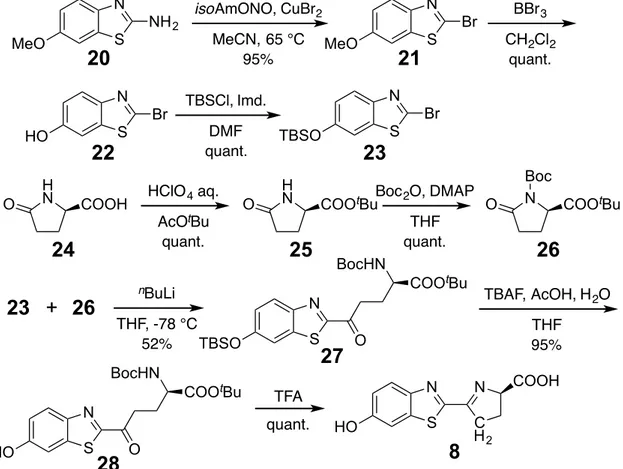
(24) In the pathway for the synthesis of carboluciferin (Figure 2-4, 8), bromide 21[52] was generated through Sandmeyer reaction of a commercially available aminothiazolidine 20. Removal of the methoxy group in 21 gave the corresponding phenol 22, which was protected by using TBSCl to give the phenylsilyl ether 23. To generate the pyrrolidinone 26, which was used in a coupling reaction with 23, the pyrrolidione carboxylate 24 was transformed into tbutyl ester 25.[53] Protection of 25 with Boc gave 26,[53] which reacted with the lithium organometallic derivative of 23, produced by treatment with. n. BuLi, to generate the glutamate-linked benzothioazole 27.. TBAF-mediated desilylation of 27 formed the free phenol 28, which then underwent TFA-promoted cyclization to produce linked pyrroline benzothiazole 8.. 19.
(25) N MeO. 9�. S. 200 °C 89%. N COOMe. K 2CO3. CN HO. 10�. S. MeOH 94%. NaH, MOMCl. N. DMF 70%. S. S. HO. N. Pyridine · HCl. CN. 11�. COOMe. MOMO. 12� COOMe. 1) H-Gly-OMe·HCl, Et 3N, MeOH. H S. HO. 2) TFA, 2 steps 46% 1) H-D-Ala-OMe·HCl, Et 3N, MeOH 2) TFA, 2 steps 84%. 1) H- L-Ala-OMe·HCl, Et 3N, MeOH 2) TFA, 2 steps 49% 1) H-D-Ser-OMe·HCl, Et 3N, MeOH. 1) H-D-Ser-OMe·HCl, Et 3N, MeOH. S. HO. HO. 2�. HO. HN. S. O COOMe. N. HN. S. O. 15�. HN. S. O. 16�. COOMe. N. HN. S. O. 17�. COOH. S. 3�. O. Me HO. N. HN. S. O. Me. COOH. HN OH. 5�. O. N. HN. S. O. OH HO. 6�. 89%�. 85%�. Figure 2-2. Synthesis of benzothiazoylamino acids 2–6.. 20. 4�. 78%�. COOH S. MeOH see table. COOMe. N. 92%� N. NaOH aq.. COOH. 96%�. HO. 14�. HN. N. O. N. OH. COOH N. COOMe. OH HO. HN. O. Me HO. 2) TFA, 2 steps 64%. 13�. Me HO. 2) TFA, 2 steps 40%. HN. N.
(26) COOMe HN. N. N. HN. THF 79%. S. O. OH S. HO. 16�. O. DAST CH2Cl2 90%. HO. AcO N. N. S. O. N. N. S. O. 19�. COOMe. Ac2O, NaHCO 3. OH AcO. COOMe. 18�. Lipase PS PS IM IM Amano Amano Lipase NH44HCO HCO33 aq. aq. DIPE EtOH NH 92% 92%. COOH. 7�. Figure 2-3. Synthesis of oxazoline benzothiazole 7.. N MeO. 20�. NH 2. S. N Br S. HO. O. 22�. H N. 23� +� 26�. S. S. TBSO H N. CH2Cl2 quant.. 23� Boc 2O, DMAP. COOtBu. 25�. COOtBu. S. BocHN. 27�. HO. COOtBu. TBAF, AcOH, H 2O THF 95%. N. N. S. C H2. 8�. O. Figure 2-4. Synthesis of synthesis of carboluciferin 8.. Boc N COOtBu. 26�. O. TFA. 21. O. THF quant.. N. quant.. 28�. S. Br. THF,-78 -78°C °C THF, TBSO 52% 52%. N. 21�. DMF quant.. nBuLi n-BuLi. BocHN. MeO. N. O. BBr 3. Br. TBSCl, Imd.. AcOtBu quant.. 24�. HO. MeCN, 65 °C 95%. HClO 4 aq.. COOH. N. isoAmONO, CuBr2. COOH.
(27) 2-2-2. Bioluminescence. Luciferase-promoted BL was evaluated by using 50 mM solutions of the luciferin analogues 2–8 in potassium phosphate buffer (500 mM, pH 6.0). The results showed that only 8 produced a BL response, whereas benzothiazoylamino acids 2–6 and oxazoline 7 analogues did not (Figure 2-5). The total photon yield of the luciferase-catalyzed reaction of 8 (1.52 × 10⁵ photons) was 0.56% of the process with 1 as a substrate (2.72 × 10⁷ photons). In addition, the maximum emission wavelength of 8 BL was blueshifted (λmax = 547 nm) relative to that of 1 (Figure 2-6). To assess the binding affinity of synthetic luciferin 8 to luciferase, the Michaelis constant (Km, defined as the concentration at half of the maximum reaction rate) was determined.[43, 54] The Km value for 8 (Figure 2-7, 82.5 ± 7.2 µM) was smaller than that of 1 (Figure 2-7, 116 ± 22.0 µM), which showed that the binding affinity of 8 to luciferase was higher than that of 1. The Km value of 1 was different from that previously reported because of the low ATP-Mg2+ concentrations.. 22.
(28) HO. N. N. S. S. COOH HO. 1�. N. N. S. S. S. O. H HO. 2�. COOH HN. S. O. COOH. Me. N. HN. S. O. COOH. COOH N S. O. HN. S. O. OH HO. 4� HN. N Me. HO. 3�. 5�. N. N. COOH. N. N. S. O. HO. S. C H2. OH HO. N. HN. L-1�. N HO. COOH. COOH. HO. 6�. 7�. 8�. COOH. 30000000. photon counts for 30 s. 250000 24000000. 200000 18000000. 150000 12000000. 100000 6000000. 50000 0. 0. compounds. 1. L-1. 2. 3. 4. 5. 6. 7. 8. Figure 2-5. The total photons of BL synthetic compounds for 30 s. L-1, which is the inhibitor of 1, was used as negative control.. 23.
(29) 120000. photon / 0.1 s. 100000 1200 80000 1000 60000. 800. 40000. 600. 20000. 400. 0 200 0. 5. 10. 15. 20. 25. 30. 35. 0 0. 5. 10. 15. 20. 25. 30. 35. time (s). 1.00. normalized. 0.80 0.60 0.40 0.20 0.00 400. 450. 500. 550. 600. 650. 700. 750. 800. wavelength (s). Figure 2-6. Change in the BL properties of 8 (blue) and 1 (green) with time and BL spectra.. 24.
(30) 1 0.0000012. 1 / [V] (photons-1). 0.000001 0.0000008 0.0000006 0.0000004 0.0000002 0 -0.02. -0.01. 0. 0.01. 0.02. 0.03. 0.04. 0.05. 0.03. 0.04. 0.05. 1 / [S] (μM-1). 8 0.000007. 1 / [V] (photons-1). 0.000006 0.000005 0.000004 0.000003 0.000002 0.000001 0.000000 -0.02. -0.01. 0. 0.01. 0.02. 1 / [S] (μM-1) Figure 2-7. Lineweaver-Burk plot for 1 and carboluciferin 8. 25.
(31) 2-2-3. Chemiluminescence. Next, the chemiluminescence properties of methyl esters of the synthetic derivatives were examined.[55] The methyl ester derivative 29 was synthesized through selective deprotection of an acetyl group in 19. The carboxylic acid group in 8 converted into the corresponding methyl ester 30 (Figure 2-8). For this purpose, 100 mM tBuOK in DMSO was added to solutions of the individual 1 mM luciferin analogue in DMSO at room temperature. Light emission was monitored for 60 s at sampling intervals of 0.1 s. The results showed that the methyl esters 13, 14, 16, 29, and 30 generated chemiluminescence with emission maxima in the 509–547 nm range. Furthermore, total photon yields from 30 (2.81 × 10⁷ photons) and 29 (8.83 × 10⁷ photons) were higher than those of the methyl ester of 1 (2.50 × 10⁷ photons) (Figure 2-9).. 26.
(32) AcO. N. N. S. O. COOMe. MeOH 99%. 19�. HO. N. N. S. C H2. 8�. K 2CO3. COOH. HO. N. S. O. COOMe. 29�. TMSCHN2 MeOH 33%. N. HO. N. N. S. C H2. 30�. COOMe. Figure 2-8. Synthesis of 29 and 30.. Figure 2-9. Chemiluminescence spectra of methyl ester derivatives and (b) decay curves of the photon counts of the signal of methyl ester of 1 (green), 13 (purple), 14 (yellow), 16 (blue), 29 (red) and 30 (brown).. 27.
(33) 2-2-4. Inhibitor activity of firefly bioluminescence. The results appear to show that the structures of the groups mimicking the thiazoline ring in 1 influence the BL yield as a consequence of their effect on binding to and/or reacting with the enzyme. Information was gained to show why 2–7 did not display BL activities, despite having chemiluminescence activities. For this purpose, the Ki (Figure 2-10) values of these substances as inhibitors of the luciferase-catalyzed reaction of 1 were determined by means of Lineweaver-Burk analysis.[56] L-Firefly luciferin (L-1) was utilized as a positive control because it was known to inhibit BL similar to that of 1.[23] The results demonstrated that alanine derivative 3, 4, and oxazoline analogue 7 bound to luciferase, whereas related substances 2, 4, and 5 did not. On the basis of these observations, the cyclic structure of the thiazoline moiety was important for the recognition of firefly luciferase (alanine derivative 3 and 4 could not be explained). I propose that the thiazoline moiety in 1 is a key structural requirement for substances that participate in the LLR leading to BL.. 28.
(34) 160 140. Ki (μM). 120 100 80. n=5. 60 40 20 0 compound. L-1�. 2�. 3�. 4�. Figure 2-10. Ki values of L-1 and 2-7.. 29. 5�. 6�. 7�.
(35) 2-2-5. Adenylation of synthetic compound. The BL activity of AMP-carboluciferin was explored next to clarify why the BL activity of 8 was lower than that of 1. AMP-carboluciferin and AMP firefly luciferin were prepared by using a procedure reported in the literature,[43, 47] and purified by means of HPLC (Figure 2-12). The luciferase solution used was diluted 10-fold because the photon emission of AMP-firefly luciferin exceeded the limit of the luminometer. The BL activity of AMP-carboluciferin (4.75 × 10⁷ photons) was 13,000 times greater than that of 8 (3.54 × 10³ photons). In contrast, the bioluminescent activity of AMP-firefly luciferin (2.11 × 10⁸ photons) was only 285 times greater than that of 1 (7.42×10⁵ photons, Figure 2-13). These observations suggested that the low BL activity of 8 was a consequence of the low efficiency of the adenylation step in the LLR pathway.. 30.
(36) HO. N. N. S. S. COOH. DCC, AMP. N. N. Acetone. S. S. HO. firefly luciferin�. HO. N. N. S. C H2. COOAMP. AMP-firefly luciferin� COOH. DCC, AMP. N. N. Acetone. S. C H2. carboluciferin�. HO. COOAMP. AMP-carboluciferin�. Figure 2-12. Synthesis and HPLC chromatograms of AMP-firefly luciferin and AMP-carboluciferin (Mightysil RP-18 GP Aqua 250-4.6 mm, H2O (include TFA0.05%) / MeCN = 9:1→1:9 (30 min), flow rate 0.5 mL/min).. 31.
(37) Figure 2-13. Change in bioluminescence activities of AMP-carboluciferin (a), AMP-firefly luciferin (b) and BL spectra (c).. 32.
(38) 2-2-6. Bioluminescence in vivo. In the final phase of this investigation, the ability of 8 to display BL in CAG-ffLuc-cp156 transgenic mice, developed in our previous research, was explored.[31] Compounds 1 and 8 (5 mM, 100 mL in sterile water) were independently injected into the abdominal cavities of mice (n = 3). The results of in vivo imaging showing the change in BL with time are displayed in Figure 2-14. Carboluciferin 8 displayed BL in a mouse that increased with time and peaked at 2,400 s following injection. In comparison, BL from 1 increased more rapidly with time and reached a maximum at 300 s after injection.. 33.
(39) Figure 2-14. In vivo imaging showing changes in BL as a function of time following injection of mice with carboluciferin 8 (blue) and firefly luciferin (1) (green).. 34.
(40) 2-3. Summary. I synthesized new luciferin analogues (2–6) containing acyclic D-aminoacid side chains and their cyclic derivatives (7 and 8) derived from D-amino acid linked to the benzothiazole ring system of the parent compound. Evaluation of these substances showed that 8 had only 0.5% of the luciferase-promoted BL activity of 1, and that the benzothiazoylamino acids and oxazoline luciferin analogues did not display BL in the presence of this enzyme. Observations made in an additional study showed that ester derivatives of these substances were chemiluminescent. An evaluation of BL of AMP-carboluciferin and AMP-firefly luciferin revealed that the activity of the former substance was 13,000 times larger than that of 8, whereas the activity of AMP-firefly luciferin was only 285 times larger than that of 1. These results indicated that the thiazoline-mimicking moiety in 8 affected the adenylation step of LLR, whereas the benzothiazole moiety played an important role in determining the luminescence intensity and wavelength. Finally, longer lived BL was displayed by 8 than that of 1 following injection into live mice. The observations made in this investigation provide information that could contribute to the design of substrates that undergo improved luciferase-promoted BL.. 35.
(41) 36.
(42) Chapter 3. Development of a luminescence-controllable firefly luciferin analogue using selective enzymatic cyclization. 37.
(43) 3-1. Introduction. A number of firefly luciferin analogues have been developed in order to detect life processes spatiotemporally.[57-61] Z-DEVD-aminoluciferin (31),[58-59,. 61]. in which the. NH2 group of aminoluciferin (32) is substituted by the DEVD peptide sequence, does not show BL in the presence of luciferase. However, the DEVD peptide is selectively cleaved in the presence of Caspase 3/7 substrates, resulting in 32 that can emit light (Figure 3-1). Similarly, 6-O-β-D-galactopyranosyl-luciferin (33),[33, 57, 60] in which the OH group of firefly luciferin is bound to the C1 position of β-D-galactose, generates BL activity in the presence of β-galactosidase (Figure 3-1). Despite such luciferin-based sensing molecules having been considered as effective tools for specific monitoring of enzyme activity, these synthetic compounds have the disadvantage of possessing a D-absolute. configuration in the thiazoline moiety,[62, 63] which easily epimerizes under. physiological conditions, and the resulting L-luciferin analogues show strong inhibitory activities against firefly luciferase.[23] Furthermore, facile enzymatic or aerobic oxidation of the thiazoline moiety[64] was suggested to promote the reaction of BL-inactive derivatives with luciferase without concomitant light emission. In this study, we report a novel linear precursor of a firefly luciferin analogue that can be activated using a specific enzyme. This study in chapter 2 reported that luciferin analogues wherein the thiazoline moiety was replaced with an acyclic structure, did not produce BL and were not recognized by firefly luciferase. The ability to convert a BL-inactive acyclic luciferin to the corresponding BL-active cyclic species by a specific 38.
(44) enzyme would provide a new ‘off’ to ‘on’ control system. To confirm the applicability of this hypothesis, we used carboluciferin 8 as the BL-active cyclic luciferin, N-Ac-γ-glutamate luciferin 34 as the BL-inactive acyclic luciferin, and aminoacylase[65, 66]. as the specific enzyme. 34 was synthesized and examined to determine if it cyclizes. and generates BL when a free NH2 group is generated by reacting 34 with aminoacylase.. BL inactive luciferin N. Z-DEVD HO HO HO. BL active luciferin COOH. N. S S N H Z-DEVD aminoluciferin (31). O O. N. N. S. S. COOH. N. caspase 3/7 H 2N. β-galactose HO. OH O-β-galactopyranosyl-luciferin (33) COOH N. COOH. S S aminoluciferin (32). N. N. S. S. COOH. luciferase. light. firefly luciferin (1). AcHN. New !!. N. N. aminoacylase HO. S O HO N-Ac-γ-glutamate luciferin 34. N. COOH. S carboluciferin 8. Figure 3-1. Novel sensing luciferins. N-Ac-γ-glutamate luciferin 34 does not contain the thiazoline, whereas the other luciferins shown here do.. 39.
(45) 3-2. Results and discussion. 3-2-1. Synthesis of N-Ac-γ-glutamate luciferin. N-Ac-γ-glutamate luciferin precursor 38 was synthesized by the condensation reaction of 23 and 37 by using nBuLi. In order to obtain 37, the α-carboxylic acid and NH2 group of precursor 35[67] were protected by tBu and Ac groups, respectively. Compound 23 was prepared using a method of chapter 2. Successive deprotection of the TBS ether and t. Bu ester of 38, afforded N-Ac-γ-glutamate luciferin 34. H 2N. HOOC D -glutamic acid. CH 2Cl 2 92%. MeOH, 0 °C MeOOC 80%. AcHN. Ac2O, Et 3N. MeOOC. N S. COOtBu. COOH. tBuOAc. MeOOC. 35�. Br. + TBSO. COOtBu. 36�. nBuLi. N. THF, -78 °C 20%. S. 23� COOtBu. H 2N. HClO 4 aq. 40%. 37�. AcHN. TBSO. H 2N. SOCl2. COOH. AcHN COOtBu. TBAF, AcOH, H 2O THF 80%. O. N S. HO. 38�. O. 39�. AcHN COOH. TFA. N. quant.. HO. S. O. 34� Figure 3-2. Synthesis of N-Ac-γ-glutamate luciferin 34.. 40.
(46) 3-2-2. Cyclization with aminoacylase. Furthermore, the cyclization of 34 was examined by the deprotection of N-Ac group with aminoacylase. The retention times of 8 and 34 were confirmed using high-performance liquid chromatography (HPLC), and the following results were obtained: 17.8–17.9 min for 8 and 25.0–25.4 min for 34 (Figure 3-3, a–c). The reaction of 3 and aminoacylase was monitored for 1 h at 37 °C using HPLC under the same solvent conditions as those used for 8 and 34 Figure 3-3, d). The results showed that 34 was converted almost completely to 8, indicating that the N-Ac group of 34 was hydrolyzed by aminoacylase, and 8 was generated by the subsequent cyclization.. 41.
(47) b.. a.. 17.8. carboluciferin 8. 25.4. N-Ac-γ-glutamate luciferin 34. time (min.). time (min.). c.. d. carboluciferin 8. carboluciferin 8. 17.9. 18.2. 25.0 N-Ac-γ-glutamate luciferin 34. 25.2. N-Ac-γ-glutamate luciferin 34. Figure 3-3. HPLC results. a) Carboluciferin 8, b) N-Ac-γ-glutamate luciferin 34, c) mixture of 8 and 34, d) mixture of 34 and aminoacylase after reaction (Develosil C30-UG-5, H2O (include 0.05% formic acid) / MeCN = 9:1→1:9 (45 min), flow rate 0.5 mL/min). All compounds were detected at 350 nm.. 42.
(48) 3-2-3. Reaction rate of cyclization. The rate of the aminoacylase-promoted cyclization reaction was obtained by evaluating the fluorescence of 8, 34, and the reaction mixture of 8 and 34. Cyclic 8 emitted fluorescence at 565 nm (green light), whereas the acyclic precursor 3 emitted fluorescence at 525 nm (yellow light) upon irradiation with an excitation light of 350 nm (Figure 3-4, a). The fluorescence intensities of 8 and 34 were detected at wavelengths of 565 nm and 525 nm, respectively, and at both wavelengths for the reaction mixture of 34 and aminoacylase (5 min at 37 °C), as shown in Figure 3-4, b. The 565 nm fluorescence of the acyclic structure decreased with increase in the enzyme concentration. In contrast, the 525 nm fluorescence of the cyclic structure increased with increase in the enzyme concentration. Removal of the N-Ac group of 34, generating an NH2 group, resulted in 34 being immediately cyclized to afford 8 as judged from the fluorescence value attained by the reaction mixture, which was same as that attained by an equivalent molar solution of 8 within 5 min.. 43.
(49) fluorescence intensity. a. 16000 14000. Excited light (350 nm). 12000 10000 8000 8. 6000 4000. 34. 525 nm. 565 nm. 2000 0 300. 400. 500. 600. 700. wavelength (nm) b.. fluorescence intensity. 35000 30000 25000 20000 15000 10000 5000. 0 aminoacylase compounds detect. 0 0.1 1 10. 0. 0 0.1 1. 34. 8. 34. 565 nm (yellow). 10. 0. mg/mL. 8. 525 nm (green). Figure 3-4. Fluorescence results. a) The fluorescence wavelength spectra of 8 (525 nm) and 34 (565 nm). b) The fluorescence intensities at 565 nm and 525 nm were measured after the reaction of 34 with aminoacylase solution for 5 min at 37 °C.. 44.
(50) 3-2-4. Bioluminescence with aminoacylase. The potential of 34 or the reaction mixture of 34 and aminoacylase to display BL was finally examined. The total photon counts are shown in Figure 3-5, a. There was no significant difference between the photon counts obtained using acyclic 34 (Figure 3-5, a, lane 2) and the photon counts of the background (Figure 3-5, a, lane 1); therefore, 34 did not exhibit BL. In contrast, the reaction mixture of 34 and aminoacylase resulted in BL, and this intensity depended on the substrate and enzyme concentrations (Figure 3-5, a, lanes 3–5). The BL curves showed similar shapes even on using different concentrations of aminoacylase (Figure 3-5, b). The maximum BL wavelength (545 nm) and waveform of the mixture of 34 and aminoacylase were found to be the same as those of 8 (Figure 3-6, b); therefore, these results indicate that the generated 8 emitted BL immediately after the BL-inactive luciferin 34 was converted to the BL-active luciferin 8 via almost instantaneous catalysis using aminoacylase, i.e., BL immediately turned “on” after injecting aminoacylase. A prolonged observation of this BL resulting from enzymatic catalysis was allowed because of the slow luminescence decay (Figure 3-6, a). Therefore, it was possible to control the BL “off-on” states by cyclization of the thiazoline moiety structure through a specific enzyme reaction.. 45.
(51) a.. lane. 1. 2. 3. 4. 5. 14000000 7000000. photon counts for 60 s. 12000000 6000000 10000000 5000000 8000000 4000000 6000000 3000000 4000000 2000000 2000000 1000000 0 0. 34 (μM). 34. 0. 0.1 1 10 aminoacylase (mg/mL). 0. 0. 0.1. 1. 10. b. 30000 25000. photon / 0.1 sec.. 20000. 34: 10 μM. 15000 5000. aminoacylase 10000 4000. 10 mg/mL. 5000 3000. 1 mg/mL 0.1 mg/mL. 0. 2000. 0. 20. 40. 60. 0 mg/mL. 1000. 0 0. 20. 40. 60. time (s). Figure 3-5. BL results. a) The total photons measured during 60 s emitted by the BL of 34 or a mixture of 34 and aminoacylase reacted for 5 min at 37 °C. b) BL change of 34 by reaction with aminoacylase solution for 5 min at 37 °C. 46.
(52) c. 250000. photon / s. 200000. 150000. 100000. 50000. 0 0. 100. 200. 300. 400. 500. 600. time (s). d. 1. normalized intensity. 0.9 0.8 0.7 0.6 0.5 0.4 0.3 0.2 350. 400. 450. 500. 550. 600. 650. 700. 750. 800. wavelength (nm). Figure 3-6. BL results. a) Aminoacylase solution was added to a mixture of 34, luciferase and ATP-Mg2+ for 50 s, and the photon count was the measured for 510 s. b) The wavelength spectrum of the reaction mixture of 34 and aminoacylase. The maximum wavelength was 545 nm.. 47.
(53) 2-3. Summary. To allow the detection of specific life processes, I synthesized a new firefly luciferin 34. Luciferin 34 did not possess a thiazoline ring, but a linear precursor that can be cyclized to give carbothiazoline 8. When the N-Ac group was removed by aminoacylase, 34 was cyclized and converted to carboluciferin 8 without requiring the isolation of the NH2 intermediate. Fluorescence studies of 8, 34, and the reaction mixture of 34 and aminoacylase showed that the enzymatic deprotection and cyclization reactions occurred very fast. Luciferin 34 did not exhibit BL, whereas it was exhibited by the reaction mixture of 34 and aminoacylase. The BL intensity made it possible to monitor the enzyme reaction spatiotemporally. These observations will contribute to the development of novel luciferin derivatives that can be used to analyze specific life processes by using a switching system between acyclic (BL “off”) and cyclic (BL “on”) structures (Figure 3-7).. enzyme. peptide NH COOH N HO. S. 1) hydrolysis 2) cyclization. N HO. N. S. O. BL inactive. BL active. Figure 3-7. Novel imaging system was proposed in this study.. 48. COOH.
(54) Chapter 4. Conclusion. 49.
(55) In this study, five new firefly luciferin (1) analogues were synthesized and their light emission properties were examined. Modifications of the thiazoline moiety in 1 were employed to produce analogues containing acyclic amino acid side chains (2–6) and heterocyclic rings derived from amino acids (7 and 8) linked to the benzothiazole moiety. Only carboluciferin 8, possessing a pyrroline-substituted benzothiazole structure, demonstrated BL activity. In contrast, acyclic luciferins did not demonstrate BL. On the basis of this result, a new firefly luciferin analogue that can switch firefly BL activity from the “off” state to the “on” state was designed and synthesized. BL-inactive N-Ac-γ-glutamate luciferin 34 contains an acyclic precursor of the thiazoline moiety. Enzymatic treatment of 34 with aminoacylase resulted in smooth removal of the acyl protecting group and concomitant cyclization to provide BL-active carboluciferin 8.. Chapter 1 is the introductory section. This section described the BL mechanisms of various luminescent organisms, the history of the study of firefly BL, and the BL applications in biological research. The SAR of firefly luciferin (1) was also summarized. Although many SAR studies of the benzothiazole ring have been reported, few SAR studies of the thiazoline ring exist.. Chapter 2 showed the SAR of the thiazoline moiety of 1; this chapter contained the molecular design, syntheses, and BL evaluations of 2–8. This section revealed that 50.
(56) 1.. only carboluciferin 8 showed BL activity, while the acyclic luciferins did not demonstrated BL;. 2.. the nature of the thiazoline-mimicking moiety affected the adenylation step of the LLR, which is required for the production of potent BL. 3.. carboluciferin 8 also emits light in vivo.. The BL activity of AMP-carboluciferin (4.75 × 10⁷) was 65 times greater than that of 1 (7.42 × 10⁵). However, AMP-carboluciferin (also AMP-firefly luciferin) was highly unstable and was not useful. If this AMP-luciferin can be stabilized, a luciferin analogue with strong BL activity can be developed. Study of 8 revealed that this compound has the advantages of greater stability and more facile chemical modification compared to 1.. Chapter 3 described the development of a new “off” to “on”-control imaging tool; this chapter. included. the. molecular. design,. synthesis,. and. BL. evaluation. of. N-Ac-γ-glutamate luciferin 34. This section revealed that 1.. 34 did not demonstrate BL;. 2.. 34 cyclized quickly upon reaction with aminoacylase to generate 8;. 3.. The cyclization of 34 resulted in BL in the presence of firefly luciferase.. With the structure of 1, this system cannot be introduced because the precursor of firefly luciferin contains a strong amide bond. For cyclization of the precursor, a strong reactant such as a Lewis acid is required. This reaction is not suitable in vivo. In the future, the Ac group of 34 will be converted to a peptide group that is recognized by a 51.
(57) specific enzyme.. Thus, the carboluciferin developed in this study has different features than firefly luciferin, although its BL activity is lower than that of firefly luciferin. This study represents a contribution to the design of new luciferin analogues with increased BL activity and to the development of new imaging systems.. 52.
(58) Experimental Section. 53.
(59) General IR spectra were recorded on a JASCO Model FT/IR-4200 spectrophotometer. 1H and 13C NMR spectra were obtained on JEOL JNM ECA-500, JEOL JNM AL-400, JEOL JNM ECX-400, and JEOL JNM α-400 spectrometers as solutions in CDCl3 and CD3OD with tetramethylsilane as the internal standard. HRMS results were obtained on a Waters LCT Premier XE (ESI) spectrometer. Preparative and analytical TLC were carried out on silica-gel plates (Kieselgel 60 F254, E. Merck AG, Germany), and UV (λ=254 nm) light and 5 % phosphomolybdic acid in ethanol were used as developers. Kanto Chemical silica gel 60N (spherical, neutral, 63–210 mm) was used for column chromatography. All reactions were carried out under an argon atmosphere. When necessary, solvents were dried prior to use. Anhydrous solvents were purchased from Kanto Chemical Co. Inc. and stored over 4 Å molecular sieves under an argon atmosphere.. Determination of luminescent activities A 10 mg/mL stock solution of commercially available luciferase (Promega (QuantiLum recombinant Photinus pyralis luciferase, E1701) and Sigma (L9506)) in 50 mM Tris-HCl buffer (pH 8.0) containing 10 % glycerol was prepared and stored at −80 °C. Prior to use, the luciferase stock solution was diluted 1, 10, 100, and 1000-fold with 50 mM potassium phosphate buffer (pH 6.0) containing 35 % glycerol. The diluted luciferase solution was cooled on ice until required. ATP-Mg2+ was purchased from Nacalai Tesque, and buffer chemicals were obtained from Wako Chemicals or Kanto 54.
(60) Chemicals. A stock solution of the synthesized analogue (5 mM), the carboluciferin (0.1, 1, and 10 mM) and synthesized analogue (0.1, 1, and 10 mM) in 50 mM potassium phosphate buffer (pH 6.0) was prepared and stored at −80 °C. Deionized water (Millipore, Milli-RX-12a) was used to prepare aqueous assay solutions. The pH values of the solutions were determined with a Horiba F-23 pH meter. Chemiluminescence and BL intensities were measured by using ATTO AB-2200 or AB-2700 luminometers (Hamamatsu, R4220 photomultiplier tube). D-Aminoacylase Amano was purchased from Wako Chemicals.. Chemiluminescence activity Methyl. ester. derivatives. were. synthesized. to. evaluate. chemiluminescence.. Chemiluminescence measurements were made by adding a solution of tBuOK (80 µL) in DMSO (100 mM) into solutions of the methyl esters of the luciferin analogues in DMSO (1 µM, 20 µL) at room temperature. Light emission was monitored for 60 s at sampling intervals of 0.1 s. Emission intensities were expressed as the light count per 0.1 s. Spectra were recorded from λ=400 to 750 nm in 1 nm increments.. BL activity (Chapter 2) Solutions for BL measurements were prepared by mixing the luciferin derivatives (20 µL; 100 µM), firefly luciferase solution (20 µL; 0.01 mg/mL), and potassium phosphate buffer (20 µL; 500 mM, pH 8.0). The enzymatic reactions were initiated by adding ATP-Mg2+ (40 µL; 200 µM) to these solutions at room temperature. A low 55.
(61) ATP-Mg2+ concentration was used because high concentrations gave a strong background signal. Light emission was monitored for 30 s at sampling intervals of 0.1 s. Measurements of emission intensities and wavelengths were made by using the same techniques as those employed for chemiluminescence measurements.. The Km values of the luciferase-catalyzed reactions of 1 and 8 were determined by using the initial maximum light intensities to estimate initial velocities. Reactions were initiated by injection of 200 µM ATP-Mg2+ into a mixture of 100 mM potassium phosphate buffer containing 1–500 µM 1 and 8 and 20 µg/mL firefly luciferase (P. pylalis, Promega Corp). For the Ki determinations, reactions were initiated by the injection of 200 µM ATP-Mg2+ into a mixture of 100 mM potassium phosphate buffer containing 1–500 µM of luciferin, 1–500 µM of analogues, and 20 µg/mL firefly luciferase.. Following a procedure reported in the literature,[36–39,. 47]. we prepared and purified. AMP-firefly luciferin and AMP-carboluciferin immediately prior to their use. For this purpose, a solution of dicyclohexylcarbodiimide (DCC; 20 mg, 0.097 mmol) in DMSO (0.8 mL). was. added. to. a. solution. of. 1. or. 8. (1 mg,. 3.81 µmol). and. (−)-adenosine-5′-monophosphoric acid (AMP; free acid, Oriental Yeast Co.; 10 mg, 0.029 mmol) in DMSO (0.5 mL) under an argon atmosphere. The mixture was stirred vigorously for 10 min at room temperature, and then acetone (1.5 mL) was added to quench the reaction. The white precipitate formed was separated by centrifugation and 56.
(62) the supernatant was discarded. The precipitates were suspended in ice-cold acetone (1 mL) and then centrifuged. The washing operation was repeated. The twice-washed precipitates were dissolved in distilled water containing 0.05 % (v/v) TFA (0.5 mL). Acetone, dissolved in the solution, was removed under reduced pressure and the resulting aqueous solution was promptly subjected to HPLC purification immediately prior to use. To evaluate their bioluminescent activities, luciferase solution (20 µL; 1 µg/mL) was added to a solution of the adenylated substrates (20 µL; 100 µM), potassium phosphate buffer (20 µL; 500 mM, pH 8.0), and water (40 µL). Light emission was monitored for 180 s at sampling intervals of 1 s. Emission intensities were expressed in light counts per second.. HPLC detection The HPLC system consisted of a Hitachi L-2130 pump (Hitachi, Tokyo, Japan), a Rheodyne 7725i injector, a 20 µL sample loop, a Hitachi L-2455 Diode Array Detector, and a reversed-phase C30 column; Develosil C30-UG-5; Nomura Chemical Co., LTD. Carboluciferin 8 (5 mM, 10 µL), N-Ac γ-glutamate luciferin 31 (2.5 mM, 10 µL), the mixture (10 µL) of 8 (5 mM) and 31 (0.5 mM) and the mixture (10 µL) of 31 (0.5 mM) and aminoacylase (2.5 mg/mL) after reaction for 1 h at 37 °C were analyzed under the condition of 20% MeCN and 80% H2O containing 0.05% formic acid. All compounds were detected at 350 nm.. 57.
(63) Fluorescence activity The fluorescent spectra and intensity were measured by TECAN SPECTRAFLUOR microplate reader (excitation wavelength; 350 nm, emission wavelength; 565 nm and 525 nm, excitation bandwidth; 7.5 nm, emission bandwidth; 7.5 nm, gain; 70, number of flashes; 10, integration time; 20 µs). The fluorescent intensities were measured after the compound 34 (1 mM in H2O) reacted aminoacylase solution (0, 0.1, 1, and 10 mg/mL in H2O) for 5 min at 37 °C.. BL activity (Chapter 3) Solutions for BL measurement (Figure 3-5) were prepared by mixing 10 µL of the luciferin derivative (0.1, 1, and 10 mM), 10 µL of firefly luciferase solution (0.1 mg/mL), 10 µL of potassium phosphate buffer (500 mM, pH 8.0), and 40 µL of amino acylase in H2O (0, 0.1, 1, and 10 mg/mL) or H2O. These solutions were reacted for 5 min at 37 °C. The luciferin luciferase reactions were initiated by adding 10 µL of ATP-Mg2+. (200 µM). to. these. solutions. at. room. temperature.. A. low. ATP-Mg2+ concentration was used, because high concentrations gave a strong background signal. Light emission was monitored for 60 s at sampling intervals of 0.1 s. Emission intensities were expressed as the light count per 0.1 s.. The change in BL of aminoacylase detection with time (Figure 3-6) was measured by using the mixing solution at 37 °C of 10 µL of the luciferin derivative (10 mM), 10 µL of firefly luciferase solution (0.1 mg/mL), 10 µL of potassium phosphate buffer (500 58.
(64) mM, pH 8.0), and 10 µL of ATP-Mg2+ (200 µM). After 50 s, 40 µL of aminoacylase in H2O (10 mg/mL) at 37 °C was added to this solution. Light emission was monitored for 600 s at sampling intervals of 1 s. Spectra were recorded from 400 nm to 750 nm in 1 nm increments.. In vivo imaging For BL measurements, 37-week-old CAG-ffLuc-cp156 transgenic mice were grown. A mouse was injected with a mixture of somnopentyl (30 mg/kg) and luciferin (5 mM, 100 µL in sterile water) into its abdominal cavity. Immediately after injection, the scans were performed with an in vivo imaging system (LumazoneFA, Roper Japan). Light emission was monitored for 2,700 s at exposure times of 0.5 (1) and 60 s (8). The experimental procedures and housing conditions for animals were approved by the Institutes Animal Experiments Committee at RIKEN and all animals were cared for and treated in accordance with the Institutional Guidelines for Experiments Using Animals.. 59.
(65) Synthesis of new compounds. Methyl 6-(methoxymethoxy)benzo[d]thiazole-2-carboxylate (12) N. N COOMe. HO. S 11. COOMe MOMO. S 12. NaH (60 mg, 1.50 mmol, 60 % dispersion in mineral oil) and MOMCl (0.51 mL, 6.75 mmol) were added to a stirred solution of 11 (284 mg, 1.35 mmol) in DMF (5 mL). The resulting mixture was stirred at room temperature for 30 min. The reaction was quenched by addition of H2O (10 mL). The mixture was extracted with CHCl3. The organic layers were combined, washed with brine, dried over anhydrous Na2SO4, and concentrated in vacuo. The residue was subjected to column chromatography on silica gel (EtOAc/hexane, 1/2) to give 12 (207.5 mg, 61 %) as a yellow oil: IR (film): 1743 cm-1; 1H NMR (CDCl3) δ 8.08 (1 H, d, J = 9.2 Hz), 7.57 (1 H, d, J = 2.5 Hz), 7.23 (1 H, dd, J = 2.5, 9.2 Hz), 5.23 (2 H, s), 4.03 (3 H, s), 3.48 (3 H, s);. 13. C NMR (CDCl3) δ. 161.2, 157.2, 155.9, 148.5, 138.5, 126.2, 118.7, 106.9, 94.8, 56.3, 53.6. ESI-HRMS calcd for C11H12NO4S 254.0487 (M++H), found, m/z 254.0486.. 60.
(66) Methyl 2-(6-hydroxybenzo[d]thiazole-2-carboxamido)acetate (13) COOMe. N COOMe MOMO. N. HN. S. O. H. S. HO. 12. 13. Glycine methyl ester hydrochloride (286 mg, 2.28 mmol) and Et3N (0.63 mL, 4.56 mmol) were added to a stirred solution of 12 (57.7 mg, 0.228 mmol) in MeOH (4 mL). The mixture was stirred at room temperature for 19 h, diluted with a saturated aqueous solution of NH4Cl (20 mL), and extracted with CHCl3. The organic layers were combined, washed with brine, dried over anhydrous Na2SO4, and concentrated in vacuo. The residue was dissolved in TFA (2 mL). After being stirred for 30 min., the mixture was concentrated in vacuo. The residue was subjected to column chromatography on silica gel (EtOAc/hexane, 1/1) to give 13 (28.1 mg, 46 %) as a yellow powder: mp 215-217 °C; IR (KBr): 3265, 1734, 1654 cm-1; 1H NMR (acetone-d6) δ 8.48 (1 H, br), 7.92 (1 H, d, J = 9.0 Hz), 7.53 (1 H, d, J = 2.5 Hz), 7.16 (1 H, dd, J = 2.5, 9.0 Hz), 4.23 (2 H, d, J = 6.3 Hz), 3.73 (3 H, s); 13C NMR (acetone-d6) δ 170.5, 161.1, 158.2, 157.4, 147.7, 139.5, 126.0, 118.2, 107.7, 52.4, 41.8. ESI-HRMS calcd for C11H11N2O4S 267.0440 (M++H), found, m/z 267.0437.. 61.
(67) 1 -(6-Hydroxybenzo[d]thiazole-2-carboxamido)acetic acid (2) COOMe N. HN. S. O. COOH N. HN. S. O. H HO. H HO. 13. 2. A 25 mM aqueous solution of NaOH (5 mL) was added to a stirred solution of 13 (12.0 mg, 0.0432 mmol) in MeOH (1 mL). The resulting mixture was stirred at room temperature for 25 min, diluted with 2 M HCl (0.1 mL), and extracted with EtOAc. The organic layers were combined, washed with brine, dried over anhydrous Na2SO4, and concentrated in vacuo to give 2 (11.3 mg, 96%) as a white powder: mp 210-212 °C; IR (KBr): 3379, 3093, 1736, 1644 cm-1; 1H NMR (CD3OD) δ 7.93 (1 H, d, J = 9.3 Hz), 7.36 (1 H, d, J = 2.4 Hz), 7.07 (1 H, dd, J = 2.4, 9.3 Hz), 4.15 (2 H, s);. 13. C NMR. (CD3OD) δ 172.5, 162.6, 160.4, 158.7, 148.2, 140.0, 126.3, 118.3, 107.4, 42.0. ESI-HRMS calcd for C10H9N2O4S 253.0283 (M++H), found, m/z 253.0291.. 62.
(68) (R)-Methyl. 2-(6-hydroxybenzo[d]thiazole-2-carboxamido)propanoate. (14) COOMe. N COOMe MOMO. N. HN. S. O. Me. S. HO. 12. 14. Compound 12 (69.6 mg, 0.274 mmol) in MeOH (4 mL) was treated with D-alanine methyl ester hydrochloride (279 mg, 2.00 mmol) and Et3N (0.55 mL, 4.00 mmol) in the same procedure as that described for 13 to give 14 (60.2 mg, 84 %) as a white powder: mp 172-175 °C; [α]25D -15.6 (c 1.00, CH3OH); IR (KBr): 3286, 1733, 1652 cm-1; 1H NMR (CD3OD) δ 7.90 (1 H, d, J = 9.0 Hz), 7.33 (1 H, d, J = 2.5 Hz), 7.06 (1 H, dd, J = 2.5, 9.0 Hz), 4.65 (1 H, q, J = 7.2 Hz), 3.77 (3 H, s), 1.53 (3 H, d, J = 7.2 Hz); 13C NMR (CD3OD) δ 174.1, 162.0, 160.0, 159.4, 147.9, 140.0, 126.2, 118.7, 107.5, 53.0, 49.8, 17.5. ESI-HRMS calcd for C12H13N2O4S 281.0596 (M++H), found, m/z 281.0601.. (S)-Methyl 2-(6-hydroxybenzo[d]thiazole-2-carboxamido)propanoate (15) COOMe. N COOMe MOMO. N. HN. S. O. Me. S. HO. 12. 15. Compound 15 was synthesized from 12 by the same method as the preparation of 14. (yield 49%); [α]25D +19.3 (c 1.00, CH3OH).. 63.
(69) (R)-2-(6-Hydroxybenzo[d]thiazole-2-carboxamido)propanoic acid (3) COOMe. COOH. HN. N. HN. N Me. HO. S. O. Me HO. S. 14. O 3. Alkaline hydrolysis of 14 (17.3 mg, 0.0624 mmol), as in the case of 2, provided 3 (11.3 mg, 92%) as a white powder: mp 174-176 °C; [α]25D -24.4 (c 1.00, CH3OH); IR (KBr): 3388, 2942, 1729, 1652 cm-1; 1H NMR (CD3OD) δ 7.93 (1 H, d, J = 9.3 Hz), 7.35 (1 H, d, J = 2.4 Hz), 7.07 (1 H, dd, J = 2.4, 9.3 Hz), 4.62 (1 H, q, J = 7.3 Hz), 1.56 (3 H, d, J = 7.3 Hz); 13C NMR (CD3OD) δ 173.0, 161.8, 160.6, 158.7, 148.2, 139.9, 126.2, 118.3, 107.4, 49.8, 17.8. ESI-HRMS calcd for C11H11N2O4S 267.0440 (M++H), found, m/z 267.0420.. (S)-2-(6-Hydroxybenzo[d]thiazole-2-carboxamido)propanoic acid (4) COOMe N. HN. S. O. COOH N. HN. S. O. Me HO. Me HO. 15. 4. Compound 4 was synthesized from 15 by using the same method as 3. (yield 78%); [α]25D +29.6 (c 1.00, CH3OH).. 64.
(70) (R)-Methyl-3-hydroxy-2-(6-hydroxybenzo[d]thiazole-2-carboxamido) propanoate (16) COOMe. N COOMe MOMO. N. HN. S. O. OH. S. HO. 12. 16. Compound 12 (232 mg, 0.916 mmol) in MeOH (10 mL) was treated with D-serine methyl ester hydrochloride (1.01 g, 6.39 mmol) and Et3N (1.8 mL, 12.8 mmol) in the same procedure as that described for 13 to give 16 (161 mg, 64 %) as a white powder: mp 181-183 °C; [α]25D -4.7 (c 1.00, CH3OH); IR (KBr): 3373, 3262, 1749, 1653 cm-1; 1. H NMR (CD3OD) δ 7.91 (1 H, d, J = 9.0 Hz), 7.34 (1 H, d, J = 2.5 Hz), 7.07 (1 H, dd,. J = 2.5, 9.0 Hz), 4.74 (1 H, t, J = 4.0 Hz), 4.06 (1 H, dd, J = 4.0, 11.4 Hz), 3.97 (1 H, dd, J = 4.0, 11.4 Hz), 3.80 (3 H, s); 13C NMR (CD3OD) δ 171.8, 162.1, 159.8, 159.6, 147.8, 140.1, 126.2, 117.6, 62.8, 56.4, 53.1. ESI-HRMS calcd for C12H13N2O5S 297.0545 (M++H), found, m/z 297.0569.. (S)-Methyl-3-hydroxy-2-(6-hydroxybenzo[d]thiazole-2-carboxamido) propanoate (17) COOMe. N COOMe MOMO. N. HN. S. O. OH. S. HO. 12. 17. Compound 17 was synthesized from 12 by the same method as the preparation of 16. (yield 40%); [α]25D +6.3 (c 1.00, CH3OH).. 65.
(71) (R)-3-Hydroxy-2-(6-hydroxybenzo[d]thiazole-2-carboxamido) propanoic acid (5) COOMe. COOH. HN. N. N. OH HO. S. HN OH. O. HO. S. 16. O 5. Alkaline hydrolysis of 16 (12.8 mg, 0.0462 mmol), as in the case of 2, provided 5 (10.1 mg, 85%) as a white powder: mp 171-172 °C; [α]25D -8.9 (c 1.00, CH3OH); IR(KBr): 3388, 2942, 1729, 1652 cm-1; 1H NMR (CD3OD) δ 7.94 (1 H, d, J = 9.3 Hz), 7.36 (1 H, d, J = 2.4 Hz), 7.08 (1 H, dd, J = 9.3 2.4 Hz), 4.69 (1 H, t, J = 3.9 Hz), 4.07 (1 H, dd, J = 3.9, 11.2 Hz), 3.99 (1 H, dd, J = 3.9, 11.2 Hz);. 13. C NMR (CD3OD) δ 172.9, 161.9,. 160.4, 158.8, 148.1, 140.0, 126.3, 118.4, 107.5, 62.8, 56.4. ESI-HRMS calcd for C11H11N2O5S 283.0389 (M++H), found, m/z 283.0379.. (S)-3-Hydroxy-2-(6-hydroxybenzo[d]thiazole-2-carboxamido) propanoic acid (6) COOMe. COOH. HN. N. N. OH HO. S. HN OH. O. HO. 17. S. O 6. Compound 6 was synthesized from 17 by the same method as the preparation of 5. (yield 89%); [α]25D +10.2 (c 1.00, CH3OH).. 66.
(72) Methyl (R)-2-(6-acetoxybenzo[d]thiazole-2-carboxamido)-3-hydroxy propanoate (18) COOMe. COOMe. HN. N. N. HN. OH HO. S. OH. O. AcO. 16. S 18. O. Acetic anhydride (0.11 mL, 1.02 mmol) and NaHCO3 (103.0 mg, 1.23 mmol) were added to a stirred solution of 16 (132.1 mg, 0.407 mmol) in THF (10 mL). The mixture was stirred at room temperature for 1 h, diluted with a saturated aqueous solution of NH4Cl (30 mL), and extracted with CHCl3. The organic layers were combined, washed with brine, dried over anhydrous Na2SO4, and concentrated in vacuo. The residue was subjected to column chromatography on silica gel (EtOAc/hexane, 1/1) to give 18 (109.4 mg, 79 %) as a white powder: mp 138-141 °C; [α]25D -16.6 (c 1.00, CHCl3); IR (KBr): 3402, 1752, 1743, 1653 cm-1; 1H NMR (CDCl3) δ 8.26 (1 H, d, J = 7.8 Hz), 8.02 (1 H, d, J = 8.8 Hz), 7.68 (1 H, d, J = 2.0 Hz), 7.26 (1 H, dd, J = 2.0, 8.8 Hz), 4.87 (1 H, ddd, J = 3.9, 7.8 Hz), 4.16 (1 H, dd, J = 3.9, 11.7 Hz), 4.08 (1 H, dd, J = 3.9, 11.7 Hz), 3.82 (3 H, s), 2.35(3 H, s); 13C NMR (CDCl3) δ 170.4, 169.5, 163.1, 160.0, 150.1, 149.5, 137.8, 125.2, 121.9, 115.1, 63.1, 55.1, 53.1, 21.3. ESI-HRMS calcd for C14H15N2O6S 339.0626 (M++H), found, m/z 339.0651.. 67.
(73) Methyl (R)-2-(6-acetoxybenzo[d]thiazol-2-yl)-4,5-dihydrooxazole-4carboxylate (19) COOMe N. HN OH. AcO. S 18. O. AcO. N. N. S. O. COOMe. 19. DAST (0.42 mL, 3.17 mmol) was added to a stirred solution of 18 (109.4 mg, 0.323 mmol) in CH2Cl2 (110 mL). The resulting mixture was stirred at −80 °C for 20 min, diluted sequentially with saturated aqueous solutions of NaHCO3 (30 mL) and NH4Cl (50 mL), and extracted with CHCl3. The organic layers were combined, washed with brine, dried over anhydrous Na2SO4, and concentrated in vacuo. The residue was subjected to column chromatography on silica gel (EtOAc/hexane, 1/1) to give 15 (95.2 mg, 90 %) as a white powder: mp 168-172 °C; [α]25D -14.1 (c 1.00, CHCl3); IR (KBr): 1740, 1731, 1635 cm-1; 1H NMR (CDCl3) δ 8.16 (1 H, d, J = 9.0 Hz), 7.72 (1 H, d, J = 2.2 Hz), 7.28 (1 H, dd, J = 2.2, 9.0 Hz), 5.07 (1 H, dd, J = 8.3, 10.8 Hz), 4.87 (1 H, dd, J = 8.3, 8.8 Hz), 4.77 (1 H, dd, J = 8.8, 10.8 Hz), 3.84 (3 H, s), 2.51(3 H, s); 13C NMR (CDCl3) δ 170.6, 169.4, 161.1, 155.1, 151.11, 149.8, 137.0, 125.7, 121.9, 114.7, 71.1, 68.9, 53.1, 21.3. ESI-HRMS calcd for C14H13N2O5S 321.0545 (M++H), found, m/z 321.0521.. 68.
(74) (R)-2-(6-Hydroxybenzo[d]thiazol-2-yl)-4,5-dihydrooxazole-4-carboxyli c acid (7). AcO. N. N. S. O. COOMe HO. 19. N. N. S. O. COOH. 7. A 100 mM aqueous solution of NH4HCO3 (10 mL) was added to a stirred solution of 19 (32.0 mg, 0.0992 mmol) and Lipase PS IM Amano (200 mg) in isopropyl ether (10 mL). The resulting mixture was stirred at 50 °C for 6 h and concentrated in vacuo. The residue was filtered to give 5 (95.2 mg, 91 %) as a brown powder: mp 140–143 °C; [α]25D -2.89 (c 1.00, CH3OH); IR(KBr): 3287, 2960, 1729, 1647 cm-1; 1H NMR (CD3OD) δ 7.89 (1 H, d, J = 8.8 Hz), 7.34 (1 H, d, J = 2.4 Hz), 7.07 (1 H, dd, J = 2.4, 8.8 Hz), 4.91-4.67 (3 H, m);. 13. C NMR (CD3OD) δ 170.4, 169.5, 163.1, 160.0, 150.1,. 149.5, 137.8, 125.2, 121.9, 115.1, 63.1, 55.1, 53.1, 21.3. ESI-HRMS calcd for C11H9N2O4S 265.0265 (M++H), found, m/z 265.0285.. 69.
(75) Methyl 2-(6-hydroxybenzo[d]thiazol-2-yl)-4,5-dihydrooxazole-4-carboxylate (29). AcO. N. N. S. O. COOMe HO. 19. N. N. S. O. COOMe. 29. NaHCO3 (10.5 mg, 0.125 mmol) was added to a stirred solution of 19 (13.4 mg, 0.0416 mmol) in MeOH (5 mL). The resulting mixture was stirred at room temperature for 2 h, diluted with a saturated aqueous solution of NH4Cl (30 mL), and extracted with EtOAc. The organic layers were combined, washed with brine, dried over anhydrous Na2SO4, and concentrated in vacuo. The residue was subjected to column chromatography on silica gel (EtOAc/hexane, 1/1) to give 29 (10.4 mg, 90 %) as a white needle crystal: mp 155–156 °C; IR (KBr): 3260, 1743, 1620 cm-1; 1H NMR (CD3OD) δ 7.91 (1 H, d, J = 9.0 Hz), 7.36 (1 H, d, J = 2.5 Hz), 7.09 (1 H, dd, J = 2.5, 9.0 Hz), 5.07 (1 H, dd, J = 8.0, 10.3 Hz), 4.87-4.75 (2 H, m), 3.82 (3 H, s); 13C NMR (CD3OD) δ 172.3, 162.8, 159.2, 1552.3, 148.0, 139.2, 126.0, 118.6, 107.2, 72.4, 69.6. ESI-HRMS calcd for C12H10N2O4S278.0361 (M++H), found, m/z 279.0442.. 70.
(76) 3 -Bromo-6-(tert-butyldimethylsilyloxy)benzo[d]thiazole (23) N. N. Br. Br HO. S. S. TBSO. 22. 23. Imidazole (1.41 g, 20.7 mmol) was added to a stirred solution of 22 (794 mg, 3.45 mmol) and TBSCl (1.75 g, 11.6 mmol) in DMF (42 mL). The resulting mixture was stirred at room temperature for 24 h, diluted with a saturated aqueous solution of NH4Cl (50 mL), and extracted with CHCl3. The organic layers were combined, washed with brine, dried over anhydrous Na2SO4, and concentrated in vacuo. The residue was subjected to column chromatography on silica gel (EtOAc/hexane, 1/3) to give 23 (1.21 g, quant.) as a brown oil: IR (film): 1597 cm-1; 1H NMR (CDCl3) δ 7.79 (1 H, d, J = 8.8 Hz), 7.19 (1 H, d, J = 2.5 Hz), 6.94 (1 H, dd, J = 2.5, 8.8 Hz), 0.97 (9 H, s), 0.20 (6 H, s); 13C NMR (CDCl3) δ 154.1, 147.3, 138.4, 135.9, 123.2, 120.3, 111.1, 25.7, 18.2, -4.4. ESI-HRMS calcd for C13H1979BrNOSSi 344.0140 (M++H), found, m/z. 344.0168.. 71.
(77) (R)-tert-Butyl-2-(tert-butoxycarbonylamino)-5-[6-(tert-butyldimethylsil yloxy)benzo[d]thiazol-2-yl]-5-oxopentanoate (27) N Br S. TBSO. +. O. BocHN. Boc N COOtBu. N TBSO. 23. COOtBu. S. 26. O 27. A 2.7 M solution of nBuLi in hexane (0.63 mL) was added to a stirred solution of 23 (528 mg, 1.53 mmol) in dry THF (15.3 mL). The resulting mixture was stirred at -78 °C for 10 min and diluted by the addition of 26 (558 mg, 1.96 mmol) in dry THF (10 mL) at -78 °C. The resulting mixture was stirred at -40 °C for 2 h, diluted with a saturated aqueous solution of NaHCO3 (50 mL), and extracted with CHCl3. The organic layers were combined, washed with brine, dried over anhydrous Na2SO4, and concentrated in vacuo. The residue was subjected to column chromatography on silica gel (EtOAc/hexane, 1/5) and gel permeation chromatography (AIGEL-1H, CHCl3, flow rate 3.5 mL/min, UV detection at λ=254 nm) to give 27 (491 mg, 52 %) as a yellow oil: [α]25D -7.16 (c 1.00, CHCl3); IR (film): 2930, 1717, 1710, 1703 cm-1; 1H NMR (CDCl3) δ 7.99 (1 H, d, J = 9.0 Hz), 7.34 (1 H, d, J = 2.2 Hz), 7.07 (1 H, dd, J = 2.2, 9.0 Hz), 5.18 (1 H, d, J = 8.8 Hz), 4.30 (1 H, d, J = 6.0 Hz), 3.31 (2 H, m), 2.30 (1 H, m), 2.11 (1 H, m), 1.47 (9 H, s), 1.40 (9 H, s), 1.00 (9 H, s), 0.24 (6 H, s);. 13. C NMR (CDCl3) δ. 194.3, 171.6, 164.0, 156.1, 155.5, 148.6, 139.1, 126.2, 121.7, 111.9, 82.3, 79.9, 53.6, 34.5, 28.4, 28.1, 27.2, 25.7, 18.4, -4.2. ESI-HRMS calcd for C27H44N2O6SSi 551.2611 (M++H), found, m/z. 551.2599.. 72.
(78) (R)-tert-Butyl-2-(tert-butoxycarbonylamino)-5-(6-hydroxybenzo[d]thiaz ol-2-yl)-5-oxopentanoate (28) BocHN. BocHN. COOtBu. N. N. S. TBSO. COOtBu. O. HO. 27. S. O 28. A solution of 1.0 M TBAF (0.3 mL) in THF (1 mL), AcOH (0.057 mL), and H2O (0.018 mL) were added to a stirred solution of 27 (133 mg, 0.242 mmol) in THF (3 mL). The resulting mixture was stirred at room temperature for 1 h, diluted with a saturated aqueous solution of NH4Cl (20 mL), and extracted with EtOAc. The organic layers were combined, washed with brine, dried over anhydrous Na2SO4, and concentrated in vacuo. The residue was subjected to column chromatography on silica gel (EtOAc/hexane, 1/1) to give 28 (100 mg, 95 %) as a yellow powder: mp 180–181 °C; [α]25D -17.2 (c 1.00, CH3OH); IR (KBr): 3322, 1729, 1700 cm-1; 1H NMR (CD3OD) δ 7.89 (1 H, d, J = 9.0 Hz), 7.30 (1 H, d, J = 2.2 Hz), 7.09 (1 H, dd, J = 2.2, 9.0 Hz), 5.35 (1 H, d, J = 8.1 Hz), 4.29 (1 H, d, J = 5.6 Hz), 3.29 (2 H, m), 2.28 (1 H, m), 2.10 (1 H, m), 1.46 (9 H, s), 1.43 (9 H, s);. 13. C NMR (CD3OD) δ 194.1, 171.6, 162.9, 157.1, 156.0, 147.7, 139.4, 126.4,. 117.8,106.9, 82.8, 80.6, 53.8, 34.6, 28.5, 28.1, 27.1. ESI-HRMS calcd for C21H29N2O6S 437.1746 (M++H), found, m/z. 437.1732.. 73.
(79) (R)-5-(6-Hydroxybenzo[d]thiazol-2-yl)-3,4-dihydro-2H-pyrrole-2-carbo xylic acid (8) BocHN. COOtBu. N. N S. HO. O. HO. N. COOH. S 8. 28. A solution of 28 (20.8 mg, 0.0476 mmol) in TFA (1 mL) was stirred at room temperature for 24 h. The resulting mixture was concentrated in vacuo. The residue was subjected to reverse-phase preparative column chromatography on silica gel (H2O/MeOH, 1/1) to give 8 (10.3 mg, 82 %) as a yellow-plate crystal: mp 82–84 °C dec.; [α]25D -26.5 (c = 1.00, CH3OH); IR (film): 3088, 2924, 1702, 1677, 1597 cm-1; 1H NMR (DMSO-d6) δ 7.93 (1 H, d, J = 9.2 Hz), 7.41 (1 H, d, J = 2.5 Hz), 7.03 (1 H, dd, J = 2.5, 9.2 Hz), 4.87 (1 H, dd, J = 6.7, 8.5 Hz), 3.17-3.08 (2 H, m), 2.34 (1 H, m), 2.13 (1 H, m);. 13. C NMR (CD3OD) δ 175.2, 175.0, 159.8, 159.3, 148.8, 139.6, 126.2, 118.5,. 107.5, 75.3, 36.7, 27.7. ESI-HRMS calcd for C12H11N2O3S 263.0490 (M++H), found, m/z. 263.0490.. 74.
(80) Methyl 5-(6-hydroxybenzo[d]thiazol-2-yl)-3,4-dihydro-2H-pyrrole-2-carboxyla te (30) N HO. N. COOH. N. S. HO 8. N. COOMe. S 30. A solution of 0.6 mM TMSCH2N2 in hexane (0.08 mL) was added to a stirred solution of 8 (10.0 mg, 0.0381 mmol) in MeOH (3 mL). The resulting mixture was stirred at room temperature for 12 h and concentrated in vacuo. The residue was subjected to preparative column chromatography on silica gel (EtOAc/hexane, 1/1) to give 30 (3.20 mg, 33 %) as a white powder: mp 237–238 °C dec.; IR (KBr): 3088, 1745, 1640 cm-1; 1. H NMR (DMSO-d6) δ 7.93 (1 H, d, J = 8.8 Hz), 7.41 (1 H, d, J = 2.5 Hz), 7.03 (1 H,. dd, J = 2.5, 8.8 Hz), 4.87 (1 H, dd, J = 6.7, 8.5 Hz), 3.22-3.08 (2 H, m), 2.35 (1 H, m), 2.14 (1 H, m);. 13. C NMR (CD3OD) δ 181.7, 181.3, 168.7, 166.6, 156.2, 146.6, 134.2,. 126.2, 116.2, 83.6, 61.5, 44.8, 35.7. ESI-HRMS calcd for C13H13N2O3S 277.0647 (M++H), found, m/z. 277.0649.. 75.
(81) 1-(tert-Butyl) 5-methyl acetyl-D-glutamate (37) H 2N MeOOC. AcHN. COOtBu. MeOOC. 36. COOtBu. 37. To a stirred solution of 36 (500 mg, 1.97 mmol) in tBuOAc (5 mL) was added 70% HClO4 aq. (1 mL) at 0 °C. The resulting mixture was stirred at room temperature for 3 h, quenched with satd. NaHCO3 aq. (100 mL), and extracted with EtOAc. The organic layers were combined, washed with brine, dried over anhydrous Na2SO4, and concentrated in vacuo to give 37 (171.2 mg, 40%). The residue in CH2Cl2 (3 mL) was added Ac2O (0.93 mL, 9.83 mmol) and Et3N (2.75 mL, 19.7 mmol) at 0 °C. The resulting mixture was stirred at room temperature overnight, quenched with satd. NH4Cl aq. (100 mL), and extracted with CHCl3. The organic layers were combined, washed with brine, dried over anhydrous Na2SO4, and concentrated in vacuo. The residue subjected to silica-gel column chromatography (EtOAc/hexane, 1/1) to give 34 (188.5.9 mg, 92%) as a brown oil: [α]25D -14.72 (c 0.50, CHCl3); IR (film): 1734, 1652 cm-1; 1H NMR (CDCl3) δ 6.15 (1 H, d, J = 8.0 Hz), 4.51 (1 H, dt, J = 5.3, 8.0 Hz), 3.67 (1 H, s), 2.38 (2 H, m), 2.17 (1 H, m), 2.01 (3 H, m), 1.94 (1 H, m), 1.43 (9 H, s);. 13. C NMR. (CDCl3) δ 173.5, 171.3, 170.0, 82.6, 52.2, 51.9, 30.2, 28.1, 27.9, 23.3. ESI-HRMS calcd for C12H22NO5 261.1498 (M+H)+, found, m/z 261.1512.. 76.
(82) tert-Butyl-(R)-2-acetamido-5-(6-((tert-butyldimethylsilyl) oxy)-benzo[d]thiazol-2-yl)-5-oxopentanoate (38) AcHN AcHN. MeOOC. 37. COOtBu. N. N. + TBSO. COOtBu. Br S. TBSO. 23. S. O 38. To a stirred solution of 23 (243.8 mg, 0.71 mmol) in dry THF (7 mL) was added 1.6 M nBuLi in hexane (0.5 mL, 0.8 mmol). The resulting mixture was stirred at -78 °C for 10 min and diluted by the addition of 37 (260.9 mg, 1.01 mmol) in dry THF (5 mL) at -78 °C. The resulting mixture was stirred at -78 °C for 2 h, diluted with satd. NH4Cl aq (100 mL), and extracted with CHCl3. The organic layers were combined, washed with brine, dried over anhydrous Na2SO4, and concentrated in vacuo. The residue was subjected to silica-gel column chromatography (EtOAc/hexane, 1/1) to give 38 (89.6 mg, 27%) as a yellow oil: [α]D25 -6.12 (c 0.50, CHCl3); IR (film): 2954, 1734, 1684, 1652, 1545 cm−1; 1H NMR (CDCl3) δ 8.00 (1 H, d, J = 9.0 Hz), 7.34 (1 H, d, J = 2.5 Hz), 7.08 (1 H, dd, J = 2.5, 9.0 Hz,), 6.21 (1 H, d, J = 7.5 Hz), 4.62 (1 H, dd, J = 5.5, 7.5 Hz), 3.31 (2 H, m), 2.34 (1 H, m), 2.15 (1 H, m), 2.01 (3 H, s), 1.48 (9 H, s), 1.01 (9 H, s), 0.25 (6 H, s); 13C NMR (CDCl3) δ 194.1, 171.3, 169.9, 163.7, 156.0, 148.5, 139.0, 126.1, 121.6, 111.7, 82.5, 52.2, 34.3, 28.0, 26.8, 25.6, 23.2, 18.2, -4.4. ESI-HRMS calcd for C24H37N2O5SSi 493.2192 (M+H)+, found, m/z. 493.2212.. 77.
(83) tert-Butyl-(R)-2-acetamido-5-(6-hydroxybenzo[d]thiazol-2-yl)-5-oxopen tanoate (39) AcHN. AcHN COOtBu. COOtBu N. N TBSO. S. O. HO. 38. S. O. 39. To a stirred solution of 38 (9.0 mg, 0.0183 mmol) in THF (1 mL) was added a solution (0.1 mL) of 1.0 M TBAF in THF (1 mL), AcOH (0.057 mL), and H2O (0.018 mL). The resulting mixture was stirred at room temperature for 1 h, diluted with satd. NH4Cl aq (20 mL), and extracted with EtOAc. The organic layers were combined, washed with brine, dried over anhydrous Na2SO4, and concentrated in vacuo. The residue was subjected to preparative thin layer chromatography (CHCl3/MeOH, 20/1) to give 39 (6.0 mg, 87%) as a yellow powder: mp 197–198 °C; [α]D25 -1.02 (c 0.50, CH3OH); IR (KBr): 3364, 1708, 1684, 1653, 1545 cm−1; 1H NMR (CD3OD) δ 7.97 (1 H, d, J = 9.0 Hz), 7.36 (1 H, d, J = 2.5 Hz), 7.11 (1 H, dd, J = 2.5, 9.0 Hz), 4.38 (1 H, dd, J = 5.5, 8.0 Hz), 3.33 (2 H, m), 2.23 (1 H, m), 2.09 (1 H, m), 1.98 (3 H, s), 1.46 (9 H, s); 13C NMR (CD3OD) δ 195.3, 173.7, 172.9, 164.3, 159.9, 148.9, 140.6, 127.4, 119.1, 107.7, 83.3, 54.2, 35.7, 28.5, 27.0, 22.6. ESI-HRMS calcd for C18H23N2O5S 379.1304 (M+H)+, found, m/z. 379.1299.. 78.
(84) (R)-2-Acetamido-5-(6-hydroxybenzo[d]thiazol-2-yl)-5-oxopentanoic acid (34) AcHN. AcHN. COOtBu. COOH. N HO. N. S. O. HO. 39. S. O. 34. A solution of 39 (6.0 mg, 0.0159 mmol) in TFA (1 mL) was stirred at room temperature for 24 h. The resulting mixture was concentrated in vacuo. The residue was subjected to HPLC (using preparative reversed-phase C30 column; Develosil C30-UG-5; Nomura Chemical Co., LTD) (H2O/MeOH, 1/1) to give 34 (3.9 mg, 76%) as a yellow-plate powder: mp 123–124°; [α]D25 -17.9 (c 0.50, CH3OH); IR (KBr): 3361, 2926, 1700, 1673, 1605 cm−1; 1H NMR (CD3OD) δ 7.95 (1 H, d, J = 9.0 Hz), 7.34 (1 H, d, J = 2.5 Hz), 7.09 (1H, dd, J = 2.5, 9.0 Hz), 4.40 (1 H, dd, J = 5.5, 8.0 Hz), 3.33 (2 H, m), 2.34 (1 H, m), 2.10 (1 H, m), 1.95 (3 H, s); 13C NMR (CD3OD) δ 196.5, 181.4, 169.7, 164.8, 159.9, 149.3, 140.6, 127.6, 119.1, 107.3, 54.7, 35.4, 28.4, 23.1. ESI-HRMS calcd for C14H15N2O5S 323.0702 (M+H)+, found, m/z. 323.0702.. 79.
(85) 80.
(86) References. 81.
(87) [1] K. Imai, Y. Ohmiya, “Handbook of Bio-Chemiluminescence”, Maruzen, 2006. [2] J. Lee, Biology 2008, 3, 194-205. [3] A. Roda, “Chemiluminescence and Bioluminescence : Past, Present and Future”, The Royal Society of Chemistry 2011. [4] R. Dubois, Compt. Rend. Soc. Biol. 1885, 37, 559-562. [5] H. H. Sellger, W. D. McElroy, E. H. White, G. F. Field, Proc. Natl. Acad. Sci. U. S. A. 1961, 47, 1129-1134. [6] Y. Kishi, T. Goto, Y. Hirata, O. Shimomura, F. H. Johnson, Tetrahedron Lett. 1966, 29, 3427-3436. [7] H. Nakamura, Y. Kishi, O. Shimomura, D. Morse, J. W. Hastings, J. Am. Chem. Soc. 1989, 111, 7607-7611. [8] O. Shimomura, F. H. Johnson, H. Morise, Biochemistry 1974, 13, 994-998 [9] O. Shimomura, F. H. Johnson, Biochemistry 1968, 7, 2574-2580. [10] H. Ohtsuka, N. G. Rudie, J. E. Wampler, Biochemistry 1976, 15, 1001-1004. [11] N. Harvey, “Bioluminescence”, Academic Press 1952. [12] Y. Ohmiya, Japanese Journal of Applied Physics 2005, 44, 6368-6379. [13] O. Shimomura, J. Microsc. 2005, 217, 1-15. [14] D. C. Prasher, V. K. Eckenrode, W. W. Ward, F. G. Prendergast, M. J. Cormier, Gene 1992, 111, 229-233. [15] M. Chalfie, Y. Tu, G. Euskirchen, W.W. Ward, D.C. Prasher, Science 1994, 263, 802-805. [16] R. Heim, A. B. Cubitt, R. Y. Tsien, Nature 1995, 373, 663-664. 82.
(88) [17] G. U. Nienhaus, Angew. Chem. Int. Ed. 2008, 47, 8992-8994. [18] R. Y. Tsien, Annu. Rev. Biochem. 1998, 67, 509-544. [19] A. B. Cubitt, R. Heim, S. R. Adams, A. E. Boyd, L. A. Gross, R. Y. Tsien, Trends Biochem. Sci., 1995, 20, 448-455. [20] B. Bitler, M. D. McEloy, Arch. Biochem. Biophys. 1957, 72, 358-368. [21] E. Conti, N.P. Franks, P. Brick, Structure 1996, 4, 287-298. [22] Y. Ando, K. Niwa, N. Yamada, T. Enomoto, T. Irie, H. Kubota, Y. Ohmiya, H. Akiyama, Nat. Photonics 2008, 2, 44-47. [23] S. Inouye, Cell. Mol. Life Sci. 2010, 67, 387-404. [24] N. Thorne, J. Ingles, D. S. Auld, Chem. Biol. 2010, 17, 646-657. [25] A.. Roda, M.. Guardigli, E.. Michelini, M.. Mairasoli, TrAC. Trends. Anal.. Chem. 2009, 28, 307-322. [26] S. T. Adams, S. C. Miller, Curr. Opin. Chem. Biol. 2014, 21, 112-120. [27] D. M. Mofford, S. T. Adams, G. S. K. K. Reddy, G. R. Reddy, S. C. Miller, J. Am. Chem. Soc. 2015, 137, 8684-8687. [28] http://biochemifa.kikkoman.co.jp/products/kit/atpamp/ [29] https://www.thermofisher.com/jp/ja/home/life-science/protein-biology/protein-biolog y-learning-center/protein-biology-resource-library/pierce-protein-methods/luciferasereporters.html [30] T. Yoshida, M. Sato, T. Ozawa, Y. Umezawa, Anal. Chem. 2000, 72, 5918-5924. [31] C. Hara-Miyauchi, O. Tsuji, A. Hanyu, S. Okada, A. Yasuda, T. Fukano, C. Akazawa, M. Nakamura, T. Imamura, Y. Matsuzaki, J. H. Okano, A. Miyawaki, H. 83.
Gambar

+7


Dokumen terkait
Pada tahun 2000 menurut WHO diperkirakan sedikitnya 171 orang diseluruh dunia menderita Diabetes Melitus, atau sekitar 2.8% dari total populasi, insidennya terus meningkat
Pertama, relevansi kebijakan Kementerian Agama untuk memberikan akses pendidikan beasiswa bagi keluarga tidak mampu di mana pada tataran implementasi sudah mengakomodasi
Menulis merupakan suatu keterampilan berbahasa yang digunakan untuk berkomunikasi secara tidak langsung. Pemilihan kata, penempatan tanda baca, pemilihan diksi,
Penrapan Media Pembelajaran (Audio Visual) terhadap Peningkatan Hasil Belajar Senam Lantai.. Universitas Pendidikan Indonesia | repository.upi.edu | perpustakaan.upi.edu
Setelah mempelajari subbab ini, Anda diharapkan dapat (1) menentukan isi atau amanat, (2) menjelaskan secara lisan nilai-nilai dalam cerita rakyat dengan memerhatikan
Ada beberapa manfaat jika metode para sufi yaitu metode irfani’ diamalkan dalam kehidupan sehari-hari terutama dekat dengan Allah Swt sehingga kita bisa hidup
Tahapan terakhir adalah mbprocess , yaitu proses untuk menggabungkan seluruh kalibrasi yang telah dilakukan, meliputi : merging navigation, perhitungan kembali
