AUTHOR COPY ONLY
REPRODUCTION
RESEARCHRegucalcin is broadly expressed in male reproductive tissues and
is a new androgen-target gene in mammalian testis
S S Laurentino, S Correia, J E Cavaco, P F Oliveira, L Rato, M Sousa
1,2, A Barros
2,3and S Socorro
CICS-UBI Health Sciences Research Center, University of Beira Interior, Avenida Infante D. Henrique, 6200-506 Covilha˜, Portugal,1Laboratory of Cell Biology, Department of Microscopy, Institute of Biomedical Sciences Abel Salazar (ICBAS), UMIB, University of Porto, Largo Prof. Abel Salazar 2, 4099-003 Porto, Portugal,2Centre for Reproductive Genetics Alberto Barros, Avenida do Bessa, 240, 18Dto Frente 4100-009 Porto, Portugal and
3Department of Genetics, Faculty of Medicine, University of Porto, Alameda Prof. Hernaˆni Monteiro 4200 – 319 Porto,
Portugal
Correspondence should be addressed to S Socorro; Email: [email protected]
S S Laurentino and S Correia contributed equally to this work
Abstract
Regucalcin (RGN) is a calcium (Ca2C
)-binding protein which regulates intracellular Ca2C
homeostasis by modulating the activity of enzymes regulating Ca2C
concentration and enhancing Ca2C
-pumping activity. Several studies have described the pivotal role of proper Ca2C
homeostasis regulation to spermatogenesis and male fertility. Recently,RGNwas identified as a sex steroid-regulated gene in prostate and breast; however, a possible role of RGN in spermatogenesis has not been examined. In this study, the expression and localization of RGN in rat and human testis, and other rat reproductive tissues was analyzed. Moreover, we studied whether RGN protein was present in seminiferous tubule fluid (STF). Finally, we examined the effect of 5a-dihydrotestosterone (DHT) on the expression ofRgn mRNA in rat seminiferous tubules (SeT) culturedex vivo. The results presented in this study show that RGN is expressed in Leydig and Sertoli cells, as well as in all types of germ cells of both rat and human testis. RGN is also expressed in rat prostate, epididymis, and seminal vesicles. Moreover, RGN protein is present in rat STF. The results also demonstrate thatRgnexpression is age dependent in rat testis, and is upregulated by the non-aromatizable androgen DHT in rat SeT culturedex vivo. Taken together, these findings indicate that Rgnis a novel androgen-target gene in rat testis and that it may have a role in male reproductive function, particularly in the control of spermatogenesis.
Reproduction(2011)142447–456
Introduction
Regucalcin (RGN) was first identified in the late 1970s by the Yamaguchi group in Japan as a calcium (Ca2C
)-binding protein that does not contain EF-hand motif (Yamaguchi & Yamamoto 1978). Independently, another research group identified and named it senescence marker protein 30, based on its characteristic down-regulated expression with aging in rat liver (Fujitaet al. 1992). RGN plays an important role in intracellular Ca2C
homeostasis by modulating the activity of enzymes regulating Ca2C
concentration, and enhancing Ca2C
-pumping activity through the plasma membrane, endoplasmic reticulum and mitochondria of several cell types (Yamaguchi 2005). In turn,Rgnexpression is upregulated by increased Ca2C
concentration in liver and kidney cells (Shimokawa & Yamaguchi 1992,1993, Yamaguchi & Kurota 1995).
Although there are no studies characterizing expression of RGN in testis, several evidences have
highlighted the importance of Ca2C
to spermatogenesis. Calcium is essential for the maintenance of Sertoli cell (SC) tight junctions forming the blood–testis barrier (Grima et al. 1998) and modulates the activity of enzymes interfering in SC architecture (Franchi & Camatini 1985). The tight regulation of Ca2C influx and outflux maintaining intracellular Ca2C
homeostasis also seems to be essential for Leydig cells (LC) steroidogenesis, for example by controlling the expression of STAR protein (Mannaet al. 1999,Pandey
et al. 2010). Moreover, it has been shown that
administration of Ca2C
channel blockers has deleterious effects on mammalian spermatogenesis, being associ-ated with reversible infertility (Junejaet al. 1990,Benoff et al. 1994,Almeidaet al. 2000,Leeet al. 2006,2011). Recently, we have identified Rgn as a sex steroid-regulated gene in rat reproductive organs such as breast and prostate (Maia et al. 2008, 2009). Also, we have shown that 5a-dihydrotestosterone (DHT) treatment
AUTHOR COPY ONLY
decreases RGN expression in human prostate cancer cells (LnCaP; Maia et al. (2009)). However, the effects of sex steroids controlling RGN testicular expression are unknown.
The first aim of the present work was to study the expression and cellular localization of RGN in rat and human testis and other male reproductive tissues, such as prostate, epididymis, and seminal vesicles. RGN has been shown to be secreted to biological fluids, namely saliva (Carolan et al. 2009) and plasma (Isogai et al. 1994a,1994b,Lvet al. 2007,2008). Thus, secondly, we investigated whether RGN is present in seminiferous tubule fluid (STF). Our third aim was to determine the effect of DHT on Rgn expression in rat seminiferous tubules (SeT) culturedex vivo.
Results
RGN expression and localization in rat and human cell types of the testis
RGN mRNA and protein expression was analyzed in rat and human whole testis and isolated SC by RT-PCR, in situ hybridization (ISH), western blot (WB), immuno-histochemistry (IHC), and/or immunocytochemistry (ICC).
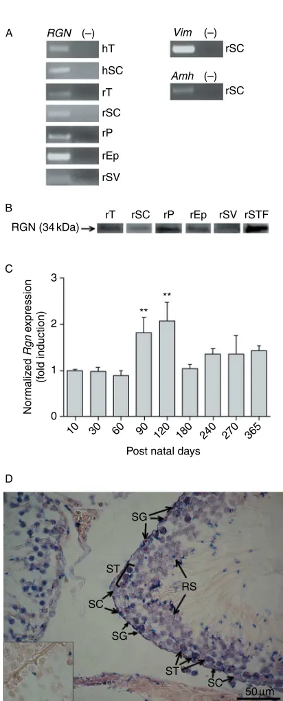
Analysis by RT-PCR demonstrated RGN mRNA
expression in rat and human testis (Fig. 1A). Through real-time quantitative PCR (qPCR) analysis at different postnatal ages it was shown thatRgnmRNA expression in rat testis reaches a maximum at 120 days, and decreases during aging process (Fig. 1C).
The localization of Rgn mRNA in rat testis was assessed using a specific digoxigenin-labeled probe (Fig. 1D). We were able to localize Rgn hybridization signal in SC and several germ cells, namely spermato-gonia (SG), spermatocytes (ST), and round spermatids (RS). Specificity of ISH staining was evaluated by hybridization with sense probe, which resulted in absence of signal (Fig. 1D insert).
In rat testis RGN protein was localized to LC and SC and also in a variety of germ cells, more specifically SG, ST, RS, elongating spermatids (ES), and spermatozoa (Sz; Fig. 2A and C). IHC in human testicular tissue showed a similar expression pattern for RGN, as observed by the staining of the same cell types as in rat testis (Fig. 2B and D). Although cell localization was essentially cytoplasmic, some nuclear staining is visible particularly in rat sections (Fig. 2A and C). Specificity of the IHC staining was assessed by omission of primary antibody, resulting in complete absence of immunological staining (inserts in corresponding panels,Fig. 2).
The expression of RGN mRNA and protein in SC was confirmed using rat and human primary SC cultures. PCR amplification of SC-specific markers vimentin (Vim) and anti-Mu¨llerian hormone (Amh; Fig. 1A), and ICC detection of VIM (Fig. 2G and H) were used to confirm the isolation of SC. After 96 h of culture contaminant cells
RGN
A (–)
rSC
rSC
rSC hT
rT hSC
rP
rEp
rSV
Vim (–)
Amh (–)
rSC rT RGN (34 kDa) B
C
D
SG
ST
SC
SG
RS
ST SC
50µm 3
2
Post natal days ** **
Nor
maliz
ed
Rgn
e
xpression
(f
old induction) 1
10 30 60 90 120 180 240 270 365 0
rSTF rP rEp rSV
Figure 1Expression of regucalcin (RGN) in reproductive tract tissues and seminiferous tubule fluid (STF). (A) RT-PCR of RGN, Sertoli cells (SC)-specific markers vimentin (Vim), and anti-Mu¨llerian hormone (Amh). (K), Negative control (no cDNA template). (B) Western blot detection of RGN in protein extracts. hT, human testis; rT, rat testis; hSC, human Sertoli cells; rSC, rat Sertoli cells; rP, rat prostate; rEp, rat epididymis; rSV, rat seminal vesicles; rSTF, rat seminiferous tubule fluid. (C) Expression ofRgnin rat testis at different postnatal ages, determined by quantitative PCR, normalized with cyclophilin A andb 2-micro-globulin as internal reference genes.nR5 in each group. **P!0.005 relative to ten postnatal days. Error bars representS.E.M. (D) Localization ofRgnmRNA transcript in adult rat testis byin situhybridization using a digoxigenin-labeled antisense probe. Insert – hybridization with sense probe resulting in no staining. SC, Sertoli cell; SG, spermatogonia; ST, spermatocyte; RS, round spermatid. Magnification indicated as scale bar.
AUTHOR COPY ONLY
were fewer than 5% for both cultures. By RT-PCR,RGN mRNA expression was detected in both rat and human SC (Fig. 1A). These cells also expressed RGN protein, which was detected mainly to the cytoplasm (Fig. 2E and F). Specificity of the immunostaining was assessed by the omission of the primary antibody, which resulted in complete absence of immunological staining (inserts in corresponding panels,Fig. 2). The presence of RGN in rat SC was further confirmed by WB analysis (Fig. 1B).
RGN expression in rat reproductive tissues and STF
The expression of RGN in other rat reproductive tissues besides testis was investigated. We detectedRgnmRNA expression in rat prostate, epididymis, and seminal vesicles (Fig. 1A). Subsequently, we confirmed the expression of RGN protein in these tissues by detecting
an immunoreactive band of expected size in WB (Fig. 1B). Moreover, we detected the same immuno-reactive band in rat STF (Fig. 1B).
The cellular localization of RGN in rat prostate, epididymis, and seminal vesicles was determined by IHC. In all three tissues RGN was localized mainly in the epithelial cells (Fig. 3A–C); in epididymis it was also localized to smooth muscle cells and connective tissue (Fig. 3B). Specificity of staining was assessed by omission of the primary antibody, which resulted in complete absence of staining (inserts in corresponding panels,Fig. 3).
DHT regulation of Rgn expression in rat SeT
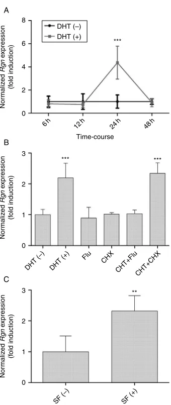
The effect of DHT (10K7M) on the expression of Rgn mRNA in cultured rat SeT was evaluated by qPCR. First, a time-course experiment was performed showing that DHT induced a sharp increase in the expression levels of Rgn at 24 h (4.37G0.64; P!0.001) compared with control (1.00G0.26), while at all other experimental timesRgnexpression levels remained similar to control group (Fig. 4A). The 24 h experimental time was therefore selected to explore the mechanisms involved in DHT regulation of Rgn expression by incubating rat SeT with 10K7M DHT, and with antiandrogen flutamide
(Flu) and protein synthesis inhibitor cycloheximide (CHX), alone or in presence of DHT (Fig. 4B). Treatment
with DHT produced an increase in Rgn mRNA
expression (2.20G0.27; P!0.01) compared with the control (1.00G0.08). Administration of Flu neutralized the hormone’s effect (1.04G0.06 vs 1.00G0.08 control), while incubation with CHX (2.34G0.15 vs 1.00G0.08 control;P!0.001) did not change the upregulating effect of DHT onRgnexpression. Incubation with Flu or CHX alone had no significant effect on the expression ofRgn.
Rgn expression is upregulated in rat SeT cultured in presence of survival factors
Rat SeT were cultured in a hormone-free medium with or without survival factors, and the expression of Rgn quantified by qPCR (Fig. 4C). Under survival-promoting conditions there was an upregulation ofRgnexpression (2.23G0.22 vs 1.00G0.23 without survival factors; P!0.005).
Discussion
Although several studies have highlighted the importance of Ca2C
homeostasis and signaling for normal spermato-genic process, a possible role of RGN in testicular physiology had not been explored. In this study, we report the expression and localization of RGN in rat and human testis and the effect of DHT on its expression.
Expression of RGN mRNA was detected in rat and human testicular tissue (Fig. 1A) and transcripts were
Sz
ES
ES
(–) (–)
SC
SC
SC
SC SG
SG
ST LC
RS
ST
ES
SC ST
SG ST
ST
ST
50µm
LC SC
A B
C D
E F
G H
(–) (–)
(–) (–)
10µm
100µm
Figure 2Immunochemical localization of regucalcin in testis paraffin-sections and Sertoli cell (SC) cultures. (A and C) Rat testis, (B and D) human testis, (E) rat SC, and (F) human SC. Purity of the rat and human SC cultures was assessed by staining with anti-vimentin antibody (G and H respectively). (K), Negative control obtained by omission of the primary antibody (inserts in corresponding panels). SC, Sertoli cell; LC, Leydig cell; SG, spermatogonia; ST, spermatocyte; RS, round spermatid; ES, elongated spermatid; Sz, spermatozoa. Magnification indicated as scale bar (B similar to A; D similar to C; F, G, and H similar to E).
AUTHOR COPY ONLY
localized both to somatic and germ cells in adult rat testis (Fig. 1D). A developmentally regulated expression pattern, where a peak is reached after which levels decrease with aging, has been described forRgn in rat kidney and liver tissues. In rat kidney the expression of Rgn mRNA starts to increase at 21 postnatal days and reaches a peak at 35 days, levels are maintained high until 3 months when it starts to decrease, returning to the low levels observed before 21 days (Fujita et al. 1996). In a comparable manner,Rgnexpression in liver increases until 10-day-old, reaching a plateau that is maintained until 6.5 months, decreasing in senescent rats (Fujitaet al. 1996). The authors hypothesized that the
age-dependent increase ofRgn expression in liver and kidney was coupled with periods of maturation and differentiation for both tissues and suggested it as a senescence marker (Fujitaet al. 1996). Developmental analysis shows that in rat testis the characteristic downregulation of Rgn expression during aging is also observed. The expression ofRgnmRNA increases until it reaches a maximum at 120 days of age, a period which corresponds to rat adulthood, decreasing afterwards with rat aging (Fig. 1C).
The results presented in this study demonstrate that RGN protein is broadly expressed in rat (Fig. 2A) and human (Fig. 2B) testis, being localized to all cell types of the SeT epithelium, somatic as well as germ cells. Relative intensity of RGN staining appears to be weaker in human sections, a pattern we have consistently observed when analyzing the localization of other proteins in human and rat testis sections (data not shown). We think that the use of different fixation protocols may be causing this histological artifact. RGN immunostaining is visible in cytoplasm as well as in nucleus, which is in accordance with reports showing that RGN is able to translocate to the nucleus regulating DNA synthesis and gene expression (Inagaki & Yamaguchi 2001,Tsurusaki & Yamaguchi 2004,Maia
et al. 2009). This is the first report describing RGN
expression and localization in testis of any vertebrate. Tight control of intracellular Ca2C
homeostasis has been shown to be of uttermost importance to LC steroidogenesis (Mannaet al. 1999,Pandeyet al. 2010) and maintenance of SC function (Franchi & Camatini 1985,Grimaet al. 1998,Gorczynska-Fjalling 2004). The deleterious effects of Ca2C channel blockers on male fertility emphasize even more the importance of tight Ca2C
regulation to spermatogenesis (Junejaet al. 1990, Benoffet al. 1994,Almeidaet al. 2000,Leeet al. 2006, 2011). The common cellular localization pattern observed in rat and human testis, together with the wide cellular distribution of RGN indicate a relevant role in testicular physiology suggesting that RGN may play a role in spermatogenesis as a Ca2C
homeostasis regulator in both somatic and germ cells.
Knowing that RGN was identified as a secreted protein in a pea aphid saliva (Carolanet al. 2009), and murine (Lv et al. 2007,2008), rat (Isogaiet al. 1994a,1994b), and human plasma (Lvet al. 2008) we decided to investigate its presence in STF, which could be confirmed by WB analysis (Fig. 1B). The STF is produced essentially due to the secretory activity of SC (Fisher 2002), which we demonstrated, by several approaches, to express RGN. Therefore, it is highly expected that RGN present in STF may be a secretion product of SC. Exogenous RGN has been shown to translocate to the nucleus being capable of altering gene expression and modulating enzyme activity in osteoblasts (Otomo & Yamaguchi 2006) and liver cells (Omura & Yamaguchi 1999). Nevertheless, to A
(–)
SM
50µm
CT
SM
(–)
(–) B
C
Figure 3Immunohistochemical localization of regucalcin in rat prostate (A), epididymis (B), and seminal vesicles (C). (K), Negative
control obtained by omission of the primary antibody (inserts in corresponding panels). Arrowhead, epithelium; CT, connective tissue; SM, smooth muscle cells. Magnification indicated as scale bar (similar in all panels).
AUTHOR COPY ONLY
this point, the role of SeT secreted RGN remains to be determined.
In addition to testis we also analyzed the expression and localization of RGN in other rat reproductive tissues: prostate, epididymis, and seminal vesicles (Fig. 3). This is the first time RGN expression and immunolocalization have been reported in rat epididymis and seminal vesicles. In seminal vesicles, RGN immunostaining is confined to epithelial cells, while in epididymis it is also present in connective tissue and smooth muscle cells.
In rat prostate RGN protein was detected in epithelial cells, which is in accordance with published results in rat and human prostatic tissue (Maia et al. 2008, 2009). RGN has been proposed to have a physiological function in prostate, as its expression is downregulated in prostate cancer tissues, and RGN immunoreactivity correlates with the grade of adenocarcinoma cellular differen-tiation (Maia et al. 2009). However, further studies are required to detail RGN function in these tissues.
Administration of 17b-estradiol (E2) to rats causes an
increase in the expression of Rgn mRNA in the liver (Yamaguchi & Oishi 1995). The same effect is observed in cultured rat hepatoma cells (Nakajima et al. 1999). Also, in osteoblastic cells, incubation with E2causes an
upregulation of Rgn expression, while treatment with 1,25-dihydroxyvitamin D3 causes downregulation of
Rgn expression (Yamaguchi et al. 2008). Contrarily, administration of E2 to rats decreases the expression of
Rgn in renal cortex (Kurota & Yamaguchi 1996). The hormonal regulation of Rgn expression has also been described in sex hormone target organs. Our group has described the downregulation of Rgn expression in rat prostate and mammary gland by E2(Maia et al. 2008).
Moreover,RGNis underexpressed in breast and prostate cancer cases and E2 upregulated while DHT
down-regulatedRGNmRNA expression in MCF-7 and LNCaP cell lines, respectively (Maia et al. 2009). DHT, a non-aromatizable androgen, has been shown to stimulate spermatogenesis in a similar way to testosterone (Singh
et al. 1995,Meachemet al. 2007,O’Shaughnessyet al.
2010), therefore it was used to analyze the effect of androgens onRgnexpression in rat SeT culturedex vivo. qPCR analysis showed that DHT upregulatesRgnmRNA expression in rat cultured SeT at 24 h of exposure (Fig. 4A). DHT upregulation of Rgn expression is completely reversed by incubation with antiandrogen Flu, but not with CHX, an inhibitor of protein synthesis (Fig. 4B). These data suggest that the involvement of a classical genomic mechanism of gene expression regulation through androgen receptor, which seems not to depend onde novoprotein synthesis.In silicoanalysis of the RGN promoter region has in fact enabled the identification of androgen response elements upstream from transcription initiation site at positions K906,
K915, K4126, and K5822 bp (Maia et al. 2009). Nevertheless, androgens are known to increase intra-cellular [Ca2C
] in a wide array of cells, namely SC (Gorczynska & Handelsman 1995), human prostatic stromal cells (Oliver et al. 2010), rat thoracic aorta (Montanoet al. 2008), and human lymphocytes (Popova
et al. 2007). It is also known that Rgn expression
is upregulated by increased [Ca2C
] (Shimokawa & Yamaguchi 1992, 1993). Therefore, we do not exclude that the DHT-induced rise in Rgn expression may be partly due to a secondary increase in intracellular Ca2C. Maintenance of spermatogenic epithelium homeo-stasis requires a fine-tuning between germ cell
A
Figure 4Effect of DHT and survival factors on regucalcin (Rgn) expression in rat seminiferous tubules (SeT) culturedex vivo determined by quantitative PCR.Rgnexpression was normalized with b-actin andGapdhas internal reference genes.nZ5 in each
experimental condition. Error bars representS.E.M. (A) Time-course experiment in which rat SeT were cultured in the absence [DHT (K)] or presence of 10K7M DHT [DHT (C)] for 6, 12, 24, or 48 h. (B) Rat SeT
AUTHOR COPY ONLY
proliferation and death. Apoptosis is an essential mechanism that enables the elimination of abnormal and exceeding germ cells and therefore its regulation is vital for normal spermatogenesis (Print & Loveland 2000). Up to 75% of germ cells undergo death by apoptosis in testis during the pubertal maturation process (Giampietri et al. 2005). On the other hand, androgens are known to inhibit apoptosis of male germ cells (Erkkila
et al. 1997, Pentikainen et al. 2000) and testosterone
withdrawal stimulates their apoptosis (Nandiet al. 1999, Tesarik et al. 2002). Germ cell apoptosis induced by androgen deprivation seems to be associated with caspases activation (Vera et al. 2006, Johnson et al. 2008). The role of RGN controlling apoptotic cell death has been established. RGN overexpression inhibits apoptosis induced by several factors, namely tumor necrosis factor-aand thapsigargin, dibucaine, and Bay K (Izumi & Yamaguchi 2004a, 2004b), and it was suggested that it may regulate AKT activity (Matsuyama
et al. 2004). In addition, hepatocytes of RGN knockout
mice are more susceptible to apoptotic cell death than their wild-type counterparts (Ishigami et al. 2002). We hypothesize that the upregulated expression of Rgn in SeT in response to DHT might be a mechanism by which androgens regulate apoptosis in testis. This is further supported by the observation that culture of SeT under hormone-free conditions although in presence of survival factors induces an upregulation of Rgn expression similar to the one caused by treatment with DHT (Fig. 4C).
In conclusion, we demonstrate that RGN is expressed in rat and human testis and other tissues of male reproductive tract, namely prostate, epididymis, and seminal vesicles. In addition, the presence of RGN in STF was identified. Our results also indicateRgnas a novel androgen-target gene in rat testis that may have an important role in the control of spermatogenesis. This opens new lines of research to explore the role of RGN and Ca2C homeostasis in male spermatogenic process and fertility.
Materials and Methods
Animals and tissues
Wistar male rats (Rattus norvegicus) were obtained from Charles River (Barcelona, Spain) and housed under a 12 h light:12 h darkness cycle, with food and water available ad libitum during the course of all experiments. Housing, maintenance and handling of animals was in compliance with the NIH guidelines and the rules for the care and handling of laboratory animals (Directive 86/609/EEC). All rats were killed under anesthesia (Clorketam1000, Vetoquinol, Lure, France).
Human testicular samples with conserved spermatogenesis were obtained by testicular sperm extraction from men undergoing treatment testicular biopsy due to obstructive azoospermia (aged 35–38, with normal karyotypes). Samples
were obtained under informed consent according to the local ethics committee guidelines, and treated as described pre-viously (Sousaet al. 2002a).
Reagents
All chemical reagents were purchased from Sigma–Aldrich and all antibodies were obtained from AbCam (Cambridge, UK) unless stated otherwise.
Primary SC culture
Testes were removed from 22-day-old rats and washed in ice-cold Hanks balanced salt solution without Ca2C
and Mg2C (HBSSf). Testes were decapsulated and washed in HBSSf. Rat SC were then isolated by a method adapted byOliveiraet al. (2009a). Briefly, decapsulated testicular tissue was placed in a glycine-containing medium (HBSSf, 1 M glycine (Merck), 0.005% (w/v) DNase, 2 mM EDTA and 0.002% (w/v) soybean trypsin inhibitor, pH 7.2) for 10 min at room temperature (RT) to remove peritubular cells. The dispersed tubules were forced through a large-bore Pasteur pipette and digested with 0.015% (w/v) type I collagenase and 0.005% (w/v) DNase in HBSSf for 20 min at RT. The SC suspension was collected by centri-fugation at 300gfor 3 min, washed three times in HBSSf and resuspended in SC culture medium (1:1 mix of Ham’s F12 and DMEM supplemented with 15 mM HEPES, 50 IU/ml penicillin, 50 mg/ml streptomycin sulfate, 0.5 mg/ml Fungizone, 50mg/ml gentamicin, and 5% (v/v) heat-inactivated FBS (Biochrom, Berlin, Germany)). The cell suspension was forced through a 20G needle, plated in culture flasks (CellC; Sarstedt, Nu¨mbrecht, Germany) at a concentration of 5000 clusters/ml and incubated at 378C in an atmosphere of 5% CO2:95% O2.
To obtain a culture of human SC, testicular biopsies were treated according to a method described by Sousa et al. (2002b). Isolation of SC was made by a method described elsewhere (Oliveiraet al. 2009b) with some modifications. The tubules were centrifuged at 500gfor 5 min and washed in HBSSf followed by another centrifugation. The pellet was redissolved in SC culture medium with a composition similar to the one used for rat SC culture except for the addition of 10% (v/v) heat-inactivated FBS, and cultured in a similar way.
STF collection
STF was collected from 90-day-old rats following a method described by Turner et al. (1984) with some modifications. Briefly, testes were removed and trimmed free of fat and connective tissue. A small incision was made at the caudal end of each testis, which was placed inside a syringe barrel within a centrifuge tube. The apparatus was centrifuged at 54g for 15 min at 08C to remove interstitial fluid. The testis was removed from the barrel, the tunica was cut and peeled back, and tubules were washed four times in saline to remove remaining interstitial fluid and blotted onto gauze. Tubules were extruded through the hub of a syringe into a centrifuge tube and centrifuged for 30 min at 08C. Supernatant containing STF was collected into a fresh tube.
AUTHOR COPY ONLY
Ex vivoculture of rat SeT
Rat SeT were used for culture instead of individual cell types, since this model has been shown to be suitable to mimic the testicular cellular environment ex vivo by several groups (Gilleron et al. 2009, Kaisman-Elbaz et al. 2009, Geoffroy-Siraudinet al. 2010). Testes (90-day-old rats) were removed, trimmed free of fat, washed in cold PBS and placed in DMEM-F12 culture medium at 328C. Tunica was cut and peeled back to expose tubules. Ten SeT fragments of about 1 cm and 2 ml of culture medium were used per well (Nunclon D 12 well multidishes; Nunc, Roskilde, Denmark). Experimental groups according to the medium composition were (nZ5 in each group): control, DHT (10K7M), Flu (10K7M), DHTCFlu
(10K7M DHT and 10K7M Flu), CHX (10mg/ml), and DHTC
CHX (10K7
M DHT and 10mg/ml of CHX). Control medium, to which DHT/Flu/CHX were added was DMEM-F12 supple-mented with 20 mg/l gentamicin sulfate, 0.1 mM 3-isobutyl-1-methylxanthine, and 1 mg/l BSA. In groups with DHT plus Flu/ CHX, the hormone was added 30 min after Flu/CHX. Tubules were incubated for 6, 12, 24, and 48 h in time-course experiments, and for 24 h in experiments with Flu and CHX. Tubules were also cultured in control medium supplemented with survival factors (10% (v/v) heat-inactivated FBS, 1 mM Na pyruvate, 4 mM glutamine, 100 ng/ml vitamin A, 200 ng/ml vitamin E, 50 ng/ml vitamin C, and 12mg/ml insulin) for 24 h. SeT remained viable during the course of the experiment as assessed by morphological analysis of hematoxylin-and-eosin-stained tissue sections. At the end of each experiment tubules were recovered from medium, snap–frozen in liquid nitrogen and stored atK808C until RNA isolation.
RNA isolation and cDNA synthesis
RNA was isolated from rat and human testis, rat SeT, rat prostate, rat epididymis, rat seminal vesicles, and rat and human SC with TRI reagent according to the manufacturer’s instructions. RNA concentration was measured in a
spectrophotometer (NanoPhotometer, Implen, Munich, Germany) and integrity was assessed by agarose gel electro-phoresis. Total RNAs were decontaminated from genomic DNA by digestion with DNase I (amplification grade) according to the manufacturer’s instructions. cDNA was synthesized in a final volume of 20ml using 160 IU M-MLV reverse transcriptase (Promega), 0.5mg random primers (Invitrogen), 10 mM each dNTP (GE Healthcare, Buckingham-shire, UK) and 1mg each RNA sample according to the protocol supplied by the manufacturer. Synthesized cDNA was stored at
K208C until further use.
RT-PCR
For the amplification of human and ratRGN, ratVimand rat
Amh specific intron-spanning primer sets were designed (Table 1). cDNA (1ml) was amplified in a final volume of 25ml containing 1! DreamTaq buffer with 20 mM MgCl2
(Fermentas, Burlington, Ontario, Canada), 0.5 IU of DreamTaq DNA polymerase (Fermentas), 10 mM each dNTP (GE Healthcare), and 0.2mM each primer (StabVida, Oeiras, Portugal). Every set of PCR included a no-template control.
In situhybridization
Detection ofRgnmRNA in rat testis (90-day-old) 4% PFA-fixed, paraffin-embeded sections was performed by hybridization with digoxigenin-labeled probes according to a protocol as described previously (Maiaet al. 2008).
Western blot
Total protein was isolated from rat SC, testis, prostate, epididymis, and seminal vesicles using RIPA buffer supple-mented with protease inhibitors (1 mM PMSF; 5 mM EDTA; 1! protease inhibitor cocktail). Protein content in STF was concentrated by acetone precipitation. Protein pellet was
Table 1PCR primer sequences, amplicon size, and cycling conditions.
Gene and accession numbers Primer sequences(50–30) Amplicon size(bp) Annealing temperature(8C)
Rat vimentin S: AGATCGATGTGGACGTTTCC 198 50
NM_031140.1 AS: TCCGGTATTCGTTTGACTCC
Rat anti-Mu¨llerian hormone S: GGCTGTGTTACAGGCTGACA 210 54
NM_012902.1 AS: GACTCTTGGACAGCCTCCAG
HumanRGN S: GCCTGTCCTACTCCGTGGATGC 143 55
NM_152869.2 AS: GGCCACCCAGAGCTTCCCCT
RatRgn S: TCAAAGACTGTCTGCCGATG 93 56
NM_031546.1 AS: GACTGTCGAAGTGCCACTGA
RatRgna S: GGAGGAGGCATCAAAGTG 155 60
NM_031546.1 AS: CAATGGTGGCAACATAGC
B-actina S: ATGGTGGGTATGGGTCAG 97 60
NM_031144.2 AS: CAATGCCGTGTTCAATGG
Gapdha S: GTTCAACGGCACAGTCAAG 115 60
NM_017008.3 AS: CTCAGCACCAGCATCACC
Cyclophilin Aa S: CAAGACTGAGTGGCTGGATGG 163 60
NM_017101.1 AS: GCCCGCAAGTCAAAGAAATTAGAG
B2-microglobulina S: CCGTGATCTTTCTGGTGCTTGTC 150 60
NM_012512.1 AS: CTATCTGAGGTGGGTGGAACTGAG
S, sense primer; AS, antisense primer. aPrimer pairs used in quantitative PCR.
AUTHOR COPY ONLY
dissolved in RIPA buffer with inhibitors. Protein concentration was determined by Bio-Rad protein assay.
Proteins (100mg) were mixed with sample buffer, denatured for 10 min at 1008C and resolved by SDS–PAGE on 12.5% gels. Proteins were blotted onto PVDF membranes (GE Healthcare) by wet transfer using 10 mM pH 11 CAPS with 20% (v/v) methanol. Blotted membranes were blocked with Tris-buffered saline with 0.05% (v/v) Tween-20 and 5% (w/v) dry skimmed milk for 1 h at RT and then incubated overnight with primary monoclonal anti-RGN antibody (1:250; ab81721). Membranes were incubated with secondary antibody conjugated with alkaline phosphatase (1:15 000; anti-mouse IgG–AP, ab7069), developed for 5 min with ECF substrate (GE Healthcare) and scanned with Molecular Imager FX (Bio-Rad).
Immunohistochemistry
Sections (5mm) of rat (90-day-old) testis, prostate, epididymis, and seminal vesicles (4% PFA-fixed) and human testis (10% formalin-fixed) were deparaffinized in xylene and rehydrated in graded alcohols. Heat-induced antigen retrieval was performed in 10 mM citric acid pH 6.0 for 30 min, at 80–858C. Endogenous peroxidase was blocked by incubating samples in 3% (v/v) H2O2 (Panreac, Barcelona, Spain) for
10 min at RT and unspecific staining was blocked by incubation with 1:20 normal goat serum for 30 min at RT. Sections were incubated overnight at 48C with primary monoclonal anti-RGN antibody (ab81721) diluted 1:50 in PBS with 1% (w/v) BSA (PBA). Sections were then incubated with secondary goat anti-mouse biotinylated antibody (ab7067) diluted 1:200 in PBA for 1 h at RT, followed by incubation with ExtrAvidin Peroxidase diluted 1:20 in PBA. Antibody binding was detected using HRP substrate solution (Dako, Glostrup, Denmark). Sections were slightly counter-stained with Harris’ hematoxylin (Merck), dehydrated, cleared, and mounted. Specificity of the staining was assessed by the omission of primary antibody.
Immunocytochemistry
Human and rat cultured SC were washed with PBS and fixed with cold 4% PFA. Permeabilization was performed by incubation with 0.01% (w/v) digitonin for 10 min at RT. Endogenous peroxidase was blocked by incubation with 0.1% (v/v) H2O2for 10 min at RT and unspecific staining was
blocked with PBA for 30 min at RT. Cells were incubated overnight at 48C with primary monoclonal anti-RGN antibody (1:50 in PBA, ab81721) or ready-to-use polyclonal anti-Vim antibody (V9 clone, Invitrogen). Cells were then incubated with secondary horse anti-mouse biotinylated antibody (BA-200; Vector Labs, Burlingame, CA, USA) diluted 1:400 in PBA for 1 h at RT, followed by incubation with ExtrAvidin Peroxidase diluted 1:20 in PBA. Antibody binding was detected using HRP substrate solution. After color development sections were slightly counterstained with Harris’ hematoxylin. Specificity of the staining was assessed by omission of primary antibody.
qPCR
Quantification of Rgn expression in rat cultured SeT and rat testis at different postnatal ages (10, 30, 60, 90, 120, 180, 240, 270, and 365 days, nR5 in each group) was performed by qPCR. An intron-spanning primer set was designed for the quantification of ratRgnexpression (Table 1). In addition two internal reference genes (b-actin andGapdhfor cultured SeT;
b2-microglobulin and cyclophilin A for rat testis at different postnatal ages) were selected from a panel for the normal-ization of the expression based on their stability in the experimental conditions used according to two methods described elsewhere (Vandesompele et al. 2002, Andersen
et al. 2004; data not shown). Reactions were carried out in an
iQ5 system (Bio-Rad) and efficiency of the reactions was determined for all primer sets using serial dilutions of cDNA samples (1:1, 1:10, and 1:100). Primer concentration and annealing temperature were optimized before the assay and specificity of the amplicons was determined by melting curve analysis. Reaction mixtures consisted of SYBR Green master mix (Bio-Rad), sense and antisense primers (500 nM for RGN and 200 nM for all other primer pairs), and 1ml of cDNA in a final volume of 20ml. Also, a no-template control was included for each reaction and all reactions were carried out in triplicate. Normalized expression values of Rgn were calculated according to a published mathematical model proposed by the Vandesompele group (Hellemanset al. 2007).
Statistical analysis
Statistical significance of differences in Rgn expression between groups was evaluated by one-way ANOVA followed by Bonferroni’s multiple comparison test or unpairedt-test with Welch’s correction, using GraphPad Prism v5.00 (GraphPad Software, San Diego, CA, USA). Statistically significant differences were considered for P!0.05. Experimental data are shown as meanGS.E.M.
Declaration of interest
The authors declare that there is no conflict of interest that could be perceived as prejudicing the impartiality of the research reported.
Funding
This work was supported by Foundation for Science and Technology (FCT; Grant numbers SFRH/BD/30173/2006 and SFRH/BD/60945/2009), Plurianual Program of FCT, and Program Cieˆncia 2008.
Acknowledgements
The authors thank Catarina Ferreira for technical assistance in tissue processing for histological analysis.
AUTHOR COPY ONLY
References
Almeida SA, Teofilo JM, Anselmo Franci JA, Brentegani LG & Lamano-Carvalho TL2000 Antireproductive effect of the calcium channel blocker amlodipine in male rats. Experimental and Toxicologic Pathology52
353–356.
Andersen CL, Jensen JL & Orntoft TF2004 Normalization of real-time quantitative reverse transcription-PCR data: a model-based variance estimation approach to identify genes suited for normalization, applied to bladder and colon cancer data sets.Cancer Research645245–5250. (doi:10.1158/0008-5472.CAN-04-0496)
Benoff S, Cooper GW, Hurley I, Mandel FS, Rosenfeld DL, Scholl GM, Gilbert BR & Hershlag A 1994 The effect of calcium ion channel blockers on sperm fertilization potential. Fertility and Sterility 62
606–617.
Carolan JC, Fitzroy CI, Ashton PD, Douglas AE & Wilkinson TL2009 The secreted salivary proteome of the pea aphid Acyrthosiphon pisum characterised by mass spectrometry.Proteomics92457–2467. (doi:10. 1002/pmic.200800692)
Erkkila K, Henriksen K, Hirvonen V, Rannikko S, Salo J, Parvinen M & Dunkel L 1997 Testosterone regulates apoptosis in adult human seminiferous tubules in vitro. Journal of Clinical Endocrinology and
Metabolism822314–2321. (doi:10.1210/jc.82.7.2314)
Fisher D2002 New light shed on fluid formation in the seminiferous tubules of the rat.Journal of Physiology542445–452. (doi:10.1113/jphysiol. 2002.018648)
Franchi E & Camatini M1985 Evidence that a Ca2C chelator and a
calmodulin blocker interfere with the structure of inter-Sertoli junctions.
Tissue & Cell1713–25. (doi:10.1016/0040-8166(85)90012-6)
Fujita T, Uchida K & Maruyama N1992 Purification of senescence marker protein-30 (SMP30) and its androgen-independent decrease with age in the rat liver.Biochimica et Biophysica Acta1116122–128. (doi:10.1016/ 0304-4165(92)90108-7)
Fujita T, Shirasawa T, Uchida K & Maruyama N1996 Gene regulation of senescence marker protein-30 (SMP30): coordinated up-regulation with tissue maturation and gradual down-regulation with aging.Mechanisms
of Ageing and Development 87 219–229. (doi:10.1016/0047-6374
(96)01711-3)
Geoffroy-Siraudin C, Perrard MH, Chaspoul F, Lanteaume A, Gallice P, Durand P & Guichaoua MR2010 Validation of a rat seminiferous tubule culture model as a suitable system for studying toxicant impact on meiosis effect of hexavalent chromium. Toxicological Sciences 116
286–296. (doi:10.1093/toxsci/kfq099)
Giampietri C, Petrungaro S, Coluccia P, D’Alessio A, Starace D, Riccioli A, Padula F, Palombi F, Ziparo E, Filippini Aet al.2005 Germ cell apoptosis control during spermatogenesis. Contraception72 298–302. (doi:10. 1016/j.contraception.2005.04.011)
Gilleron J, Carette D, Carpentier F, Segretain D & Pointis G 2009 Three-dimensional analysis of connexin 43 gap junction in theex vivo rat seminiferous tubules: short-term effects of hormonal effectors.
Microscopy Research and Technique72 845–855. (doi:10.1002/jemt.
20731)
Gorczynska E & Handelsman DJ1995 Androgens rapidly increase the cytosolic calcium concentration in Sertoli cells. Endocrinology 136
2052–2059. (doi:10.1210/en.136.5.2052)
Gorczynska-Fjalling E2004 The role of calcium in signal transduction processes in Sertoli cells.Reproductive Biology4219–241.
Grima J, Wong CC, Zhu LJ, Zong SD & Cheng CY1998 Testin secreted by Sertoli cells is associated with the cell surface, and its expression correlates with the disruption of Sertoli–germ cell junctions but not the inter-Sertoli tight junction. Journal of Biological Chemistry 273
21040–21053. (doi:10.1074/jbc.273.33.21040)
Hellemans J, Mortier G, De Paepe A, Speleman F & Vandesompele J2007 qBase relative quantification framework and software for management and automated analysis of real-time quantitative PCR data. Genome
Biology8R19. (doi:10.1186/gb-2007-8-2-r19)
Inagaki S & Yamaguchi M2001 Regulatory role of endogenous regucalcin in the enhancement of nuclear deoxyribonuleic acid synthesis with proliferation of cloned rat hepatoma cells (H4-II-E).Journal of Cellular
Biochemistry82704–711. (doi:10.1002/jcb.1193)
Ishigami A, Fujita T, Handa S, Shirasawa T, Koseki H, Kitamura T, Enomoto N, Sato N, Shimosawa T & Maruyama N2002 Senescence
marker protein-30 knockout mouse liver is highly susceptible to tumor necrosis factor-alpha- and Fas-mediated apoptosis.American Journal of
Pathology1611273–1281. (doi:10.1016/S0002-9440(10)64404-5)
Isogai M, Oishi K & Yamaguchi M1994aSerum release of hepatic calcium-binding protein regucalcin by liver injury with galactosamine adminis-tration in rats.Molecular and Cellular Biochemistry13685–90. (doi:10. 1007/BF00931609)
Isogai M, Shimokawa N & Yamaguchi M1994bHepatic calcium-binding protein regucalcin in released into the serum of rats administered orally carbon tetrachloride. Molecular and Cellular Biochemistry 131
173–179. (doi:10.1007/BF00925954)
Izumi T & Yamaguchi M 2004a Overexpression of regucalcin suppresses cell death and apoptosis in cloned rat hepatoma H4-II-E cells induced by lipopolysaccharide, PD 98059, dibucaine, or Bay K 8644.Journal of Cellular Biochemistry93598–608. (doi:10.1002/jcb. 20214)
Izumi T & Yamaguchi M2004bOverexpression of regucalcin suppresses cell death in cloned rat hepatoma H4-II-E cells induced by tumor necrosis factor-alpha or thapsigargin.Journal of Cellular Biochemistry92
296–306. (doi:10.1002/jcb.20056)
Johnson C, Jia Y, Wang C, Lue YH, Swerdloff RS, Zhang XS, Hu ZY, Li YC, Liu YX & Hikim AP2008 Role of caspase 2 in apoptotic signaling in primate and murine germ cells.Biology of Reproduction79806–814. (doi:10.1095/biolreprod.108.068833)
Juneja R, Gupta I, Wali A, Sanyal SN, Chakravarti RN & Majumdar S1990 Effect of verapamil on different spermatozoal functions in guinea pigs – a preliminary study. Contraception 41 179–187. ( doi:10.1016/0010-7824(90)90146-M)
Kaisman-Elbaz T, Sekler I, Fishman D, Karol N, Forberg M, Kahn N, Hershfinkel M & Silverman WF2009 Cell death induced by zinc and cadmium is mediated by clusterin in cultured mouse seminiferous tubules.Journal of Cellular Physiology220222–229. (doi:10.1002/jcp. 21754)
Kurota H & Yamaguchi M1996 Steroid hormonal regulation of calcium-binding protein regucalcin mRNA expression in the kidney cortex of rats.
Molecular and Cellular Biochemistry 155 105–111. (doi:10.1007/
BF00229307)
Lee JH, Kim H, Kim DH & Gye MC 2006 Effects of calcium channel blockers on the spermatogenesis and gene expression in peripubertal mouse testis. Archives of Andrology 52 311–318. (doi:10.1080/ 01485010600664024)
Lee JH, Ahn HJ, Lee SJ, Gye MC & Min CK2011 Effects of L- and T-type Ca(2C) channel blockers on spermatogenesis and steroidogenesis in the prepubertal mouse testis.Journal of Assisted Reproduction and Genetics
2823–30. (doi:10.1007/s10815-010-9480-x)
Lv S, Wei L, Wang JH, Wang JY & Liu F 2007 Identification of novel molecular candidates for acute liver failure in plasma of BALB/c murine model. Journal of Proteome Research 6 2746–2752. (doi:10.1021/ pr0701759)
Lv S, Wang JH, Liu F, Gao Y, Fei R, Du SC & Wei L2008 Senescence marker protein 30 in acute liver failure: validation of a mass spectrometry proteomics assay. BMC Gastroenterology 8 17. ( doi:10.1186/1471-230X-8-17)
Maia CJ, Santos CR, Schmitt F & Socorro S2008 Regucalcin is expressed in rat mammary gland and prostate and down-regulated by 17beta-estradiol. Molecular and Cellular Biochemistry 311 81–86. (doi:10. 1007/s11010-007-9697-x)
Maia C, Santos C, Schmitt F & Socorro S 2009 Regucalcin is under-expressed in human breast and prostate cancers: effect of sex steroid hormones.Journal of Cellular Biochemistry107667–676. (doi:10.1002/ jcb.22158)
Manna PR, Pakarinen P, El-Hefnawy T & Huhtaniemi IT1999 Functional assessment of the calcium messenger system in cultured mouse Leydig tumor cells: regulation of human chorionic gonadotropin-induced expression of the steroidogenic acute regulatory protein.Endocrinology
1401739–1751. (doi:10.1210/en.140.4.1739)
Matsuyama S, Kitamura T, Enomoto N, Fujita T, Ishigami A, Handa S, Maruyama N, Zheng D, Ikejima K, Takei Yet al.2004 Senescence marker protein-30 regulates Akt activity and contributes to cell survival in Hep G2 cells.Biochemical and Biophysical Research Communications
321386–390. (doi:10.1016/j.bbrc.2004.06.161)
AUTHOR COPY ONLY
Meachem SJ, Schlatt S, Ruwanpura SM & Stanton PG2007 The effect of testosterone, dihydrotestosterone and oestradiol on the re-initiation of spermatogenesis in the adult photoinhibited Djungarian hamster.
Journal of Endocrinology192553–561. (doi:10.1677/JOE-06-0136)
Montano LM, Calixto E, Figueroa A, Flores-Soto E, Carbajal V & Perusquia M 2008 Relaxation of androgens on rat thoracic aorta: testosterone concentration dependent agonist/antagonist L-type Ca2C
channel activity, and 5beta-dihydrotestosterone restricted to L-type Ca2C
channel blockade. Endocrinology 149 2517–2526. (doi:10.1210/en. 2007-1288)
Nakajima M, Murata T & Yamaguchi M 1999 Expression of calcium-binding protein regucalcin mRNA in the cloned rat hepatoma cells (H4-II-E) is stimulated through Ca2C
signaling factors: involvement of protein kinase C.Molecular and Cellular Biochemistry198101–107. (doi:10. 1023/A:1006996506238)
Nandi S, Banerjee PP & Zirkin BR1999 Germ cell apoptosis in the testes of Sprague–Dawley rats following testosterone withdrawal by ethane 1,2-dimethanesulfonate administration: relationship to Fas? Biology of
Reproduction6170–75. (doi:10.1095/biolreprod61.1.70)
Oliveira PF, Sousa M, Barros A, Moura T & Rebelo da Costa A 2009a Membrane transporters and cytoplasmatic pH regulation on bovine Sertoli cells.Journal of Membrane Biology 22749–55. (doi:10.1007/ s00232-008-9139-z)
Oliveira PF, Sousa M, Barros A, Moura T & Rebelo da Costa A2009b Intracellular pH regulation in human Sertoli cells: role of membrane transporters.Reproduction137353–359. (doi:10.1530/REP-08-0363)
Oliver VL, Anderson C, Ventura S & Haynes JM2010 Androgens regulate adenylate cyclase activity and intracellular calcium in stromal cells derived from human prostate. Prostate70 1222–1232. (doi:10.1002/ pros.21157)
Omura M & Yamaguchi M 1999 Effect of anti-regucalcin antibody on neutral phosphatase activity in rat liver cytosol: involvement of endogenous regucalcin. Molecular and Cellular Biochemistry 197
25–29. (doi:10.1023/A:1006979606225)
O’Shaughnessy PJ, Verhoeven G, De Gendt K, Monteiro A & Abel MH
2010 Direct action through the Sertoli cells is essential for androgen stimulation of spermatogenesis.Endocrinology1512343–2348. (doi:10. 1210/en.2009-1333)
Otomo Y & Yamaguchi M2006 Regulatory effect of exogenous regucalcin on cell function in osteoblastic MC3T3-E1 cells: involvement of intracellular signaling factor. International Journal of Molecular
Medicine18321–327.
Pandey AK, Li W, Yin X, Stocco DM, Grammas P & Wang X2010 Blocking L-type calcium channels reduced the threshold of cAMP-induced steroidogenic acute regulatory gene expression in MA-10 mouse Leydig cells.Journal of Endocrinology20467–74. (doi:10.1677/JOE-09-0206)
Pentikainen V, Erkkila K, Suomalainen L, Parvinen M & Dunkel L2000 Estradiol acts as a germ cell survival factor in the human testisin vitro.
Journal of Clinical Endocrinology and Metabolism 85 2057–2067.
(doi:10.1210/jc.85.5.2057)
Popova NY, Dukhanin AS & Shimanovskii NL2007 Nongenomic effect of androgens on Ca(2C) concentration in human lymphocytes.Bulletin of
Experimental Biology and Medicine 143 605–607. (doi:10.1007/
s10517-007-0193-9)
Print CG & Loveland KL 2000 Germ cell suicide: new insights into apoptosis during spermatogenesis.BioEssays22423–430. (doi:10.1002/ (SICI)1521-1878(200005)22:5!423::AID-BIES4O3.0.CO;2-0)
Shimokawa N & Yamaguchi M1992 Calcium administration stimulates the expression of calcium-binding protein regucalcin mRNA in rat liver.
FEBS Letters305151–154. (doi:10.1016/0014-5793(92)80884-J)
Shimokawa N & Yamaguchi M1993 Expression of hepatic calcium-binding protein regucalcin mRNA is mediated through Ca2C
/calmodulin in rat liver.FEBS Letters31679–84. (doi:10.1016/0014-5793(93)81740-Q)
Singh J, O’Neill C & Handelsman DJ1995 Induction of spermatogenesis by androgens in gonadotropin-deficient (hpg) mice. Endocrinology 136
5311–5321. (doi:10.1210/en.136.12.5311)
Sousa M, Cremades N, Silva J, Oliveira C, Ferraz L, Teixeira da Silva J, Viana P & Barros A2002aPredictive value of testicular histology in secretory azoospermic subgroups and clinical outcome after microinjec-tion of fresh and frozen–thawed sperm and spermatids. Human
Reproduction171800–1810. (doi:10.1093/humrep/17.7.1800)
Sousa M, Cremades N, Alves C, Silva J & Barros A2002bDevelopmental potential of human spermatogenic cells co-cultured with Sertoli cells.
Human Reproduction17161–172. (doi:10.1093/humrep/17.1.161)
Tesarik J, Martinez F, Rienzi L, Iacobelli M, Ubaldi F, Mendoza C & Greco E
2002In-vitroeffects of FSH and testosterone withdrawal on caspase
activation and DNA fragmentation in different cell types of human seminiferous epithelium.Human Reproduction171811–1819. (doi:10. 1093/humrep/17.7.1811)
Tsurusaki Y & Yamaguchi M 2004 Role of regucalcin in liver nuclear function: binding of regucalcin to nuclear protein or DNA and modulation of tumor-related gene expression.International Journal of
Molecular Medicine14277–281.
Turner TT, Jones CE, Howards SS, Ewing LL, Zegeye B & Gunsalus GL1984 On the androgen microenvironment of maturing spermatozoa.
Endocrinology1151925–1932. (doi:10.1210/endo-115-5-1925)
Vandesompele J, De Preter K, Pattyn F, Poppe B, Van Roy N, De Paepe A & Speleman F 2002 Accurate normalization of real-time quantitative RT-PCR data by geometric averaging of multiple internal control genes.
Genome Biology 3 RESEARCH0034. (
doi:10.1186/gb-2002-3-7-research0034)
Vera Y, Erkkila K, Wang C, Nunez C, Kyttanen S, Lue Y, Dunkel L, Swerdloff RS & Sinha Hikim AP 2006 Involvement of p38 mitogen-activated protein kinase and inducible nitric oxide synthase in apoptotic signaling of murine and human male germ cells after hormone deprivation. Molecular Endocrinology 20 1597–1609. (doi:10.1210/ me.2005-0395)
Yamaguchi M2005 Role of regucalcin in maintaining cell homeostasis and function (review). International Journal of Molecular Medicine 15
371–389.
Yamaguchi M & Kurota H1995 Expression of calcium-binding protein regucalcin mRNA in the kidney cortex of rats: the stimulation by calcium administration. Molecular and Cellular Biochemistry 146 71–77. (doi:10.1007/BF00926884)
Yamaguchi M & Oishi K1995 17 beta-Estradiol stimulates the expression of hepatic calcium-binding protein regucalcin mRNA in rats.Molecular
and Cellular Biochemistry143137–141. (doi:10.1007/BF01816947)
Yamaguchi M & Yamamoto T 1978 Purification of calcium binding substance from soluble fraction of normal rat liver. Chemical &
Pharmaceutical Bulletin261915–1918.
Yamaguchi M, Otomo Y, Uchiyama S & Nakagawa T2008 Hormonal regulation of regucalcin mRNA expression in osteoblastic MC3T3-E1 cells.International Journal of Molecular Medicine21771–775.
Received 20 March 2011 First decision 18 April 2011
Revised manuscript received 8 June 2011 Accepted 16 June 2011