Int. J. Trop. Vet. Biomed. Res. Vol. 2 (2) : 1-6; November 2017
www.jurnal.unsyiah.ac.id/IJTVBR E-ISSN : 2503-4715
The Administration of Epididymis Extract Increased the Testosterone Concentration without Affects the Dihydrotestosterone Concentration in Local Male Goat
Nanda Yuliansyah, Muslim Akmal, Tongku Nizwan Siregar, Sri Wahyuni, Mahdi Abrar, Syafruddin, Gholib and Farida Athaillah
Faculty of Veterinary Medicine, Syiah Kuala University, Banda Aceh.
Email for correspondence:[email protected];[email protected]
Abstract
This study was aimed to determine the effect of epididymis extract (EE) on the testosterone and dihydrotestosterone (DHT) level of local male goat. An experimental study was performed using a completely randomized design (CRD) pattern of one-way analysis of variance (ANOVA). 15 local male goats aged 1.5 years with body weight 14-16 kg were used in this study. The K0 group as a control group, injected with only 1 ml physiological saline, while each KP1, KP2, KP3, and KP4 groups treated with multilevel EE dose, ie 1, 2, 3, and 4 ml / goat for 13 consecutive days. At the end of treatment (day 14th), testes, epididymis (caput, corpus, and cauda)
and ductus deferens samples were taken through the close-castration method for examining the testosterone and DHT concentration by using enzyme-linked immunosorbent assay (ELISA) technique. Data gathered were later
analyzed using ANOVA followed by Tukey’s HSD in SPSS 16.0 for Windows. The result showed that the average
concentration of testosterone on K0, KP1, KP2, KP3, and KP 4 in testis respectively were 10.00±2.64 ng/ml; 7.66±2.51 ng/ml; 10.00±6.55 ng/ml; 0.66±0.57 ng/ml; 11.66±7.37 ng/ml; caput epididymis; 5.00±1.73 ng/ml; 2.33±1.52 ng/ml; 5.00±2.64 ng/ml; 1.33±0.57 ng/ml; 5.66±1.15 ng/ml; corpus epididymis; 1.33±0.57 ng/ml; 0.66±0.57 ng/ml; 4.00±2.64 ng/ml; 0.66±0.57 ng/ml; 4.33±2.30 ng/ml; cauda epididymis: 1.00±0.00 ng/ml; 0.66±0.57 ng/ml; 1.66±0.57 ng/ml; 1.00 ± 0.00 ng/ml; 2.00±1.73 ng/ml; ductus deferens: 3.66±2.51 ng/ml; 0.66±0.57 ng/ml; 3.00±1.00 ng/ml; 1.00±0.00 ng/ml and 3.66±1.15 ng/ml. While the average concentration of DHT on K0, KP1, KP2, KP3, and KP 4 in testis respectively; 10.00±2.64 ng/ml; 7.66±2.51 ng/ml; 10.00±6.55 ng/ml; 0.66±0.57 ng/ml; 11.66±7.37 ng/ml; caput epididymis; 5.00±1.73 ng/ml; 2.33±1.52 ng/ml; 5.00±2.64 ng/ml; 1.33±0.57 ng/ml; 5.66±1.15 ng/ml; corpus epididymis; 1.33±0.57 ng/ml; 0.66±0.57 ng/ml; 4.00±2.64 ng/ml; 0.66±0.57 ng/ml; 4.33±2.30 ng/ml; cauda epididymis: 1.00±0.00 ng/ml; 0.66±0.57 ng/ml; 1.66±0.57 ng/ml; 1.00 ± 0.00 ng/ml; 2.00±1.73 ng/ml; ductus deferens: 3.66±2.51 ng/ml; 0.66±0.57 ng/ml; 3.00±1.00 ng/ml; 1.00±0.00 ng/ml and 3.66±1.15 ng/ml. Statistical analysis showed that the administration of EE only increased testosterone concentration in testes had significant effect (P< 0.05). From this study, it can be concluded that the EE has the potential to improve spermatogenesis and sperm quality through increasing the testosterone concentration in the local male goats.
Key words : epididymis extract, testosterone, dihydrotestosterone, spermatogenesis, fertility
Background
Spermatogenesis im mammals is a complex process when the spermatogonia undergone differentiation in their step to produce spermatozoa to be released into tubule seminiferous lumen at stage VIII from the cycle of seminiferous epithel. The process is started from a spermatogonium and finalised with the production of 256 spermatozoa within 74 days in human and 58 days in mice (Mruk & Cheng, 2011).
Testosterone (androgen) is a hormon that very vital for spermatogenesis process (Ruwanpura et al., 2010). Androgen is a steroid hormon which important in
determining the expression of male phenotype, the development of secondary sex, initiation, and maintaining spermatogenesis (Wang et al., 2009a). Androgenis is one of family of main steroids hormone in human, they are progestogen, androgen, oestrogen, glucocorticoids, and mineralocorticoids. The group of androgen is conprising of four main hormon, they are dihydrotestosterone (DHT), testosterone,
androstenedione, and
failure for maturation of germinal cells, as well as spermiation, that finally resulted in impairment of spermatogenesis and infertility in male (Walker, 2010).
Dihydrotestosterone is a metabolite of testosterone (Meachem et al., 2007) and nature androgen that the most potent in human (Marchetti & Barth, 2013), that plays important role for the development of male reproductive tract (Ketelslegers et al., 1978). Nowadays, DHT is obtaining a high attention for research of male reproductive aspect, especially since the role of DHT for the development of of primary and secondary of sexual characteristic. Then, it is also importance for indicator of the development of initial prostate and breast cancer as well as for Alzheimer disease (Marchetti & Barth, 2013).
Testicular spermatozoa is functionally immature and infertile. During the transit of spermatozoa in epididymis, then resulted in a changes of specific maturity for their fertilitization. Among the changes are
morphology, motility, chemistry,
permeability, density, and metabolism (Cosentino & Cockett, 1986).
The latest research is showed that the epididymis containing proteins or molecules that important for spermatozoa maturity. Sipilä et al. (2009) was found G protein-coupled receptor 64 (GPR64), Human sperm-associated antigen 11e (SPAG11e), Cysteine-rich secretory proteins-1 (CRISP1), carbonyl reductase P34H, cluster of differentiation 52 (CD52), and Beta-defensin126 (DEFB126) in the epididymis. Then, Tanii et al. (2011) reported that the acrosome of spermatozoa is containing of pituitary adenylate cyclase-activating polypeptide (PACAP). Therefore, that means the epididymis is rich with PACAP and related with its role as the location for spermatozoa maturation.
Furthermore, Akmal et al., (2014) was found that the administration of Epididymis Extract (EE) could increased the concentration of testosterone, follicle stimulating hormone (FSH), and luteinizing hormone (LH) in the blood serum as well as the expression of follicle-stimulating hormone receptor (FSHR) in the tissue of tubuli seminiferi of testis (Akmal et al.,
2015a), followed by estrogen concentration (Akmal et al., 2016). Finally, it will affect to the increase of quality of the spermatozoa of male local goat (Akmal et al., 2015b). This research was aimed to study the effect of administration of EE to the increase of testosteron and DHT in the testis, caput, corpus, and cauda epididymis, as well as ductus deferens in male local goat.
Materials and Methods Location
This experiment was conducted in Lubok Sukon Village of Subdistrict of Ingin Jaya, District of Aceh Besar. The sample analysis was conducted in Research Laboratory of The Faculty of Veterinary Medicine of Syiah Kuala University, Darussalam, Banda Aceh.
Animal Model
Goats of age of 1.5 years old were used as animal model in this research. The average body weight of the animals was at 14-16 Kg. During the experimental period, the goats were given fresh forage and water ad lib.
Experiment Materials
The materials used in this study were comprised of plastic wrap, knife, centrifuge machine, gloves, mask, cool box, ice pack, hot plate, digital weighing, baker glass, label, multi tube vortexer, digital pippette, shaker, automatic pippette, measurement glass, staining jar, microtip, microtube, storage for organs, ELISA washer, ELISA reader, and computer package. The subtances were used comprised of NaCl 0,9%, aquadest, aquabidest, solution of phosphate buffered saline (PBS), testosterone ELISA Testosteron(e) DRG
EIA1559, DRG instruments Gmbh,
Marburg. Then, DHT ELISA DRG. EIA-4132, USA, which comprised of standart, enzyme conjugate, control, subtrate solution, stop solution (H2SO4), and wash solution.
Research Procedure
Preparation of Epidydimis Extract (EE).
follow: the testis of male local goat collected from the Abbatoir of City of Banda Aceh was taken to the Laboratory of Histology of The Faculty of Veterinary Medicine of Syiah Kuala University. Then, this sample was soaked in the water for ease in separation between the testis and epididymis. The obtained epididimys then was sliced to a smaller size and weighed. Then, it was grinded and added with aquabides of 10 ml per gram of epididymis. Next, it was filtered with a filter paper. The obtained solution of EE, then was centrifugated at a speed of 3000 RPM for 20 minutes. Finally, the supernatant was taken and stored into freezer.
Animal Treatment
This experiment were used a number of 15 male local goats of 1.5 years old. The average weight of them were at 14-16 kg. Their were divided into 5 groups, namely K0, KP1, KP2, KP3, and KP4. K0 as a control group was only given of 1 ml NaCl. Then, the groups of KP1, KP2, KP3, and KP4 were administered with different doses of EE at 1 ml, 2 ml, 3 ml, and 4 ml per goat for 13 days, consecutively. After completed the treatment, at day 14 all animals were castrated to collect testis, epididymis epididimis (capute, corpus, and cauda), and ductus defferens.
Procedure for Extraction of the Culture of Testis, Epididymis, and Ductus Defferens
Then, for all collected samples were
extracted and measured for the
concentration of testosterone and DHT using a method of enzyme-linked immunosorbent assay (ELISA). The procedure for extraction of of these samples was a modification of a method by Cusabio (2015). Firstly, the tissue from each sample was collected and taken at 100 mg, and homogenised as well as washed using 1 ml phosphate buffer saline (PBS) for one time. Next, the processed samples were stored into freezer at -20oC. Then, those samples were thawed using freezer thawing method for twice and centrifugated for 10 minutes using a speed of 1200 RPM at a temperature of 2-8oC. The obtained supernatants were stored at
-2oC. After that, the concentration of testosterone and DHT were measured.
Measurement of concentration of testosterone and DHT
The available sample were diluted using aqubidestillata at ratio of 1:4. Then, the standard solution of 0.2 ng/ml to 16 ng/ml were prepared and put into microplate ELISA well of 25 µL, followed by adding conjugate enzym, except to blank well. Next, all samples were covered with cling film and homogenized by a slow shaking for 10 seconds as well as incubated for 60 minutes under room temperature. Each microplate was washed using a solution of 300 µL for 3 to 4 times. The substrate of 200 µL was added into each of well and covered with cling film as well as incubated for 15 minutes under room temperature. The enzymatic reaction was stopped by adding a stop solution of 0.5 M H2SO4 at 100 µL to each of well. The reading of absorbance was conducted using ELISA at wavelength of 450 nm
Data Analysis
Data of measurement of estrogen concentration was analysed using analysis of varians (ANOVA). The analysis for significant different of treatment was continued with test of Tukey HSD. Both statistical analyses were conducted using program software SPSS 16.0 for windows.
Results and Discussion
The results of measurement of testosterone concentration in local male goats after treatment with EE is showed in Table 1. Then, the results of measurement for DHT concentration is in Table 2.
Table 1. Average (±SD) of
Treatment groups
Testis Caput
Epididymis
Corpus Epididymis
Cauda Epididymis
Ductus Deferens
K0 23.33 ± 15.94a 3.66 ± 3.51a 2.66 ± 2.50a 2.33 ± 2.08a 7.66 ± 2.50a
KP1 20.66 ± 14.84ab 3.00 ± 0.00a 3.66 ± 0.57a 3.33 ± 0.57a 3.00 ± 1.00a
KP2 19.33 ± 8.62ab 5.33 ± 1.52a 6.33 ± 3.21a 4.66 ± 0.57a 7.66 ± 2.88a
KP3 4.33 ± 3.21ab 3.00 ± 1.00a 4.00 ± 1.00a 3.66 ± 2.08a 4.33 ± 1.52a
KP4 65.00 ± 42.79c 5.33 ± 2.51a 5.66 ± 4.04a 4.00 ± 1.00a 8.66 ± 6.42a
Values with different superscript (a-c) in the same column differ significantly (p < 0.05).
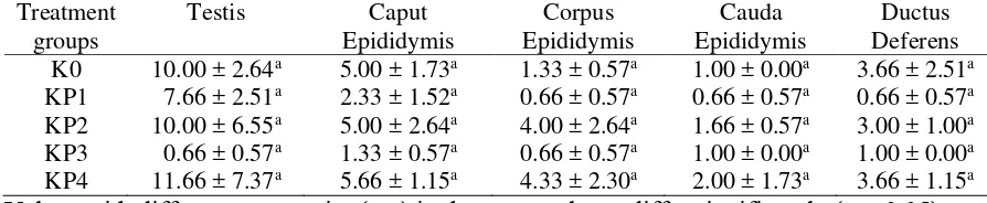
Table 2. Average (±SD) of DHT concentration (ng/ml) in the testis, epididymis, and ductus deferens after treatment with EE in local male goats.
Treatment groups
Testis Caput
Epididymis
Corpus Epididymis
Cauda Epididymis
Ductus Deferens
K0 10.00 ± 2.64a 5.00 ± 1.73a 1.33 ± 0.57a 1.00 ± 0.00a 3.66 ± 2.51a
KP1 7.66 ± 2.51a 2.33 ± 1.52a 0.66 ± 0.57a 0.66 ± 0.57a 0.66 ± 0.57a
KP2 10.00 ± 6.55a 5.00 ± 2.64a 4.00 ± 2.64a 1.66 ± 0.57a 3.00 ± 1.00a
KP3 0.66 ± 0.57a 1.33 ± 0.57a 0.66 ± 0.57a 1.00 ± 0.00a 1.00 ± 0.00a
KP4 11.66 ± 7.37a 5.66 ± 1.15a 4.33 ± 2.30a 2.00 ± 1.73a 3.66 ± 1.15a
Values with different superscript (a-c) in the same column differ significantly (p < 0.05).
It can be seen in the Table 1 that the treatment with EE was significantly increased testosterone concentration in the testis (P<0,05), but not in all part of epididymis and ductus deferens. However, the treatment of EE were not affect the concentration of DHT in all locations (P>0,05) (Table 2).
The increased testosterone
concentration after administration of EE in the goats in this study is in agreement with the report by Akmal et al. (2014). Perhaps, that effects of EE is related with its content of PACAP, which plays an important role regulation of steroidogenesis in Leydig cells to produce testosterone (El-Gehani et al., 1998; Yanaihara et al., 1998).
The continues production of testosterone is very important for spermatogenesis. Therefore, when the stimulation for testosterone is impaired, then will interrupt spermatogenesis process at stadium of meiosis. In turn, this condition will results in the death of germinal cells. The disturbance during testosterone secretion will cause a problem in spermiation process and spermatogenesis, which could induces infertility in man (Nieschlag et al., 1979; Walker, 2010).
Furthermore, testosterone as an androgen hormone is play a pivotal role in maintaining a continues production of
spermatozoa and the growt of secondary sex organ of prostate gland (Yong et al., 1998). It has been suggested by Verhoeven et al. (2010) that testis, as an organ that produces testosterone, is a target organ for androgen action. This action is importance for initiation of spermatogenesis and the growt of secondary sex organ (Zirkin et al., 1989).
Androgen is a family of steroidal hormones that comprised of DHT, testosterone, androstenedione, and dehydroepiandrosterone (Morales et al., 2004; Marchetti & Barth, 2013). DHT is a natural androgen that very potent (300%) in man (Wang et al., 2004; Marchetti & Barth, 2013). According to Wang et al. (2009b), the androgen action is mediated by androgen receptor (AR), which is the member of nuclear receptor superfamily. The function of AR is mediated by ligand-dependent transcription factor that regulated expression of androgen-responsive genes.
binding protein (ABP) as well as other proteins to enter lumen of seminiferus tubule. ABP is believed as a paracrice factor that refulated infranuclear, which is the location of enzyme 5α-reductase. This enzyme is found along of epididymis and controlled by androgen circulation (Robaire et al., 2006). Therefore, a high synthesis of DHT in the caudal epididymis is to support the circulation of androgen-dependent genes (Robaire et al., 2006).
However, in the present study, although the production of DHT was trend to increased, but it was not significant. A further study is needed to clarify this findings.
Conclusion
In conclusion, the administration of EE was increased the concentration of testosterone in the testis of local male goats.
Acknowledgments
We would like to grateful for all support from Directorate Research and Community Service of the Ministry of Research, Technology, and Higher Education of Republic of Indonesia, Research and Community Service Institute of Syiah Kuala University.
References
Akmal M, Siregar TN, Wahyuni S. 2014. Eksplorasi potensi ductus epididimis sebagai induktor peningkatan kualitas spermatozoa: upaya peningkatan populasi dan mutu genetik kambing lokal. Laporan Penelitian Hibah Tim Penelitian Pascasarjana Tahun I. Lembaga Penelitian Universitas Syiah Kuala, Darussalam, Banda Aceh.
Akmal M, Syafruddin, Abrar M. 2015a.
Eksplorasi potensi ductus epididimis sebagai induktor peningkatan kualitas spermatozoa: upaya peningkatan populasi dan mutu genetik kambing lokal. Laporan Penelitian Hibah Tim Penelitian Pascasarjana Tahun II. Lembaga Penelitian Universitas Syiah Kuala, Darussalam, Banda Aceh.
Akmal M, Siregar TN, Wahyuni S, Hambal M, Sugito, Amiruddin, Syafruddin, Roslizawaty, Zainuddin, Adam M, Gholib, Iskandar CD, Rinidar, Asmilia N, Hamny, Joharsyah, Suriadi, 2015b. Pemberian ekstrak
epididimis berpotensi meningkatkan kualitas sperma kambing lokal jantan. Jurnal Kedokteran Hewan 9(2): 168-173.
Akmal M, Siregar TN, Wahyuni S, Nasution MK, Abrar M, Syafruddin. 2016. Pemberian ekstrak epididimis berpotensi meningkatkan konsentrasi estrogen kambing lokal jantan. Pemakalah POSTER dalam Seminar Nasional “Seminar Hasil Penelitian dan Pengabdian kepada Masyarakat Tahun 2016” dengan Tema “Inovasi untuk Kedaulatan Pangan”. LPPM IPB, Bogor.
Cosentino MJ, Cockett AT. 1986. Structure and function of the epididymis. Urological Research 14(5):229-240. Cusabio. 2015. Fish Insulin-Like Growth
Factors 1 (IGF-1) ELISA Kit. http://cusabio.com/ELISA-Kit/Fish-InsulinINS-ELISA-Kit. 12 Januari 2016.
El-Gehani F, Tena-Sempere M, Huhtaniemi I. 1998. Vasoactive intestinal peptide stimulates testosterone production by cultured fetal rat testicular cells.
Molecular and Cellular
Endocrinology 140: 175-178.
Ketelslegers JM, Hetzel WD, Sherins RJ, Catt KJ. 1978. Developmental changes in testicular gonadotropin receptors: plasma gonadotropins and plasma testosterone in the rat. Endocrinology 103: 212-222.
Marchetti PM, Barth JH. 2013. Clinical biochemistry of dihydrotestosterone. Annals of Clinical Biochemistry 50:
95-107. DOI:
10.1258/acb.2012.012159.
Journal of Endocrinology. 192: 553-561.
Morales A, Buvat J, Gooren LJ, Guay AT, Kaufman JM, Tan HM, Torres LO. 2004. Endocrine aspects of sexual dysfunction in men. The Journal of Sexual Medicine 1: 69-81.
Mruk DD, Cheng CY. 2011. The
myotubularin family of lipid phosphatases in disease and in spermatogenesis. Biochemical
Journal 433: 253-262.
doi:10.1042/BJ20101267.
Nieschlag E, Wickings EJ, Mauss J. 1979. Endocrine testicular function in vivo and in vitro in infertile men. Acta Endocrinologica (Copenhagen) 90: 544-551.
Robaire B, Hinton BT, Marie-Claire Orgebin-Crist. The Epididymis. 2006. Knobil and Neill’s Physiology of Reproduction,Third Edition edited by Jimmy D. Neill, Elsevier © 2006. Chapter 22.
Ruwanpura SM, McLachlan RI, Meachem SJ. 2010. Hormonal regulation of male germ cell development. Journal of Endocrinology 205: 117-131. Sipilä P, Jalkanen J, Huhtaniemi IT,
Poutanen M. 2009. Novel
epididymal proteins as targets for the development of post-testicular male contraception. Reproduction 137: 379-389.
Tanii I, Aradate T, Matsuda K, Komiya A, Fuse H. 2011. PACAP-mediated sperm-cumulus cell interaction promotes fertilization. Reproduction 141: 163-171.
Verhoeven G, Willems A, Denolet E, Swinnen JV, De Gendt K. 2010.
Review: Androgens and
spermatogenesis: lessons from
transgenic mouse models.
Philosophical Transactions of the Royal Society B 365: 1537-1556. doi:10.1098/rstb.2009.0117.
Walker WH. 2010. Non-classical actions of testosterone and spermatogenesis. Philosophical Transactions of the Royal Society B 365: 1557-1569. doi:10.1098/rstb.2009.0258.
Wang C, Catlin DH, Demers LM, Starcevic
B, Swerdloff RS. 2004.
Measurement of total serum testosterone in adult men: comparison of current laboratory
methods versus liquid
chromatography-tandem mass
spectrometry. The Journal of Clinical Endocrinology & Metabolism 89: 534-43.
Wang RS, Yeh S, Tzeng CR, Chang C. 2009a. Androgen receptor roles in spermatogenesis and fertility: lessons from testicular cell-specific androgen receptor knockout mice. Endocrine Review. 30(2): 119-132.
Wang G, Weng CCY, Shao SH, Zhou W, De Gendt K, Braun RE, Verhoeven G, Meistrich ML. 2009b. Androgen receptor in Sertoli cells is not required for testosterone-induced suppression of spermatogenesis, but contributes to Sertoli cell organization in Utp14bjsd mice. Journal of Andrology 30: 338-348. (doi:10.2164/jandrol.108. 006890). Yanaihara H, Vigh S, Kozicz T,
Somogyvàri-Vigh A, Arimura A.
1998. Immunohistochemical
demonstration of the intracellular localization of pituitary adenylate cyclase activating polypeptide-like immunoreactivity in the rat testis using the stamp preparation. Regulatry Peptides 78:83-88.
Yong EL, Ghadessy F, Wang Q, Mifsud A, Ng SC. 1998. Androgen receptor transactivation domain and control of spermatogenesis. Reviews of Reproduction 3: 141-144.