THE USE OF ELECTRORETINOGRAPHY
AND OPTICAL COHERENCE TOMOGRAPHY
IN PATIENTS WITH SCHIZOPHRENIA
Din Duraković1, Ante Silić1,3, Vjekoslav Peitl1,3, Rašeljka Tadić2, Kristina Lončarić2, Trpimir Glavina1, Daniela Šago1, Ljiljana Pačić Turk3 and Dalibor Karlović1,3
1Sestre milosrdnice University Hospital Centre, Department of Psychiatry, Zagreb, Croatia;
2Sestre milosrdnice University Hospital Centre, Department of Ophthalmology, Zagreb, Croatia;
3Catholic University of Croatia, Zagreb, Croatia
SUMMARY – The use of electroretinography (ERG) and optical coherence tomography (OCT) has currently expanded beyond ophthalmology alone. The aim of this review is to present the results and knowledge acquired by these two methods in patients suffering from schizophrenia. Reviewing the studies applying ERG and OCT methods in the field of psychiatry, one can conclude that results of the research imply morphological and functional changes of retina in patients with schizophrenia that are not consistent. However, in most studies there was reduction of the amplitude and changes in the implicit time related parameters on ERG and thinning of the retinal nerve fiber layer on OCT. Neurons in the eye use the same neurotransmitters as neurons in the basal brain structures that are most affected in schizophrenia, according to the dopamine hypothesis of schizophrenia. Unlike neu-rons in the basal brain structures, the neuneu-rons in the eye are in vivo available to ERG. Using the aforementioned tests together with clinical diagnostic criteria of schizophrenia, the subgroups with different prognostic and therapeutic specificities within schizophrenia as a group of diseases might be identified more precisely.
Key words: Schizophrenia; Electroretinography; Optical coherence tomography; Neuroophthalmology
Correspondence to: Assoc. Prof. Ante Silić, MD, Sestre milosrdnice University Hospital Centre, Department of Psychiatry, Vinograd-ska c. 29, HR-10000 Zagreb, Croatia
E-mail: [email protected]
Received December 10, 2019, accepted May 14, 2020
Introduction
Schizophrenia is one of the 15 most common
dis-eases that lead to disability and working incapacity1. In
Croatia, there are more than 18,000 (registered) pa-tients with schizophrenia, and 1,000 are hospitalized
on a yearly basis2. Since schizophrenia begins slowly,
with gradual onset of symptoms, it often remains
un-recognized and untreated for a certain period of time3.
The period during which the patient manifests symp-toms of the disease and has not been treated is called
duration of untreated psychosis (DUP)4. It has been
proven that the sooner psychopharmacotherapy, psy-chotherapy and sociotherapy is started, the better is prognosis of the disease. Therefore, early detection of the disease is of crucial importance for long-term
prog-nosis and favorable outcomes of the disease5. Studies
have shown that early intervention in healthy individu-als at a high risk of disease development may reduce the incidence of comorbidities such as depressive thoughts, suicide attempts, and can generally prolong duration of remission of positive and especially negative
symp-toms5,6. Interventions consist of the use of
antipsychot-ics, but also other drugs, as well as various psychothera-peutic and sociotherapsychothera-peutic techniques. Although re-duction in the duration of DUP proved useful, it did
not reduce the incidence of the disease6.
The disease usually starts between the ages of 18 and 22, and the occurrence of the disease is equal in
both genders1. We still do not have clear pathoana-tomic, pathophysiologic and etiologic criteria for defi-nition of the disease. In other words, we are missing a clear biologic marker. Therefore, at present, the diag-nosis of schizophrenia is still made solely based on clinical findings and according to the currently used diagnostic, symptom-based criteria.
None of the currently available laboratory and physical (imaging) tests provides pathognomonic find-ing for schizophrenia. New studies on electroretinog-raphy (ERG) and optical coherence tomogelectroretinog-raphy (OCT) have opened the door to the research of the central nervous system in living patients with schizo-phrenia. Namely, the retina is an integral part of the central nervous system and is derived from the same tissue as the telencephalon, i.e. the ectoderm. It does not contain myelin and is available to visualization and investigation in vivo by noninvasive methods. The ax-ons of the retinal ganglion cells form the optic nerve through which the signal is passed to the central ner-vous system. Thus, there is a premise for investigating nervous system in living patients, with the possibility of defining specific biologic indicators of schizophre-nia by noninvasive methods such as ERG and OCT.
The light passes through the cornea and the lens, then through the vitreous body to the retina. In the retina, it first passes through the ganglion cells, then through the layers of the nuclei of the amacrine, bipo-lar and horizontal cells before reaching a layer of pho-toreceptors located on the outer part of the retina. There are two types of photoreceptors, i.e., rods (for black and white viewing and viewing in the dark) and cones (for viewing colors and viewing in the light con-ditions). Photochemical substances are found in the outer section of the photoreceptor, which are rhodop-sin in the rods and different types of photoprhodop-sin in the cones (selective sensitivity for red, blue or green, which is the basis of color vision).
From the photoreceptors, the signal is transmitted across horizontal and bipolar cells to ganglion cells and through the optic nerve, and visual pathways ending in the visual cortex located in the occipital lobe. When the rods are exposed to light, the resulting receptor potential is different from the receptor potentials in almost all other sensory receptors. It increases the negativity of membrane potential, causing hyperpolarization. Under normal conditions when the photoreceptor is not stim-ulated, the electrical potential is -40 mV, and at
maxi-mum stimuli hyperpolarizes it to -80 mV. The conduc-tion of most of the signals in the retina neurons occurs electrotonically. Electrotonic conduction, unlike an ac-tion potential, is a direct flow of electrical current in the neuron cytoplasm and in the axons from the site of stimulation to the synaptic output.
The retina is particularly interesting for investiga-tions into neurophysiology because many of its cells produce neurotransmitters. For example, photorecep-tors produce glutamate and amacrine cells produce at least eight types of neurotransmitters including GABA, glycine, dopamine, acetylcholine and indol-amine. Retinal ganglion cells produce glutamate,
so-matostatin and substance P7. Today, the applicability
of ERG and OCT has extended beyond its use exclu-sively in ophthalmology. In vivo biologic indicators as-sociated with schizophrenia are of great importance in studies of the pathogenesis, screening, and early diag-nosis of disease, as well as the course of disease and
response to treatment7.
The aim of this review is to present the results and knowledge in this area acquired by the application of ERG and OCT in schizophrenic patients.
Electroretinography
Electroretinography is a noninvasive diagnostic method that measures the outbreaks of electrical po-tential that arise in response to light in different cells within the retina. It is used in ophthalmology to diag-nose retinal disease (e.g., retinal dystrophies). The sig-nals that arise are generally of low intensity and are measured in nanovolts (nV) or microvolts (mV). There are several types of ERG: flash ERG (fERG), full field ERG (ffERG), multifocal ERG (mfERG) and pattern ERG (pERG).
The electrical potential generated by light stimula-tion consists of the negative a-wave, which reflects hy-perpolarization of the photoreceptors, positive b-wave that represents depolarization of bipolar and Müller cells in the retina, and c-wave as a positive wave that arises in response to stimulation of the pigment layer and the rods. Photopic negative response (phNR) is the negative potential that follows b-wave, and is cre-ated by ganglion cells. It occurs only in fERG testing. ERG records the amplitude of the individual waves, the implicit time (time from light stimulation to the peak amplitude of waves) and latency (time from the
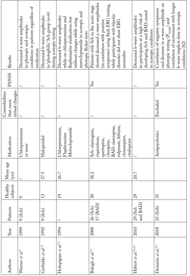
onset of stimulus to the onset of wave). Photopic vi-sion is vivi-sion in bright light conditions, which is neces-sary for color perception. Cone photoreceptors have the main role in recognizing this type of stimulation. Scotopic conditions are adaptation of vision in dark-ness, when rods are activated. Changes in the photore-ceptor function were recorded in the study by Warner et al.8. They analyzed ERG findings in nine patients with schizophrenia and nine healthy subjects, and showed changes in photoreceptor function in patients suffering from schizophrenia. Decreased amplitudes of a-waves were identified in patients regardless of the dose of antipsychotics. The authors attributed these re-sults to the absence of omega-3 fatty acids in the cell membrane of the retinal photoceptor, which caused impaired perception of light, as previously reported
from the study by Horrobin et al.9.The study was
con-ducted consequently to the research by Marmor et al., where difference in electrical potential between pa-tients with schizophrenia and control group was not
demonstrated10. The investigation by Warner et al.8 was
inspired by the observation reported by Gerbaldo et
al.11 in patients with schizophrenia who had a
tenden-cy of ‘photophilic activity’ (staring at the Sun) and re-duced sensitivity to light, with lower amplitudes of b-wave on ERG. Holopigian et al. examined the effect of the dopaminergic receptor blocker on the characteris-tics of fERG. The results showed a decrease in the
b-wave amplitude without changing the implicit time12.
Balogh et al. conducted studies on a sample of 63 middle aged subjects (26 patients with schizophrenia, 17 with bipolar affective disorder, and 20 healthy sub-jects as a control group), aiming to analyze light pro-cessing in the acute stage of the afore-mentioned
dis-eases using the ERG method13. The findings revealed a
decrease in the a-wave amplitudes in photopic condi-tions only in patients in the acute phase of schizophre-nia, and negative correlation between a-wave ampli-tudes and positive symptoms (using the Positive and Negative Syndrome Scale (PANSS) questionnaire). Negative symptoms, as well as the duration of treat-ment and doses of antipsychotics were not correlated with ERG indicators. The abnormality of the wave de-pended on the stage of the disease because normaliza-tion of the wave was observed after decrease of symp-tom intensity. Namely, after eight-week follow up and reduction in the intensity of positive symptoms of schizophrenia, there were no significant differences
between patients with schizophrenia and control group. ERG abnormalities were not found in the bipo-lar disorder group.
Hébert et al. found deviation from the normal find-ings for young, healthy individuals at a high risk of developing schizophrenia or bipolar affective disorder
using ERG14. In their study, population at a high risk
of developing schizophrenia was defined as having positive psychiatric heredity (one parent suffering from schizophrenia or bipolar disorder or multi-gen-eration heredity for these disorders). The study includ-ed 58 subjects, i.e., 29 subjects with positive herinclud-edity and 29 healthy controls without any known psychiatric heredity. Studies in the scotopic conditions recorded a significantly reduced b-wave amplitude in the high-risk group after correction for age, gender, season at the time of testing and heredity, either for
schizophre-nia or bipolar affective disorder14. This research is
par-ticularly important because it suggests that this meth-od can serve as an early and specific biomarker for the risk of developing these mental illnesses and may con-tribute to the noninvasive, rapid and simple screening, early treatment initiation, and more favorable disease
outcomes14. According to the concept of early
inter-ventions (within a period of up to 5 years from the onset of the disease), it is known that when the treat-ment is started earlier, better long-term outcomes can be achieved in terms of remission, recovery, and pa-tient functionality.
Unlike Balogh et al.13, who found a reduced a-wave
amplitude in patients with schizophrenia, Hébert et
al.14 did not prove this. Researchers believe that the
results varied due to a large sample of subjects with
bipolar disorder heredity in the study by Hébert et al.14.
The same researchers later confirmed reduction of a-wave amplitudes in photopic conditions and reduction of b-wave amplitudes in scotopic conditions in adult patients with schizophrenia, as indicated in their ear-lier study on healthy subjects at a high risk of develop-ing the disease. This was an ERG study with the larg-est sample to date (105 patients and 150 healthy
con-trols)15.
A group of American researchersused a RETeval
ERG device (LKC Technologies, Gaithersburg, MD, USA) to compare findings in 25 patients with
schizo-phrenia and 25 healthy adult subjects16. Individuals
function-Tab le 1. Elect ror etinog ra phy (E RG) r esear ch in ps ychiat ric p atients A uthors Year Patients Health y subjects Mean ag e (y rs) Medic atio n Co mor bidities that c ause retinal c hang es PANSS Results W ar ner et al . 8 1999 9 (S ch) 9 / Chlor pr omazine or no ne / / Decr eased a-wav e amplitudes in p
hotopic and scotopic
co nditio ns in patients r egar dless of medic atio n Ger baldo et al . 11 1992 9 (S ch) 13 37.5 Haloper idol / / Decr eased b-wav e amplitudes in ‘photop hilic ’ S ch gr oup (n=6) dur
ing scotopic testing
Holopigian et al . 12 1994 / 19 26.7 Chlor pr omazine , Flup henazine Metoc lopr amide / / Decr eased b-wav e amplitudes while o n c hlor pr omazine and flup henazine medic atio n, but without c hang es while using metoc lopr
amide in scotopic and
photopic flic ker tests Balogh et al . 13 2008 26 (S ch) 17 (B AD) 20 38.2 Sc h: olanz apine , risper ido ne , quetiapine , clo zapine , BAD: olanz apine , valpr oate , lithium, clo naz epam, citalopr am / Ye s Patients with S ch in the a cute stag e sho wed decr eased a-wav e amplitudes (in corr elatio n with positiv e sy mpto ms) using flash E RG testing , while par
ticipants with bipolar
disor
der did not sho
w E RG ano malies Héber t et al . 14,15 2010 29 (S ch and B AD) 29 20.7 / / / Decr eased b-wav e amplitudes in par ticipants at a high r isk of de veloping S ch and B AD , tested in scotopic co nditio ns Demmin et al . 16 2018 25 (S ch) 25 A ntipsy chotics Ex cluded Ye s Corr elatio n of negativ e sy mpto ms and decr ease in a-wav e amplitude o n photopic testing (P PhNR ) and corr elatio n of ex citement with lo ng er b-wav
e implicit time in scotopic
co nditio ns (S2) ER G= electr or etinogr ap hy ; y rs = y ears; P ANSS = P ositiv e and N egativ e S yndr ome S cale questio nnair e; S ch = sc hiz op hr enia; B AD = bipolar aff ectiv e disor der ; PPhNR = p hotopic testing
ing of the retina were not involved in the study. The PANSS questionnaire was used to evaluate the sever-ity of symptoms in patients with schizophrenia in the past two weeks.
The advantage of the RETeval ERG device is that it does not require dilation of the pupil or direct con-tact with the cornea. The device uses troland (Td) il-luminance that measures, adjusts to pupil size, and adjusts the intensity of brightness accordingly to en-sure that a constant number of photoreceptors is stim-ulated at any time. Subjects were investigated under photopic (cone activation) and scotopic (rod activated) conditions.
In the first photopic test (P1), a 100 TdˑS repetitive flash stimulation of 1 Hz frequency was used without background light. In the next photopic test (P2), stim-ulation with a flash of 100 TdˑS with background of 340 TdˑS under higher frequency (2 Hz) was used, and
in the third (PPhNR) test, red light of 58 TdˑS with blue
background of 380 TdˑS with an even higher frequen-cy (3.4 Hz) to trigger reaction of ganglion cells was used. The last photopic test was the Pf-flicker test that uses light flashing of 85 TdˑS at a frequency of 28.3 Hz to record the response of bipolar cells (used in psychi-atric studies for the first time). The scotopic tests con-sisted of white flashes of 2.8 TdˑS with a frequency 0.25 Hz for S1 test, 28 TdˑS 0.1 Hz for S2 test and 280 TdˑS with a frequency of 0.05 Hz for S3 test. All the scotopic tests were performed without background light.
In the group of patients with schizophrenia, the re-sults showed that a-wave had a decreased amplitude in
photopic tests P1 and PPhNR, while b-wave had a
de-creased amplitude in P1 and P2 tests. The a-wave also had a decreased amplitude in S3, while a decreased am-plitude of b-wave was found in S2 and S3 tests. Longer latency time of b-wave was observed in P2. In scotopic conditions, the implicit time for a-wave and b-wave was longer. Pf-flicker test showed a decrease in wave
ampli-tude in the patient group. PPhNR test showed a decrease
in the wave negativity 72 ms after stimulation.
Correlation of the results on PANSS was signifi-cant for the following parameters: negative symptoms
with decreased a-wave amplitudes (photopic PPhNR
test) and b-wave (scotopic) and with longer implicit time of a-wave (scotopic S1 test); negative symptoms with reduction of PhNR amplitude 72 ms after stimu-lus; and excitement symptoms with longer implicit
time in b-wave (S2). However, after false discovery rate corrections, statistically significant remained the correlation of negative symptoms with a-wave
ampli-tudes during photopic testing (PPhNR), and correlation
of excitement symptoms with the implicit time of b-wave in the scotopic conditions (S2). No correlation was demonstrated between the dose of antipsychotic pharmacotherapy and retinal response. Researchers believe that the correlation of negative symptoms and weakened response of the retina (reduced ERG ampli-tude) to stimulation could indicate the same rotransmitter pathophysiology based on the neu-rotransmitter hypothesis of schizophrenia, especially
the dopaminergic hypothesis16.
The results of these studies examining the charac-teristics and changes of ERG (a-wave and b-wave am-plitudes and implicit time) in patients with schizo-phrenia and the association of reductions in wave am-plitudes and changes of implicit time in correlation with stages of the disease are shown in Table 1. These results show a significant role of the ERG method in the investigation of pathophysiology of psychiatric disorders, especially schizophrenia and bipolar disor-der. Furthermore, it is a noninvasive method, which is well tolerated by adults and children, patients and con-trol group subjects. Further research should focus on better defining these indicators and examining the possible mechanisms underlying the ERG changes observed.
Optical Coherence Tomography
Optical coherence tomography is a diagnostic meth-od that uses low coherence light waves to create a high-resolution cross section image of the retina (1-10 μm). The device creates a picture by measuring the echo time delay of the light and intensity of the reflected light.
Coherence represents two waves that have a con-stant phase difference in the stage and the same fre-quency. A tomogram is a two-dimensional image that depicts the intersection of a three-dimensional image; by combining multiple tomograms, we get a three-di-mensional display. The advantage of the OCT is that it is a noninvasive and noncontact method that shows the real time condition and in vivo. Other advantages are that the examination price is relatively low, it is fast, and there is no contraindication for the application of this diagnostic technique.
The device first came into use in medicine in the early 1990s, and was used primarily in ophthalmology, to get an insight into the morphology of the posterior segment of the eye, usually for analysis of the morphol-ogy of the macula and the papilla of the optic nerve. It allows measurements of certain structures and possible pathologic changes in them, such as glaucoma, diabetic maculopathy and retinopathy, age-related macular de-generation, macular rupture and pseudorupture, central
serous maculopathy and epiretinal membranes17. Today,
its usefulness is recognized in other medical specialties, e.g., in interventional cardiology as a useful supple-mentary diagnostic device in percutaneous coronary
interventions (PCI)18, in dermatology in the diagnosis
of non-melanoma skin tumors and other inflammatory
skin diseases19,and in neurology where signs of some
diseases can be found in the retina (in Alzheimer’s
dis-ease, Parkinson’s disdis-ease, multiple sclerosis)20. Thinning
of the retinal nerve fiber layer (RNFL) has been dem-onstrated in patients with optic neuritis, multiple
scle-rosis, mild cognitive disorders21,and in patients with
Alzheimer’s disease22,where it correlates with the
se-verity of cognitive deficits.
Optical coherence tomography imaging in psychi-atric research including patients suffering from
schizo-phrenia began with Chu et al.23. These authors
con-ducted and published the first study of retinal changes using an OCT device in patients with schizophrenia and schizoaffective disorder. The study included 49 pa-tients and 40 healthy controls. Out of 49 papa-tients, 38 were diagnosed with schizophrenia and 11 with schizoaffective disorder. Forty patients had been pre-scribed antipsychotics, 10 antidepressants, four mood stabilizers along with antipsychotics, two only mood stabilizers, one had been prescribed hypnotic, and five patients were medication free. Control group consisted of 40 healthy age- and gender-matched individuals. Comparison of retinal thickness indicators between the groups was conducted using statistical multilevel analyses. Patients with schizoaffective disorder were found to have thinner RNFL in the right nasal quad-rant compared to people with schizophrenia, and the severity of positive symptoms was associated with a lower macular volume. This study was limited by low
resolution of the OCT device23.
Samani et al. used Leica Envisu TM SD-OCT high resolution device to analyze changes in macula
and fovea24. They examined 85 participants (35
diag-nosed with schizophrenia and 50 healthy control sub-jects). The PANSS questionnaire was used to evaluate the severity of symptoms in patients. The macula and the fovea (located inside the macula) are particularly interesting in research since it is the site of the sharpest vision dominated by photoreceptors (cones) and gan-glion cells.
The device was used to measure thickness of the following: RNFL; ganglion cell layer (GCL); inner plexiform layer (IPL); inner nuclear layer (INL); outer plexiform layer (OPL); outer nuclear layer (ONL); in-ner segmented layer (ISL); outer segmented layer (OSL); cone outer segment tips (COST); and retinal pigment epithelium (RPE). The derived measures in-cluded thickness of the following: whole retina = RN-FL-RPE; photoreceptor complex = ONL-COST; processing/processing complex = RNFL-OPL; and ganglionic cell complex = GCL+IPL. Sensitivity to contrast was also measured in 44 patients and 44 con-trols (selected depending on their Freiburg Acuity Test results) and showed spherical equivalent to be lower in patients. The results showed significant reduction in thickness of the photoreceptor complex in all regions, thinning of the OPL and ISL in patients with schizo-phrenia. In addition, reduction of contrast sensitivity was correlated with thinning of the temporal parafo-veal ganglion cell complex. The severity of negative symptoms was negatively correlated with thickness of the foveal photoreceptor complex and ONL.
Ascaso et al. examined 30 patients with
schizo-phrenia and 30 healthy controls25. The subjects were
examined by Time Domain OCT (TD-OCT) to es-tablish differences between the groups regarding the existence of a recent psychotic episode in the last month (recent illness episode, RIE) or the last six months (non-recent illness episode, NRIE) in relation to control. The incidence and intensity of symptoms were evaluated by the PANSS questionnaire. All pa-tients were treated with antipsychotics. Since this de-vice does not adjust to the pupil size, the pupils were dilated with 1% tropicamide. Thickness of the RNFL, thickness of the macula and macular volume were measured. All patients were found to have thinning of the peripapillary RNFL in all quadrants of both eyes. Macular thickness and macula volume were also re-duced in all patients in both eyes. Thickness of the macular inner ring, foveal thickness, and the volume of macula of the left eye were statistically significantly
re-duced. Comparison of the two patient subgroups (RIE and NRIE) yielded significant differences in the above-mentioned characteristics compared to the con-trol group, which were mainly identified in NRIE pa-tients. The authors claim that inflammation could be the reason why a significant difference was not found in the RIE subgroup, i.e., inflammatory processes in-crease thickness of the human retina, as previously
shown by Stock et al.26. Inflammatory processes have
been described in patients with schizophrenia, during acute episodes of schizophrenia, both in the first
psy-chosis episode and in chronic patients27,28. With the
use of multivariate regression models and after correc-tion for age, no significant link was found in this study between duration of the disease and thickness of the peripapillary RNFL, macular thickness and macular
density either in all subjects or in patient subgroups26.
Scientists from Kuala Lumpur examined the use of SD-OCT to analyze RNFL changes in patients with schizophrenia and their association with the duration
of the disease29. The intensity of symptoms was
as-sessed following the DSM-IV diagnostic criteria and using the PANSS questionnaire. Patients (N=30) with schizophrenia were divided into subgroups of acute, chronic and long-term chronic patients, and compared to 30 healthy age-, gender- and race-matched subjects. OCT was used to measure thickness of the peripapil-lary RNFL, average thickness of the macula, and vol-ume of the macula. Since there were no differences between the left and right eye, further research was continued solely examining the right eye of the sub-jects. Differences were found in RNFL thickness be-tween patients and healthy subjects. In patients with schizophrenia, significant differences in RNFL thick-ness were found in the peripapillary area of the retina in the superior, inferior and temporal quadrant, but not in the nasal quadrant. Significantly thinner macula in general, as well as thinning of the central part of the macula, inner and outer ring of the macula in patients compared to controls was also identified. When the inner and outer rings were divided into quadrants, a significant difference between the groups was demon-strated in all quadrants. The volume of the macula was also reduced in patients. The RNFL size and reduction in the volume of the macula were more significant in long-term chronic patients and were significantly re-lated to the duration of the disease. Such results indi-cate the value of the OCT method applied in
moni-toring of disease progression. However, correlation of the obtained parameters with rating of the PANSS questionnaire was not demonstrated. The authors at-tributed these results to the neurodegenerative nature
of the disease and to neurochemical dysregulation27. It
has been demonstrated that the dopamine neurotrans-mitter system plays a major role in visual functions
such as contrast sensitivity and color perception30.
Contrast sensitivity and color perception are affected in diseases with dopaminergic issues such as
Parkin-son’s disease, but also in schizophrenia31. Therefore,
thinning of the RNFL has been attributed to the
pos-sible dopamine dysregulation24,25.
Using the same method, Yilmaz et al.32 found
sta-tistically significant thinning of the RNFL also in the nasal quadrant in patients compared to healthy sub-jects. They analyzed the findings using SD-OCT in 34 patients with schizophrenia and 30 healthy individuals to determine changes in RNFL thickness. Thickness of the macula in the nasal quadrant and the inferior outer quadrant was also significantly thinner in pa-tients. The average macular thickness and thickness of the macula in superior external, superior internal, tem-poral external and temtem-poral internal, nasal internal and inferior interior quadrants were all reduced, but not significantly. The researchers attributed these changes to the neurodegenerative nature of schizophrenia.
Silverstein et al.33 examined the influence of
co-morbidities in patients with schizophrenia on the re-sults obtained using SD-OCT. Compared to the group of healthy subjects, there were no differences in RNFL thickness, macula and inner nuclear layer, and the de-termined thinning of the retinal structures was associ-ated with diseases such as diabetes mellitus and arte-rial hypertension in both groups of subjects. The study showed changes in the optic nerve head in patients with schizophrenia. The study also found an increase in cup-to-disc ratio of the optic nerve papilla, which was independent of comorbidities. Such results indi-cate the need for further research of the optic nerve head changes as a possible biomarker of schizophrenia.
Joe et al.34 also investigated the use of SD-OCT in
pursuit of biomarkers in psychosis. They studied macu-lar thickness and were among the first who studied thickness of the vascular layer supplying the retina, i.e., choroid. Six chronic psychiatric patients were involved (three with schizophrenia and three with bipolar dis-order) and 18 healthy age- and gender-matched
con-le 2. Optic al co he rence t omog ra phy (OC T ) r esear ch in p atients with sc hizop hr enia uthors Year Patients Health y par tici-pants Mean age (yrs) Medic atio n Co mor bidities that c an c ause retinal c hang es PANSS Results . 23 2012 38 (S ch) 11 (sc hiz o-aff ectiv e disor der) 49 29.7 40 used antipsy chotics,
4 used mood stabiliz
ers + antipsy chotics, 2 used mood stabiliz ers, 1 used noctur nal sedatio n, 5 no n-medic ated Ex cluded / Thinning of R NFL in al l patients Patients with sc hiz oaff ectiv e disor der ha d thinner R NFL in r ight nasal qua dr ant co mpar ed to patients with sc hiz op hr enia . 24 2017 35 (S ch) 50 40.6 A ntipsy chotics / Ye s Patients sho wed thinning of p hotor eceptor co mplex in al l r egio
thinning of outer plexif
or
m la
yer and inner segmented la
yer Decr eased co ntr ast sensitivit y corr
elated with thinning of tempor
par af ov eal co mplex of ganglio n cel ls The se ver ity of negativ e sy mpto ms was negativ ely r elated to thic kness of f ov eal p hotor eceptor co
mplex and outer plexif
or m la aso 25. 2015 20 (NR IE) (S ch) 10 (R IE) (S ch) 30 44.8 A ntipsy chotics Ex cluded Ye s Al l patients sho
wed thinning of per
ipapil lar y R NFL in al l qua dr ants of both e yes Ma cular thic kness and ma cular v olume w er e also decr eased in al l patients in both e yes Decr eased ma cular inner r ing , f ov eal thic kness and ma cular v olume in the lef t e ye w er e statistic all y signific ant . 29 2013 30 (S ch) 30 35.07 A ntipsy chotics Ex cluded Ye s Thinning of R NFL in the per ipapil lar y ar ea of the r etina in super ior , inf er
ior and tempor
al qua
dr
ants in patients
Thinning of the centr
al par
t of the ma
cula,
inner and outer ma
cular ring , as w ell as ma cular v olume decr ease w er e f ound in patients The amount of R
NFL thinning and decr
ease of ma
cular v
olume
corr
elated with disease dur
atio n . 32 2015 34 (S ch) 30 39.22 / Ex cluded / Thinning of R NFL in patients in al l qua dr ants Ma cular thic
kness in nasal qua
dr
ant and inf
er
ior outer qua
dr
ant
was also signific
antl y thinner ver- 33. 2018 32 (S ch) 32 40.9 A ntipsy chotics / Ye s The patient gr oup sho wed enlarg ed cup v
olume and enlarg
ed cup-to-disc r atio in both e yes These findings w er e unr elated to medic al co mor bidit y and w er e r elated to cognitiv e sy mpto ms CT = optic al co her ence to mogr ap hy ; y rs = y ears; P ANSS = P ositiv e and N egativ e S yndr ome S cale questio nnair e; S ch = sc hiz op hr enia; R IE = r ecent il lness episode; NR IE = no n-r ecent il lness episode; etinal ner ve fiber la yer
trols. As in previous studies, they found thinning of the macula, especially the inner ring of the macula (statistically significant), which they attributed to neu-rodegenerative changes. They also found thinning of the choroid in the individuals with psychosis, but it was not statistically significant, which they attributed to a small sample of study participants. The authors indicate the need for further research of these changes and their association with inflammation and degener-ative changes of the central nervous system.
From the above, we can conclude that the majority of studies using the OCT in patients with schizophre-nia determined thinning of the RNFL (Table 2). These results are undermined by methodologic limitations of the studies, such as relatively small samples and co-morbidities (e.g., diabetes and arterial hypertension) with possible influences on the retinal structures ex-amined.
Conclusion
Reviewing the research regarding the application of ERG and OCT methods in the field of psychiatry, one can conclude that the results of research conduct-ed imply morphological and functional changes in pa-tients with schizophrenia that are not consistent. However, in most studies, there was reduction in the amplitude and changes in the implicit time related pa-rameters in the ERG, and thinning of the RNFL in the OCT. Further, especially longitudinal prospective research is needed to define specific biologic indicators using these methods for the purpose of early screening, diagnostics and monitoring of patients in certain stag-es of the disease, and for therapeutic rstag-esponse in pa-tients. There is an obvious need for longitudinal pro-spective research in order to define specific biologic indicators using these methods for the possible early screening, differential diagnosis, monitoring of relapse and therapeutic response of patients in specific stages of the disease. The neurons in the eye are available in vivo, and they use the same neurotransmitters as the neurons in the basal brain structures that are most af-fected in schizophrenia according to the dopamine hypothesis of schizophrenia. The use of ERG and OCT, along with the established clinical diagnosis of schizophrenia can help define clear subgroups within schizophrenia as a group of diseases according to ERG and OCT findings. These subgroups would have
dif-ferent prognostic and therapeutic specificities35,36. We
can also expect a clear and defined biologic marker that would increase the accuracy of the disease diagno-sis and enable early initiation of treatment.
References
1. GBD 2016 Disease and Injury Incidence and Prevalence Collaborators. Global, regional, and national incidence, preva-lence, and years lived with disability for 328 diseases and inju-ries for 195 countinju-ries, 1990-2016: a systematic analysis for the Global Burden of Disease Study 2016. Lancet. 2017;390 (10100):1211-59. doi: 10.1016/S0140-6736(17)32154-2 2. Štrkalj Ivezić S, Jukić V, Štimac Grbić D, Ćelić I, Brečić P,
Silobrčić Radić M, et al. Organizacija liječenja oboljelih od mentalnih poremećaja u Republici Hrvatskoj. Acta Med Cro-atica. 2018;72:179-88. (in Croatian)
3. Šago, D, Babić, G. Roots of alexithymia. Arch Psychiatry Res. 2019;55(1):71-84. doi: 10.20471/may.2019.55.01.06
4. Karlović D, Silić A. Psychopathology. In: Karlović, D, Peitl, V, Silić A, editors. Schizophrenia. Zagreb: KBC Sestre milosrd-nice and Naklada Slap; 2019. p. 41-65. (in Croatian)
5. Kulhara P, Banerjee A, Dutt A. Early intervention in schizo-phrenia. Indian J Psychiatry. 2008;50(2):128-34. doi: 10.4103 /0019-5545.42402
6. Dama M, Shah J, Norman R, Iyer S, Joober R, Schmitz N, et al. Short duration of untreated psychosis enhances negative symp-tom remission in extended early intervention service for psy-chosis. Acta Psychiatr Scand. 2019;140(1):65-76. doi: 10.1111/ acps.13033
7. Kolb H. Neurotransmitters in the retina. In: Kolb H, Fernan-dez E, Nelson R, eds. Webvision: The Organization of the Retina and Visual System. University of Utah Health Sciences Center, 2009.
8. Warner R, Laugharne J, Peet M, Brown L, Rogers N. Retinal function as a marker for cell membrane omega-3 fatty acid depletion in schizophrenia: a pilot study. Biol Psychiatry. 1999; 45(9):1138-42. doi: 10.1016/s0006-3223(98)00379-5 9. Horrobin DF, Glen AI, Vaddadi K. The membrane hypothesis
of schizophrenia. Schizophr Res. 1994;13(3):195-207. doi: 10.1016/0920-9964(94)90043-4
10. Marmor MF, Hock P, Schechter G, Pfefferbaum A, Berger PA, Maurice R. Oscillatory potentials as a marker for dopaminergic disease. Doc Ophthalmol. 1988;69(3):255-61. doi: 10.1007/ bf00154406
11. Gerbaldo H, Thaker G, Tittel PG, Layne-Gedge J, Moran M, Demisch L. Abnormal electroretinography in schizophrenic patients with a history of sun gazing. Neuropsychobiology. 1992;25(2):99-101. doi: 10.1159/000118816
12. Holopigian K, Clewner L, Seiple W, Kupersmith MJ. The ef-fects of dopamine blockade on the human flash electroreti-nogram. Doc Ophthalmol. 1994;86(1):1-10. doi: 10.1007/ bf01224623
13. Balogh Z, Benedek G, Kéri S. Retinal dysfunctions in schizo-phrenia. Prog Neuropsychopharmacol Biol Psychiatry. 2008; 32(1):297-300. doi: 10.1016/j.pnpbp.2007.08.024
14. Hébert M, Gagné AM, Paradis ME, Jomphe V, Roy MA, Mérette C, et al. Retinal response to light in young nonaffected offspring at high genetic risk of neuropsychiatric brain disor-ders. Biol Psychiatry. 2010;67(3):270-4. doi: 10.1016/j.bio-psych.2009.08.016
15. Hébert M, Mérette C, Paccalet T, Émond C, Gagné AM, Sas-seville A, et al. Light evoked potentials measured by electroret-inogram may tap into the neurodevelopmental roots of schizo-phrenia. Schizophr Res. 2015;162(1-3):294-5. doi: 10.1016/j. schres.2014.12.030
16. Demmin DL, Davis Q, Roché M, Silverstein SM. Electroreti-nographic anomalies in schizophrenia. J Abnorm Psychol. 2018;127(4):417-28. doi: 10.1037/abn0000347
17. Mandić K, Vukojević N, Jukić T, Katušić D, Mandić JJ. Chang-es of Drusen number and central retinal thicknChang-ess in age relat-ed macular degeneration patients over two years. Acta Clin Croat. 2016;55:354-359. doi: 10.20471/acc.2016.55.03.02 18. Terashima M, Kaneda H, Suzuki T. The role of optical
coher-ence tomography in coronary intervention. Korean J Intern Med. 2012;27(1):1-12. doi: 10.3904/kjim.2012.27.1.1 19. Olsen J, Themstrup L, Jemec GB. Optical coherence
tomo-graphy in dermatology. G Ital Dermatol Venereol. 2015;150 (5):603-15.
20. Doustar J, Torbati T, Black KL, Koronyo Y, Koronyo-Hamaoui M. Optical coherence tomography in Alzheimer’s disease and other neurodegenerative diseases. Front Neurol. 2017;8:701. doi: 10.3389/fneur.2017.00701
21. Paquet C, Boissonnot M, Roger F, Dighiero P, Gil R, Hugon J. Abnormal retinal thickness in patients with mild cognitive im-pairment and Alzheimer’s disease. Neurosci Lett. 2007;420 (2):97-9. doi: 10.1016/j.neulet.2007.02.090
22. Iseri PK, Altinaş O, Tokay T, Yüksel N. Relationship between cognitive impairment and retinal morphological and visual functional abnormalities in Alzheimer disease. J Neurooph-thalmol. 2006;26(1):18-24. doi: 10.1097/01.wno.0000204645. 56873.26
23. Chu EM, Kolappan M, Barnes TRE, Joyce EM, Rona MA. A window into the brain: an in vivo study of the retina in schizo-phrenia using optical coherence tomography. Psychiatry Res. 2012;203(1):89-94. doi: 10.1016/j.pscychresns.2011.08.011 24. Samani NN, Proudlock FA, Siram V, Suraweera C,
Hutchin-son C, NelHutchin-son CP, et al. Retinal layer abnormalities as biomark-ers of schizophrenia. Schizophr Bull. 2018;44(4):876-85. doi: 10.1093/schbul/sbx130
25. Ascaso FJ, Rodriguez-Jimenez R, Cabezón L, López-Antón R, Santabárbara J, De la Cámara C, et al. Retinal nerve layer and macular thickness in patients with schizophrenia: influence of recent illness episodes. Psychiatry Res. 2015;229(1-2):230-6. doi: 10.1016/j.psychres.2015.07.028
26. Stock G, Ahlers C, Dunavoelgyi R, Kahraman G, Schauers-berger J, Schmidt-Erfurth U, et al. Evaluation of anterior-seg-ment inflammation and retinal thickness change following cataract surgery. Acta Ophthalmol. 2011;89(4):369-75. doi: 10.1111/j.1755-3768.2009.01704.x
27. García-Bueno B, Bioque M, Mac-Dowell KS, Barcones MF, Martínez-Cengotitabengoa M, Pina-Camacho L, et al. Pro-/ anti-inflammatory dysregulation in patients with first episode of psychosis: toward an integrative inflammatory hypothesis of schizophrenia. Schizophr Bull. 2014 Mar;40(2):376-87. doi: 10.1093/schbul/sbt001
28. Martínez-Gras I, Pérez-Nievas BG, García-Bueno B, Madrigal JL, Andrés-Esteban E, Rodríguez-Jiménez R, et al. The anti-in-flammatory prostaglandin 15d-PGJ2 and its nuclear receptor PPARgamma are decreased in schizophrenia. Schizophr Res. 2011;128(1-3):15-22. doi: 10.1016/j.schres.2011.01.018 29. Lee WW, Tajunisah I, Sharmilla K, Peyman M, Subrayan V.
Retinal nerve fiber layer structure abnormalities in schizophre-nia and its relationship to disease state: evidence from optical coherence tomography. Invest Ophthalmol Vis Sci. 2013; 54(12):7785-92. doi: 10.1167/iovs.13-12534
30. Cimmer C, Szendi I, Csifcsák G, Szekeres G, Ambrus Kovács Z, Somogyi I, et al. Abnormal neurological signs, visual con-trast sensitivity, and the deficit syndrome of schizophrenia. Prog Neuropsychopharmacol Biol Psychiatry. 2006;30(7): 1225-30. doi: 10.1016/j.pnpbp.2006.03.021
31. Fernandes TMP, Silverstein SM, Butler PD, Kéri S, Santos LG, Nogueira RL, et al. Color vision impairments in schizo-phrenia and the role of antipsychotic medication type. Schizophr Res. 2019;204:162-70. doi: 10.1016/j.schres.2018. 09.002
32. Yılmaz U, Küçük E, Ülgen A, Özköse A, Demircan S, Ulusoy DM, et al. Retinal nerve fiber layer and macular thickness mea-surement in patients with schizophrenia. Eur J Ophthalmol. 2016;26(4):375-8. doi: 10.5301/ejo.5000723
33. Silverstein SM, Paterno D, Cherneski L, Green S. Optical co-herence tomography indices of structural retinal pathology in schizophrenia. Psychol Med. 2018 Sep;48(12):2023-33. doi: 10.1017/S0033291717003555
34. Joe P, Ahmad M, Riley G, Weissman J, Smith RT, Malaspina D. A pilot study assessing retinal pathology in psychosis using optical coherence tomography: choroidal and macular thick-ness. Psychiatry Res. 2018;263:158-61. doiI: 10.1016/j.psy-chres.2018.03.011
35. Blažinović I, Orlović I, Karlović D, Peitl V. Comparison of clinical and sociodemographic characteristics of patients with schizophrenia treated stationary and at Day Hospital. Arch Psychiatry Res. 2019;55(2):127-37. doi: 10.20471/dec.2019. 55.02.02
36. Jurišić D, Ćavar I, Sesar A, Sesar I, Vukojević J, Ćurković M. New insights into schizophrenia: a look at the eye and related structures. Psychiatr Danub. 2020;32(1):60-69. https://doi. org/10.24869/psyd.2020.60
Sažetak
PRIMJENA ELEKTRORETINOGRAFIJE I OPTIČKE KOHERENTNE TOMOGRAFIJE U BOLESNIKA SA SHIZOFRENIJOM
D. Duraković, A. Silić, V. Peitl, R. Tadić, K. Lončarić, T. Glavina, D. Šago, Lj. Pačić Turk i D. Karlović
Primjena elektroretinografije (ERG) i optičke koherentne tomografije (OCT) danas nadilazi primjenu isključivo u oftal-mologiji. Cilj ovoga pregleda je prikazati rezultate i spoznaje dobivene primjenom ovih metoda u shizofrenih bolesnika. Pregledom dosadašnjih istraživanja primjene metoda ERG-a i OCT-a u području psihijatrije možemo zaključiti da rezulta-ti provedenih istraživanja morfoloških i funkcionalnih promjena rerezulta-tine u bolesnika sa shizofrenijom nisu konzistentni. Ipak, u većini istraživanja nalaze se smanjenje amplituda i promjene implicitnog vremena u ERG-u te stanjenje sloja mrežničnih živčanih vlakana na OCT-u. Kako se radi o neuronima dostupnima in vivo koji koriste iste neurotransmitere kao i neuroni u središnjim strukturama mozga koji su po dopaminskoj hipotezi shizofrenije najzahvaćeniji, primjenom spomenutih pretra-ga uz uobičajenu dijagnostiku shizofrenije možemo očekivati definiranje jasnijih podskupina unutar shizofrenije kao skupine bolesti koje bi imale različite prognostičke i terapijske specifičnosti.