Antioxidant responses of cucumber (
Cucumis sati
6
us
) to
photoinhibition and oxidative stress induced by norflurazon under
high and low PPFDs
Sunyo Jung
a,*, Jin Seog Kim
a, Kwang Yun Cho
a, Gun Sik Tae
b, Bin G. Kang
caScreening Research Di6ision,Korea Research Institute of Chemical Technology,P.O.Box107,Yusung,Taejon305-600,South Korea bDepartment of Biology,Dankook Uni6ersity,Chonan330-714,South Korea
cDepartment of Biology,Yonsei Uni6ersity,Seoul120-749,South Korea
Received 3 May 1999; received in revised form 8 November 1999; accepted 3 December 1999
Abstract
Photooxidative damage is exacerbated by norflurazon (NF), which blocks carotenoid biosynthesis. This study examined the influence of photosynthetic photon flux density (PPFD) on the overall responses of both non-enzymatic and enzymatic antioxidants to NF-caused oxidative damage in leaves of cucumber (Cucumis sati6us). Seven-day-old cucumber plants were exposed to NF under either low PPFD (30mmol m−2s−1) or high PPFD (300mmol m−2s−1) for 3 days. The NF plants exposed at high PPFD had lower levels ofFv/Fmratio, quantum yield of electron transport, and 33-kDa protein of photosystem II as compared with the NF plants at low PPFD. In the NF plants, there was a reduction in total chlorophylls and carotenoids except newly formed zeaxanthin in either PPFD. The NF plants at high PPFD resulted in less level of photochemical quenching,qP, and Stern – Volmer quenching, NPQ, than those of the plants at low PPFD, whereas both plants had similar level of non-photochem-ical quenching coefficient, qN. However, the level of PPFD did not significantly affect the NF-caused induction of antioxidant enzymes including peroxidase, superoxide dismutase, glutathione reductase, and ascorbate peroxidase. © 2000 Elsevier Science Ireland Ltd. All rights reserved.
Keywords:Antioxidant enzymes; Cucumber (Cucumis sati6us); Non-photochemical quenching; Norflurazon; Oxidative stress; Xanthophylls www.elsevier.com/locate/plantsci
1. Introduction
Photoinhibition and photooxidation can occur when plants are exposed to stress. High light in synergy with other stress factors such as chilling, drought, or low carbon dioxide supply reduces the capacity of photosynthetic systems to utilize inci-dent radiation, leading to a photoinhibition pro-cess [1,2]. The photosynthetic electron transport system is the major source of active oxygen species (AOS) in plant tissues [3], having the potential to generate singlet oxygen (1O
2) and superoxide (O2−),
which is favored under downregulation of
metabolic pathways. Photosystem (PS) II has long been considered the primary target for photoinhi-bition [1,2] because PSI is more stable than PSII during strong light treatments [4]. Light-induced inactivation of PSI is suggested to be caused by AOS [5,6]. AOS are eliminated efficiently by an integrated system of non-enzymatic and enzymatic antioxidants that are concentrated in the chloro-plast [3]. The capacity of the antioxidative defense system is increased under adverse stress conditions but the imbalance between AOS production and antioxidant defenses ultimately leads to oxidative damage.
The non-enzymatic reductants consist of ascor-bate, glutathione, a-tocopherol, caroteniods, and phenolic compounds [7,8]. Among those, xantho-phyll cycle-dependent energy dissipation in the
* Corresponding author. Present address: Department of Genetics, North Carolina State University, Raleigh, NC 27695-7614, USA. Tel.: +1-919-5155819; fax:+1-919-5153355.
E-mail address:[email protected] (S. Jung)
light-harvesting antennae is thought to play an important photoprotective role by mitigating oxi-dative damage. Non-radiative energy dissipation at PSII is mediated by zeaxanthin and perhaps also by antheraxanthin [9] and is proposed to occur at several sites within or around the PSII reaction center [10]. Researchers have used differ-ent terms for energy dissipation, e.g. the quench-ing coefficient qN [11] versus Stern – Volmer
quenching [9,12] that is referred to as non-photo-chemical quenching (NPQ).
In the most powerful source of AOS, chloro-plasts, O2− that is produced by photoreduction of
O2 at PSI and PSII is detoxified by the
Mehler-peroxidase pathway [3,13]. The O2− is reduced to
hydrogen peroxide (H2O2) by superoxide
dismu-tase (SOD) and then to H2O by ascorbate
peroxi-dase (APX), which is the key enzyme involved in H2O2 scavenging. These enzymes, together with
monodehydroascorbate reductase, dehydroascor-bate reductase, and glutathione reductase (GR), constitute the major defense system against AOS in the chloroplast [3,14]. Additionally, extraplastic H2O2 quenching by peroxidase (POD) and
cata-lase (CAT) is also increased in stress responses [15]. A crucial role of these enzymes in protection against oxidative processes has been shown in transgenic tobacco plants overexpressing either Mn-SOD or Fe-SOD [16,17]. In contrast, other studies with transgenic plants suggest that en-hancement of a particular antioxidant enzyme does not lead to increased protection [18,19].
Photooxidative damage is exacerbated by herbi-cides, which generate AOS either by direct in-volvement in radical production or by inhibition of biosynthetic pathways [20,21], as well as atmo-spheric pollutants and heavy metals [8]. Enhanced activities of antioxidants were associated with re-sistance to herbicides such as paraquat and oxyfluorfen [22,23]. In the present study a potent
herbicide norflurazon (NF), which blocks
carotenoid biosynthesis by non-competitively
binding to phytoene desaturase [24], was used in leaves of Cucumis sati6us. It eliminates important quenchers of the triplet chlorophyll (Chl) and1O
2,
thus initiating photooxidative processes. To assess contribution of each inductive response of antioxi-dant to overall protective strategies to NF-caused oxidative damage, photochemical efficiency of PSII, composition of photosynthetic pigments, quenching parameters and activities of antioxidant
enzymes were determined. The questions of whether the level of photosynthetic photon flux density (PPFD) influences the NF-caused oxida-tive stress was also examined, and, if so, whether the NF plants at different PPFDs have different capacities to develop antioxidant responses.
2. Materials and methods
2.1. Plant material and growth conditions
Cucumber seeds (C.sati6usL. cv Summer Long) were sown in vermiculite and transferred after 4 days into synthetic soil in plastic pots. Plants were grown in a controlled environment growth cham-ber under a temperature of 25°C, a 16-h photope-riod, and a light intensity of 200 mmol m−2 s−1
for 3 days. For NF treatment the 7-day-old plants were exposed in a surface application to 15 mM. When the treatment was initiated the first leaves were about to emerge. Following NF application plants were immediately returned to the growth chamber and exposed for 3 days under a 16-h photoperiod with an irradiance of either low PPFD (30 mmol m−2 s−1) or high PPFD (300 mmol m−2 s−1) at 25°C. Inhibition of carotenoid
biosynthesis causes a characteristic bleaching of newly developed leaves. The four treatments em-ployed were: (1) CH, control/high PPFD; (2) NH, NF treatment/high PPFD; (3) CL, control/low PPFD; and (4) NL, NF treatment/low PPFD. The first leaves were used for the measurements of Chl fluorescence, pigment contents and enzyme activi-ties. The experiments were triplicated each with three determinations.
2.2. Chl a fluorescence measurements
In vivo Chla fluorescence was measured after 5 min dark-adaptation at room temperature using a pulse amplitude modulation fluorometer
(PAM-2000, Walz, Effeltrich, Germany). Minimal
fluorescence yield, F0, was obtained upon
excita-tion with a weak measuring beam from a pulse light-emitting diode. Maximal fluorescence yield,
Fm, was determined after exposure to a saturating
pulse of white light to close all reaction centers. Determination of the quenching components qP
and qN was conducted by the saturation pulse
Schreiber et al. [25]. The quantum yield of electron transport through PSII (Y=DF/Fm%) was
calcu-lated according to Genty et al. [26].
Non-photo-chemical fluorescence quenching was also
quantified, as previously done by Bilger and Bjo¨rkman [12] according to the Stern – Volmer equation, NPQ=Fm/Fm% −1, where Fm% is the
low-ered maximal yield during illumination with pho-tosynthetically active radiation.
2.3. Immunoblot analysis
For the immunoblot of the extrinsic 33-kDa protein of the oxygen-evolving complex in PSII reaction center, the method of Tae et al. [27] was used. The thylakoid membranes isolated from chloroplasts were resuspended in 10 mM NaCl, 50 mM sucrose, and 50 mM sodium phosphate buffer, pH 7.4 and were sedimented at 10 000×g
for 10 min. The pellets were resuspended in the same buffer as mentioned above. The chlorophylls were removed with 80% acetone and the protein pellets were solubilized (1% SDS, 8 M urea, 1% 2-mercaptoethanol, 10.7 mM phosphoric acid). The protein concentrations were measured with the UV spectrophotometric method. Samples of protein to be blotted were electrophoresed in 12% SDS-polyacrylamide gel. The gel was run in 192 mM Glycine, 0.01% (w/v) SDS, and 25 mM Tris – HCl, pH 8.3. Polypeptides were transferred to nitrocellulose paper (pore size: 0.45 mm; Hybond-C, Amersham) with a semi-dry transfer blotter (130-mA constant current, 60 min) (Model TE70, Hoefer Scientific Instruments). The paper was washed in TBS buffer (500 mM NaCl, 20 mM Tris – HCl, pH 7.4), incubated in a sealed plastic bag with 10% milk casein on a rocking shaker (RK1020) for 2 h, removed from the bag, and washed in TBS buffer. After incubating with the antibody in TBS buffer containing 3% bovine serum albumin (BSA) for 2 h, and washing in TTBS buffer containing 0.05% Tween-20, 500 mM NaCl, 20 mM Tris – HCl, pH 7.4, the paper was again incubated with a second antibody [goat anti-rabbit IgG conjugated with horseradish peroxidase (Bio-Rad)] in TBS containing 3% BSA. The paper was incubated on a rocking shaker for 1 h, washed in TTBS buffer, and stained for 10 min with 0.017% 4-chloro-1-naphthol and hydrogen perox-ide in TBS buffer.
2.4. Pigment extraction and analysis
Extraction and HPLC anaysis of carotenoids and Chls were done as described previously [28].
2.5. Extraction of soluble protein
Frozen leaves (0.25 g for CAT, GR, and APX; 0.5 g for POD and SOD) were crushed to fine powder in a mortar under liquid N2. Soluble
proteins were extracted by homogenizing the pow-der in 2 ml of 100 mM potassium phosphate buffer, pH 7.5, containing 2 mM EDTA, 1% PVP-40, and 1 mM PMSF. For analysis of APX, the extraction buffer also contained 5 mM ascor-bate. Insoluble material was removed by centrifu-gation at 15 000×g for 20 min at 4°C, and the supernatant was filtered through filter papers No. 1 (Whatman, Maidstone, UK). Since maintenance of consistent CAT electrophoretic mobility and GR activity was found to require the presence of DTT, an aliquot of each sample was made to 10 mM DTT to be used for CAT and GR zy-mograms and GR spectrometric assays. For the spectrophotometric assay of SOD, extracts were passed through a PD-10 column (Pharmacia, Upp-sala, Sweden).
2.6. Enzyme assays
CAT activity was determined by using a Clark-type oxygen electrode (Rank Brothers, Cambridge, UK) according to the method of Natvig [29]. The CAT assay was performed in a 3 ml volume containing N2-bubbled 50 mM potassium
phos-phate buffer, pH 7.0, containing 20 mM H2O2.
POD activity was determined specifically with gua-iacol at 470 nm (o=25.2 mM cm−1) following the method of Egley et al. [30]. The reaction mixture contained 40 mM potassium phosphate buffer (pH 6.9), 1.5 mM guaiacol, and 6.5 mM H2O2 in a
3-ml volume. SOD activity was determined as described by Spychalla and Desborough [31]. The assay was performed at 25°C in a 3-ml volume containing 50 mM Na2CO3/NaHCO3 buffer (pH
10.2), 0.1 mM EDTA, 0.015 mM ferricytochrome
C, and 0.05 mM xanthine. APX activity was mea-sured spectrophotometrically by monitoring the decline in A290 as ascorbate (o=2.8 mM cm−1)
mM potassium phosphate buffer (pH 7.5), 0.5 mM ascorbate, and 0.2 mM H2O2at 25°C. GR activity
was measured spectrophotometrically by measur-ing the decline in A340 as NADPH (o=6.2 mM
cm−1) was oxidized, as described by Rao et al.
[33]. The 3-ml assay mixture contained 100 mM potassium phosphate buffer (pH 7.8), 2 mM EDTA, 0.2 mM NADPH, 0.5 mM GSSG, and the leaf extract. The assays were initiated by the addi-tion of NADPH at 25°C.
2.7. Nati6e PAGE and acti6ity staining
Equal amounts of protein from plants exposed to different treatments were subjected to 10% non-denaturing polyacrylamide gels at 4°C for 1.5 h with a constant current of 30 mA. After comple-tion of electrophoresis the gels were stained for the enzymatic activities. Catalase activity was detected by incubating the gels in 3.27 mM H2O2 for 25
min, rinsed in water, and stained in a solution of 1% potassium ferricyanide and 1% ferric chloride for 4 min [34]. Staining of POD isozymes was achieved by incubating the gels in sodium citrate buffer, pH 5.0, containing 9.25 mM p -phenylene-diamine and 3.92 mM H2O2 for 15 min [35]. Gels
were stained for SOD isoforms by soaking in 50 mM potassium phosphate, pH 7.8, containing 2.5 mM nitroblue tetrazolium in darkness for 25 min,
followed by soaking in 50 mM potassium phos-phate, pH 7.8, containing 28 mM nitroblue tetra-zolium and 28 mM riboflavin in darkness for 30 min [33]. Gels were then exposed to light for approximately 30 min. Following separation of APX, gels were soaked in 50 mM potassium phos-phate buffer, pH 7.0, containing 2 mM ascorbate for 30 min [33]. The gels were incubated in the same buffer containing 4 mM ascorbate and 2 mM H2O2for 20 min, and then soaked in 50 mM
potassium phosphate buffer, pH 7.8, containing 28 mM tetramethyl ethylene diamine and 2.45 mM nitroblue tetrazolium for 15 min. Gels were stained for GR activity in a solution of Tris – HCl, pH 7.5, containing 10 mg of 3-(4,5-dimethylthia-zol-2-4)-2,5-diphenyl tetrazolium bromide, 10 mg of 2,6-dichlorophenolindophenol, 3.4 mM GSSG, and 0.5 mM NADPH in darkness for 1 h [33].
3. Results
3.1. Chl a fluorescence during photooxidati6e stress
To confirm the involvement of PSII in the oxi-dative stress responses, the values of photosyn-thetic parameters were determined. Exposure of 7-day-old cucumber plants to NF caused substan-tial photoinhibition of photosynthesis, as indicated by the decline in the photochemical efficiency of photosynthesis. A significant drop in the Fv/Fm
ratio and quantum yield of electron transport through PSII was detected in both NF-treated plants, with almost no quantum yield in NH plants (Table 1). Control plants at either PPFD showed similar values ofFv/Fmand quantum yield.
The initial fluorescenceF0was the same in CH and
CL plants (Table 1). NH plants exhibited a lower
F0 in contrast to a greater F0 in NL plants,
com-pared with control plants.
3.2. Quantification of PSII
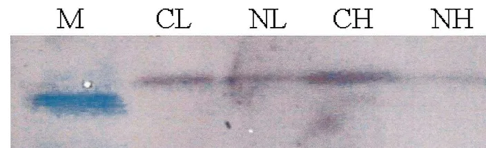
Quantification of the PSII reaction center protein 33-kDa following NF exposure showed pronounced differences among treatments (Fig. 1). The 33-kDa protein level of CH plants was greater than that of CL plants. Exposure of leaves to NF caused the loss of PSII reaction centers, as indi-cated by the loss of the extrinsic 33-kDa protein of
Table 1
Photosynthetic parameters measured in leaves exposed to norflurazon (NF) under different photosynthetic photon flux densities (PPFDs)a photoperiod. The 7-day-old plants were sprayed with 15mM NF and exposed to either high PPFD (300mmol m−2s−1) or low PPFD (30mmol m−2s−1). Treatment notations indicate the presence/absence of NF and PPFD level in order. The
Fig. 1. Influence of norflurazon (NF) and photosynthetic photon flux density (PPFD) on the extrinsic 33-kDa protein of oxygen evolving complex in photosystem (PS) II reaction center. The plants were subjected to the same treatments as in Table 1. Treatment notations are the same as in Table 1. M, a 30-kDa molecular weight marker. Measurements were made 3 days after NF treatment. Data represent the mean9S.E. of three replicates.
Table 2
Effects of norflurazon (NF) exposure at different photosynthetic photon flux densities (PPFDs) on carotenoids (mmol mol−1Chl
a) and Chl pigments (mg g FW−1) ofC.sati6usa
Treatment Pigments
NH CL
CH NL
17.091.4 (4.3) 24.391.2 (92.4)
25.290.7 (99.2) 15.891.5 (15.0)
Neoxanthin
45.892.3 (179.8)
Violaxanthin 60.290.3 (15.1) 26.791.2 (101.7) 16.992.2 (16.0)
7.690.2 (1.9) ND
Antheraxanthin 2.790.2 (10.6) 3.390.5 (3.2)
69.493.4 (17.4) 48.892.3 (185.8)
58.792.3 (231.1) 41.391.5 (39.5)
Lutein
ND
Zeaxanthin 7.891.2 (2.0) ND 6.290.3 (5.9)
b-Carotene 48.198.0 (186.6) 2.791.0 (0.7) 41.192.5 (156.5) 18.991.4 (18.1) 75.691.6 (19.0) 26.791.2 (101.7)
48.492.4 (190.5) 26.491.6 (25.1)
V+A+Z
Chla+b 1779.89162.9 112.599.5 1642.6926.1 387.9915.9
aThe values in parentheses indicate the pigment contents on nmol per g fresh weight basis. The plants were subjected to the
same treatments as in Table 1. Treatment notations are the same as in Table 1. Measurements were made 3 days after NF treatment. ND, not detected. Data represent the mean9S.E. of three replicates.
the oxygen-evolving complex, with a greater mag-nitude in NH plants.
3.3. Composition of photosynthetic pigments
Table 2 shows that there was a pronounced decline in total Chls with NF, especially under high PPFD. The carotenoid content per unit of Chl a was determined in cucumber plants upon exposure to NF that can produce oxidative stress (Table 2). In CL plants there was an approxi-mately 45% decline in violaxanthin+ antheraxan-thin+zeaxanthin and also a slight decline in lutein and b-carotene, compared with CH plants, with no difference between the two controls in the quantity of neoxanthin. NH plants had increases in violaxanthin, antheraxanthin, lutein and anthin compared with CH plants, especially zeax-anthin newly formed, coinciding with a drastic
decline in b-carotene. NL plants resulted in lower levels of the xanthophyll cycle pigments and lutein but a higher b-carotene than those of NH plants. However, NL plants also caused a great increase in antheraxanthin and zeaxanthin.
Fig. 2. Levels of qP, qN and non-photochemical quenching (NPQ) in leaves exposed to norflurazon (NF) under different photosynthetic photon flux densities (PPFDs). The plants were subjected to the same treatments as in Table 1. Treat-ment notations are the same as in Table 1. MeasureTreat-ments were made 3 days after NF treatment. Data represent the mean9S.E. of three replicates. In some cases the error bar is obscured by the symbol.
3.5. Responses of antioxidant enzyme acti6ities
The influence of NF on the activities of antioxi-dant enzymes participating in the scavenging of oxidative stress is shown in Table 3. In addition to the three primary enzymes of the Mehler-peroxi-dase pathway (SOD, GR and APX), we measured activities of CAT and guaiacol-POD. Activity of CAT was much lower in NH plants relative to the other plants, whereas POD and SOD activities in NH, CL, and NL plants were greater when com-pared with CH plants. APX activity exhibited a considerable increase in relation to NF, but only slight increase in control plants grown at low PPFD. Neither NF nor PPFD level resulted in a
discernible change in GR activity among
treatments.
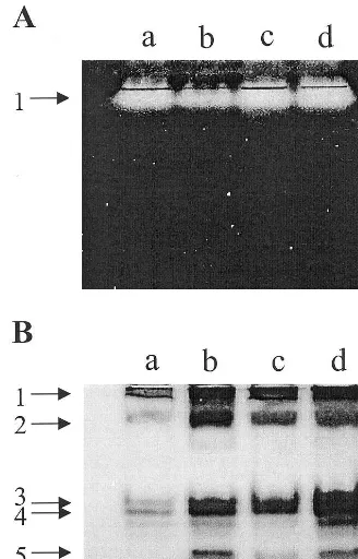
The isoform composition of different enzymes was analyzed by native PAGE. Gels stained for CAT revealed no isoform (Fig. 3A), and were in accordance with the induction profile of CAT specific activity, with the lowest activity in NH plants. POD and SOD exist as multiple isoforms (Figs. 3B and 4A). In the profile of POD activity in control plants, four bands (1, 2, 3 and 4) were detected, with staining intensity increased both with NF and with low PPFD (Fig. 3B, lanes b, c and d). NF induced a new band, which has a quite strong staining intensity (Fig. 3B, lanes b and d). The induction of POD isoform 5 was quite NF-specific. Three isoforms were observed when na-tive gels were stained for SOD activity, and no major change was observed in the activity of iso-form 3 in the leaves treated with NF (Fig. 4A). SOD isoform 1 was identified as Mn-SOD by its insensitivity to KCN and H2O2, whereas SOD
isoforms 2 and 3 were inhibited by both KCN and H2O2, suggesting that they represented
Cu,Zn-3.4. Photochemical quenching and energy dissipation
The photochemical quenching (qP), an estimate
of the fraction of open PSII centers, was lower in NF plants, with a greater magnitude of decline in NH plants, compared with control plants (Fig. 2). The lowest value ofqNwas observed in CL plants.
NL plants exhibited much greater level of NPQ than that of NH plants whereas both plants had similar level of highqN. A considerable amount of
NPQ existed in control plants at high PPFD, compared with ones at low PPFD. The combined quenching of qP, qN and NPQ was the lowest in
NH plants.
Table 3
Effects of norflurazon (NF) exposure at different photosynthetic photon flux densities (PPFDs) on antioxidant enzyme activities, catalase (CAT) (mmol O2min−1mg−1protein), SOD (units mg−1protein) and other enzymes (mmol min−1mg−1protein)a
Treatment Enzyme activities
APX SOD
POD
CAT GR
0.06390.002 1.0490.02
CH 84.591.7 0.6790.08 0.10090.001
0.4890.01 0.17090.003
NH 137.5916.1 1.0690.02 0.09890.003
0.7590.05
1.0090.06 0.14390.009 128.898.0 0.10590.002
CL
0.9890.06
1.0590.02 0.16490.009 124.9911.8 0.10590.003
NL
aThe plants were subjected to the same treatments as in Table 1. Treatment notations are the same as in Table 1. Measurements
Fig. 3. Influence of norflurazon (NF) and photosynthetic photon flux density (PPFD) on the isozyme profiles of cata-lase (CAT) (A) and peroxidase (POD) (B) ofC.sati6us. The plants were subjected to the same treatments as in Table 1. Non-denaturing activity gels were prepared and run as de-scribed in Section 2. Lane a, CH; lane b, NH; lane c, CL; lane d, NL. Treatment notations are the same as in Table 1.
photodestruction of plastid components in the deficiency of protecting carotenoids. Both Fv/Fm
ratio and quantum yield were significantly differ-ent over high and low PPFDs by NF treatmdiffer-ents, indicating that the level of PPFD affects structural and functional modifications of the photosynthetic
Fig. 4. Influence of norflurazon (NF) and photosynthetic photon flux density (PPFD) on the isozyme profiles of super-oxide dismutase (SOD) (A), ascorbate peroxidase (APX) (B), and glutathione reductase (GR) (C) ofC.sati6us. The plants
were subjected to the same treatments as in Table 1. Non-de-naturing activity gels were prepared and run as described in Section 2. Incubation of gels with KCN or H2O2 prior to staining SOD activity suggested that SOD-1 is Mn-SOD, whereas SOD-2 and -3 are Cu,Zn-SOD (data not shown). Gels stained for GR activity revealed a GSSH-specific band (GR-1) and a non-specific GR band (arrowhead). Lane a, CH; lane b, NH; lane c, CL; lane d, NL. Treatment notations are the same as in Table 1.
SOD activity. No Fe-SOD isoform was observed in all treatments. Both NF and low PPFD en-hanced the intensity of the existing isoform 2, and Mn-SOD also increased slightly. The overall in-crease of total SOD activity in NF plants and CL plants may result from the increase of isozyme 2. NF enhanced significantly the intensity of the existing APX compared with CH plants, with maximum activity in NH plants (Fig. 4B). How-ever, low PPFD slightly increased the intensity of APX in control plants. A GSSG-specific GR band was present inC. sati6us(Fig. 4C). The intensities of GR in all treatments remained unaffected by NF or PPFD.
4. Discussion
Exposure of C. sati6us to NF substantially in-hibited photochemical efficiency of photosynthe-sis, indicated as a decrease inFv/Fm and quantum
apparatus. The relatively small decline in Fv/Fm
and quantum yield in NL plants shows that NF-induced photoinhibition was alleviated during NF exposure in lower PPFD. It has been reported that the decrease inFv/Fm results from change of
reac-tion centers to quenchers by excess light [36]. In NF plants, however, the decrease is probably due to the loss of PSII reaction centers as indicated by the loss of 33-kDa protein of oxygen evolving complex (Fig. 1). The F0 level is known to be
affected by environmental stress that causes struc-tural alterations in the PSII complex [36]. The F0
decrease in NH plants (Table 1) might arise from markedly low level of Chls. On the other hand, the
F0 increase in NL plants is probably associated
not only with low PSII photochemistry (KP) but
also with low KT, which is related to excitation
energy transfer to non-fluorescent pigments, al-though the precise reasons for the decline remain to be determined.
The decrease in Fv/Fm is also due to the
forma-tion of a new quencher of excitaforma-tion energy, the
xanthophyll cycle pigment zeaxanthin. Less
amount of violaxanthin+antheraxanthin+ zeax-anthin accounts for less requirement for qN and
NPQ in CL plants due to low photoinhibitory stress, compared with CH plants (Table 2 and Fig. 2). The treatment with NF caused a significant accumulation of antheraxanthin and zeaxanthin, but a concomitant reduction in b-carotene on a Chl a basis and in Chls with a greater magnitude under high PPFD (Table 2). The profile of pig-ment levels on a fresh weight basis in NF plants was obviously different from that of Chl a basis, with an overall reduction in the other pigments except zeaxanthin. The newly formed zeaxanthin in the presence of carotenoid biosynthesis in-hibitor (Table 2) confirms the possibility that
zeax-anthin plays a role in the photoprotective
mechanism [9,28]. A decline of photochemical effi-ciency in NF plants is associated with a decrease in qPand an increase inqN (Table 1 and Fig. 2). A
lower NPQ of NH plants, as compared with NL plants, exhibited a close correlation with level of antheraxanthin and zeaxanthin on a fresh weight basis, not per Chl a content. Interestingly, the irradiance environment during NF-caused oxida-tive stress dramatically affects zeaxanthin-related NPQ, not qN. In contrast to qN, NPQ is
propor-tional to the effective rate constant for energy dissipation in the antennae as well as the
concen-tration of quenching centers [37]. The results provide evidence that energy dissipation through the xanthophyll cycle does not completely explain for the acclimatory process of the plants to oxida-tive stress caused by NF. Thus the plants necessi-tate other components of dissipative process.
Antioxidant enzymes are likely to play a consid-erable part of defense mechanism against NF-in-duced oxidative stress. CAT takes part in an efficient protective role to oxidative stress [38], however, in this study NH plants exhibited a significant decline in CAT activity (Table 3), con-sistent with reports of CAT inactivation in leaves exposed to high light [39,40]. The increase in the activities of POD, SOD and APX might occur against oxidative stress or serve to compensate for low CAT activity (Table 3). The ascorbate-glu-tathione cycle has been known to be activated under oxidative stress conditions [38]. However, GR, a rate-limiting enzyme in the H2O2
-scaveng-ing cycle [41], was not altered in all treatments by different PPFDs (Table 3), suggesting that the functioning of the cycle was not activated effi-ciently in NF plants. The Mehler-peroxidase path-way has been postulated to cause not only qP, but
also the build-up of DpH and consequent zeaxan-thin formation [11,13]. Considering similar levels of the antioxidant enzymes except CAT in NH and NL plants, the response of these enzymes to NF may not depend on the level of PPFD. The increase of APX activity appears to be NF-depen-dent, but the activities of POD and SOD are considerably increased even in CL plants (Table 3).
enzymes to oxidative stress. Both NF and PPFD exert differential effects on the expression of the multiple forms of POD (Fig. 3B). The NF-treated plants were capable of synthesizing a new isoform of POD, which could be considered as a response to NF-caused oxidative damage. The results sug-gest that enzymatic removal of H2O2by POD and
APX is the dominant pathway during NF-induced enzymatic scavenging system.
In the NF-treated leaves of C. sati6us, the detoxification of AOS is undertaken through func-tionally interrelated antioxidant mechanisms. The
qP and DpH-dependent qN through the
Mehler-peroxidase pathway, in addition to zeaxanthin for-mation, take part in mitigating oxidative stress induced by NF. NF-induced photooxidation in combination with high PPFD causes severe pho-toinhibition, which may arise from an insufficient capacity to dissipate excess excitation energy through qP and NPQ. The plants, however, seem
to develop a component of photoinhibitory quenching, qI, which is related to light-dependent
alterations in the PSII reaction center complex and frequently indicated as a decrease in Fv/Fm [36].
Interestingly, the level of PPFD influences qP and
NPQ in response to NF, not the induction of antioxidant enzymes. This might be explained by the fact that PSI is more stable than PSII upon strong light [4] and that the AOS-scavenging en-zymes of the chloroplast may serve to protect PSI.
References
[1] B. Andersson, S. Styring, Photosystem II: molecular organisation, function, and acclimation, Curr. Top Bioenerg. 16 (1991) 1 – 81.
[2] E.M. Aro, I. Virgin, B. Andersson, Photoinhibition of photosystem II. Inactivation, protein damage and turnover, Biochim. Biophys. Acta 1143 (1993) 113 – 134. [3] K. Asada, Production and action of active oxygen spe-cies in photosynthetic tissues, in: C.H. Foyer, P.M. Mullineaux (Eds.), Causes of Photooxidative Stress and Amelioration of Defence Systems in Plants, CRC Press, Boca Raton, FL, 1994, pp. 77 – 104.
[4] S.B. Powles, Photoinhibition of photosynthesis induced by visible light, Annu. Rev. Plant Physiol. 35 (1984) 15 – 44.
[5] K. Inoue, Y. Fujii, E. Yokoyama, K. Matsuura, K. Hiyama, H. Sakurai, The photoinhibition site of photo-system I in isolated chloroplasts under extremely reduc-ing conditions, Plant Cell Physiol. 30 (1989) 65 – 71. [6] S.E. Tjus, B. Andersson, Loss of the trans-thylakoid
proton gradient is an early event during photoinhibitory
illumination of chloroplast preparations, Biochim. Bio-phys. Acta 1183 (1993) 315 – 322.
[7] R.G. Alscher, J.L. Hess (Eds.), Antioxidants in Higher Plants, CRC Press, Boca Raton, FL, 1993.
[8] C.H. Foyer, M. Lelandais, K.J. Kunert, Photooxidative stress in plants, Physiol. Plant 92 (1994) 696 – 717. [9] A.M. Gilmore, H.Y. Yamamoto, Linear models relating
xanthophylls and lumen acidity to non-photochemical fluorescence quenching. Evidence that antheraxanthin explains zeaxanthin-independent quenching, Photosynth. Res. 35 (1993) 67 – 78.
[10] E. Weis, J.A. Berry, Quantum efficiency of photosystem II in relation to energy-dependent quenching of chloro-phyll fluorescence, Biochim. Biophys. Acta 894 (1987) 198 – 208.
[11] U. Schreiber, W. Bilger, C. Neubauer, Chlorophyll fluorescence as a non-intrusive indicator for rapid assess-ment of in vivo photosynthesis, in: E.D. Schulze, M.M. Caldwell (Eds.), Ecology of Photosynthesis, Springer-Verlag, Berlin, 1994, pp. 49 – 70.
[12] W. Bilger, O. Bjo¨rkman, Role of the xanthophyll cycle in photoprotection elucidated by measurements of light-in-duced absorbance changes, fluorescence and photosyn-thesis in leaves of Hedera canariensis, Photosynth. Res. 25 (1990) 173 – 185.
[13] A. Polle, Mehler reaction: friend or foe in photosynthe-sis?, Botanica Acta 109 (1996) 84 – 89.
[14] C.H. Foyer, B. Halliwell, The presence of glutathione and glutathione reductase in chloroplasts: a proposed role in ascorbic acid metabolism, Planta 133 (1976) 21 – 25.
[15] J.G. Scandalios, Regulation and properties of plant cata-lases, in: C.H. Foyer, P.M. Mullineaux (Eds.), Causes of Photooxidative Stress and Amelioration of Defense Sys-tems in Plants, CRC Press, Boca Raton, FL, 1994, pp. 275 – 314.
[16] L. Slooten, K. Capiau, W. Van Camp, M. Van Mon-tagu, C. Sybesma, D. Inze´, Factors affecting the en-hancement of oxidative stress tolerance in transgenic tobacco overexpressing maganese superoxide dismutase in the chloroplasts, Plant Physiol. 107 (1995) 737 – 750. [17] W. Van Camp, K. Capiau, M. Van Montagu, D. Inze´, L.
Slooten, Enhancement of oxidative stress tolerance in transgenic tobacco plants overproducing Fe-superoxide dismutase in chloroplasts, Plant Physiol. 112 (1996) 1703 – 1714.
[18] L.H. Pitcher, E. Brennan, A. Hurley, P. Dunsmuir, J.M. Tepperman, B.A. Zilinskas, Overproduction of petunia copper/zinc superoxide dismutase does not confer ozone tolerance in transgenic tobacco, Plant Physiol. 97 (1991) 452 – 455.
[19] P. Payton, R.D. Allen, N. Trolinder, A.S. Holiday, Over-expression of chloroplast-targeted Mn superoxide dismutase in cotton (Gossypium hirsutum L., cv. (Coker 312) does not alter the reduction of photosynthesis after short exposures to low temperature and high light inten-sity, Photosynth. Res. 52 (1997) 233 – 244.
[21] T.A. Ezhova, O.P. Soldatova, U.N. Ondar, L.B. Ma-manova, N.L. Radyukina, A.V. Sof’in, V.I. Romanov, S.V. Shestakov, Norflurazon-tolerant dwarf mutants of
Arabidopsis as a model for studying plant resistance to oxidative stress, Russ. J. Plant Physiol. 44 (1997) 575 – 579.
[22] D.B. Harper, B.M.R. Harvey, Mechanism of paraquat tolerance in perennial ryegrass. II. Role of superoxide dismutase, catalase, and peroxidase, Plant Cell Environ. 1 (1978) 211 – 215.
[23] O.C. Kno¨rzer, J. Durner, P. Bo¨ger, Alterations in the antioxidative system of suspension-cultured soybean cells (Glycine max) induced by oxidative stress, Physiol. Plant 97 (1996) 388 – 396.
[24] P.M. Bramley, Inhibition of carotenoid biosynthesis, in: A. Young, G. Britton (Eds.), Carotenoids in Photosyn-thesis, Chapman and Hall, London, 1993, pp. 127 – 159. [25] U. Schreiber, U. Schliwa, W. Bilger, Continuous record-ing of photochemical and non-photochemical chloro-phyll fluorescence quenching with a new type of modulation fluorometer, Photosynth. Res. 10 (1986) 51 – 62.
[26] B. Genty, J.M. Briantais, N. Baker, The relationship between the quantum yield of photosynthetic electron transport and quenching of chlorophyll fluorescence, Biochim. Biophys. Acta 990 (1989) 87 – 92.
[27] G.S. Tae, W.A. Cramer, Truncation of the COOH-termi-nal domain of the psbE gene product inSynechocystissp. PCC 6803: requirements for Photosystem II assembly and function, Biochemistry 31 (1992) 4066 – 4074. [28] S. Jung, K.L. Steffen, Influence of photosynthetic photon
flux densities before and during long-term chilling on xanthophyll cycle and chlorophyll fluorescence quench-ing in leaves of tomato (Lycopersicon hirsutum), Physiol. Plant 100 (1997) 958 – 966.
[29] D.O. Natvig, Comparative biochemistry of oxygen toxic-ity in lactic acid-forming aquatic fungi, Arch. Microbiol. 132 (1982) 107 – 114.
[30] G.H. Egley, R.N. Paul Jr, K.C. Vaughn, S.O. Duke, Role of peroxidase in the development of water-imper-meable seed coats in Sida spinosa L, Planta 157 (1983) 224 – 232.
[31] J.P. Spychalla, S.L. Desborough, Superoxide dismutase, catalase and alpha tocopherol content of stored potato tubers, Plant Physiol. 94 (1990) 1214 – 1218.
[32] G.X. Chen, K. Asada, Ascorbate peroxidase in tea leaves: occurrence of two isozymes and the differences in their enzymatic and molecular properties, Plant Cell Physiol. 30 (1989) 987 – 998.
[33] M.V. Rao, G. Paliyath, D.P. Ormrod, Ultraviolet-B- and ozone-induced biochemical changes in antioxidant en-zymes ofArabodopsis thaliana, Plant Physiol. 110 (1996) 125 – 136.
[34] W. Woodbury, A.K. Spencer, M.A. Stahman, An im-proved procedure for using ferricyanide for detecting catalase isozymes, Anal. Biochem. 44 (1971) 301 – 305. [35] P.D. Olson, J.E. Varner, Hydrogen peroxide and
lignifi-cation, Plant J. 4 (1993) 887 – 892.
[36] G.H. Krause, E. Weis, Chlorophyll fluorescence and photosynthesis: the basics, Annu. Rev. Plant Physiol. Plant Mol. Biol. 42 (1991) 313 – 349.
[37] B. Demmig-Adams, W.W. Adams III, D.H. Barker, B.A. Logan, D.R. Bowling, A.S. Verhoeven, Using chloro-phyll fluorescence to assess the fraction of absorbed light allocated to thermal dissipation of excess excitation, Physiol. Plant 98 (1996) 253 – 264.
[38] A. Chaoui, S. Mazhoudi, M.H. Ghorbal, E.E. Ferjani, Cadmium and zinc induction of lipid peroxidation and effects on antioxidant enzyme activities in bean (Phaseo
-lus6ulgarisL.), Plant Sci. 127 (1997) 139 – 147.
[39] J. Volk, J. Feierabend, Photoinactivation of catalase at low temperature and its relevance to photosynthetic and peroxide metabolism in leaves, Plant Cell Environ. 12 (1989) 701 – 712.
[40] N.P. Mishra, R.K. Mishra, G.S. Singhal, Changes in the activities of antioxidant enzymes during exposure of intact wheat leaves to strong visible light at different temperatures in the presence of different protein synthe-sis inhibitors, Plant Physiol. 102 (1993) 867 – 880. [41] P.P. Jablonski, J.W. Anderson, Light-dependent
reduc-tion of dehydroascorbate by ruptured pea chloroplasts, Plant Physiol. 67 (1981) 1239 – 1244.
[42] J. Kurepa, D. He´rouart, M.V. Montagu, D. Inze´, Differ-ential expression of CuZn- and Fe-superoxide dismutase genes of tobacco during development, oxidative stress, and hormonal treatments, Plant Cell Physiol. 38 (1997) 463 – 470.
[43] D.J. Thomas, T.J. Avenson, J.B. Thomas, S.K. Herbert, A cyanobacterium lacking iron superoxide dismutase is sensitized to oxidative stress induced with methyl violo-gen but is not sensitized to oxidative stress induced with norflurazon, Plant Physiol. 116 (1998) 1593 – 1602.