Research Article
Molecular Identification of
Mycobacterium
Species of
Public Health and Veterinary Importance from Cattle in
the South State of México
Adrian Zaragoza Bastida,
1,2Nallely Rivero Pérez,
2Benjamín Valladares Carranza,
3Keila Isaac-Olivé,
1Pablo Moreno Pérez,
1Horacio Sandoval Trujillo,
4and Ninfa Ramírez Durán
11Facultad de Medicina, Universidad Aut´onoma del Estado de M´exico, Paseo Tollocan/Jes´us Carranza s/n, 50180 Toluca, MEX, Mexico 2Area Acad´emica de Medicina Veterinaria y Zootecnia, Instituto de Ciencias Agropecuaria,´
Universidad Aut´onoma del Estado de Hidalgo, Av. Universidad Km 1, Ex-Hda. de Aquetzalpa, 43600 Tulancingo, HGO, Mexico
3Centro de Investigaci´on y Estudios Avanzados en Salud Animal, Facultad de Medicina Veterinaria y Zootecnia,
Universidad Aut´onoma del Estado de M´exico, Km 15.5 Carretera Panamericana Toluca-Atlacomulco, 50200 Toluca, MEX, Mexico
4Departamento de Sistemas Biol´ogicos, Universidad Aut´onoma Metropolitana-Xochimilco, Calzada del Hueso 1100,
04960 Ciudad de M´exico, Mexico
Correspondence should be addressed to Ninfa Ram´ırez Dur´an; [email protected]
Received 9 March 2017; Revised 12 May 2017; Accepted 21 May 2017; Published 14 June 2017
Academic Editor: Nahuel Fittipaldi
Copyright © 2017 Adrian Zaragoza Bastida et al. his is an open access article distributed under the Creative Commons Attribution License, which permits unrestricted use, distribution, and reproduction in any medium, provided the original work is properly cited.
Mycobacteriumgenus causes a variety of zoonotic diseases. he best known example is the zoonotic tuberculosis due toM. bovis. Much less is known about “nontuberculous mycobacteria (NTM),” which are also associated with infections in humans. he
Mexican standard NOM-ZOO-031-1995 regulates the presence ofM. bovisin cattle; however, no regulation exists for the NTM
species. he objective of this study was to isolate and identify nontuberculous mycobacteria species from cattle of local herds in the south region of the State of Mexico through the identiication and detection of the 100 bp molecular marker in the 23S rRNA gene with subsequent sequencing of the 16S rRNA gene. Milk samples (35) and nasal exudate samples (68) were collected. From the 108 strains isolated, 39 were selected for identiication. hirteen strains isolated from nasal exudates ampliied the 100 bp molecular
marker and were identiied asM. neoaurum(six strains),M. parafortuitum(four strains),M. moriokaense(two strains), andM.
conluentis(one strain). ExceptM. parafortuitum, the other species identiied are of public health and veterinary concern because they are pathogenic to humans, especially those with underlying medical conditions.
1. Introduction
he genusMycobacteriumcauses a wide variety of zoonotic diseases. he best known example is zoonotic tuberculosis due toM. bovis, for which cattle is the main reservoir.M. bovis is part of the “tuberculosis complex,” which also includes the speciesM. tuberculosis, M. africanum, M. caprae, andM. microti[1].
Within the mycobacterial group are the “nontuberculous mycobacteria (NTM),” which are also associated with infec-tions in humans. he NTM are found in various environ-mental sources such as soil, water, vegetation, animals, dairy
products, and feces and may be transmitted inadvertently by inhalation, ingestion, or skin penetration [2].
he Mexican standard NOM-ZOO-031-1995 regulates the presence ofM. bovisin cattle to control and eradicate bovine tuberculosis (bTB); however, no regulation exists for the NTM species. he oicial diagnosis of bovine tuberculosis due to the presence ofM. bovisat the ield level is based on the intradermal test using a puriied protein derivative (tuber-culin) [3]. Although used for several years, this test does not provide good sensitivity and speciicity. Approximately 20% of the animals with tuberculosis do not react to the test [4], and the presence of other mycobacterial species, both
tuberculosis complex and NTM species, causes interference that leads to false-positive and false-negative diagnoses.
Although Mexico has a regulatory standard, bTB preva-lence in excess of 2% is reported in some areas [5]. Given the poor speciicity and sensitivity of the tuberculin test, the actual presence ofM. bovisis likely to be lower and the infec-tion rate of cattle by other mycobacteria is likely to be higher, respectively. hus, cattle breeders, veterinarians, technicians, and employees working in the livestock industry might be occupationally exposed to infections byM. bovisand NTM. Very little is known about occupational exposure to zoonoses due to NTM species because the identiication of these species was a rather diicult task prior to the development of identiication techniques based on molecular biology.
Currently, the molecular biology techniques most com-monly used for the diagnosis of diseases caused by mycobac-teria are restriction fragment length polymorphism (RFLP) for the diagnosis ofM. tuberculosis[6], spoligotyping for the diagnosis ofM. bovis [7], and the detection of a 100-base pair (bp) “speciic insertion” located on the 23S rRNA gene characteristic of Gram-positive bacteria with a high guanine-cytosine (HGC) content, which is considered a molecular marker for this group of bacteria [8, 9], followed by sequence analysis of the 16S rRNA gene for the identiication of bacteria at the species level [10].
Among the NTM species identiied by the aforemen-tioned techniques areM. balnei, M. marinum, andM. platy-poecilus, which have caused supericial and deep skin lesions [11];M. kansasiifrom lung lesions [12];M. simiaefrom gener-alized infections [13];M. scrofulaceumfrom infections of the skin and internal organs [14]; M. szulgai associated with pulmonary infections, osteomyelitis, tenosynovitis, and lym-phadenitis [15]; M. ulcerans associated with subcutaneous granulomas [16]; M. fortuitum and M. chelonaeassociated with vasculitis, endocarditis, osteomyelitis, mediastinitis, meningitis, keratitis, and hepatitis [17];M. abscessus, associ-ated with erythematous lesions that progressed to ulcerassoci-ated nodules [18]; and other species.
he largest percentage of the state inventory for heads of cattle in the State of Mexico in Mexico is concentrated in the southern region, and one of the main economic activities is cattle ranching [19]. he Mexican regulation for cattle control NOM-ZOO-031-1995 only focuses on the tuberculin test for the diagnosis ofM. bovis. Little is known about the presence of NTM in the cattle of the region. Given the possibility of identifying species of actinobacteria by detection of the 100-base pair molecular marker on the 23S rRNA gene and the subsequent sequencing of the 16S rRNA gene, it is possible to identify the aforementioned NTM species.
he objective of the present study was to isolate and iden-tify NTM species from cattle of the south region of the State of Mexico. he Mycobacterium species were isolated from samples of nasal exudate and bovine milk and identiied by detecting the 100-base pair molecular marker in the 23S rRNA gene with subsequent sequencing of the 16S rRNA gene.
2. Materials and Methods
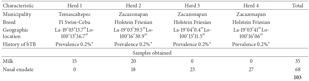
2.1. Sampling. A sampling was performed based on the spa-tial distribution of herds positive for bovine tuberculosis in the state of Mexico conducted by Zaragoza et al. 2015 [20]. Four herds of cattle were selected in the south region of the State of Mexico, one herd belonging to the Municipality of Temascaltepec and three herds belonging to the municipality of Zacazonapan. A total of 103 samples, 35 milk samples and 68 samples of nasal exudate, were collected. he distribution of the number and type of samples collected in each herd is shown in Table 1.
2.2. Obtaining Samples of Milk and Nasal Exudate. he udder and nipples were cleansed with puriied water and soap and then dried with paper towels, and nipple asepsis was subse-quently performed using swabs soaked in 70% alcohol. Five milliliters of milk was collected directly from the nipple in sterile 20 mL vessels, discarding the initial low. Nasal exudate was collected directly from the inside of the nasal oriice using a 10 cm long sterile swab, which was then submerged in an isotonic saline solution (0.85%). Samples of milk and nasal exudate were stored at 4∘C until processing.
2.3. Sample Processing
2.3.1. Isolation of Mycobacteria. he milk samples were cen-trifuged at 2500 revolutions per minute (rpm) for 10 minutes. he pellets from the milk and nasal exudate samples were inoculated into the following culture medium selective for mycobacteria: Stonebrink (BD BBL 220504), Middlebrook (BD BBL 254521), and Middlebrook (BD BBL 254521) supple-mented with 6 g of sodium pyruvate per liter (Middlebrook-P). he inoculated media were incubated at 37∘C for 8 weeks and were assessed every 3 days.
2.3.2. Classiication of Isolated Strains. he isolated strains were distributed in groups according to the following char-acteristics: colony pigmentation, growth time, and colony characteristics (shape, consistency, texture, and pigment pro-duction). Isolated strains were stained with Ziehl-Neelsen to conirm the presence of acid-fast bacilli (AFB) [21].
Table 1: Samples collected in cattle herds in the south region of the State of Mexico.
Characteristic Herd 1 Herd 2 Herd 3 Herd 4 Total
Municipality Temascaltepec Zacazonapan Zacazonapan Zacazonapan
Breed F1 Swiss-Cebu Holstein Friesian Holstein Friesian Holstein Friesian
Geographic
History of bTB Prevalence0.2%∗ Prevalence0.2%∗ Prevalence0.2%∗ Prevalence0.2%∗
Samples obtained
Milk 15 20 0 0 35
Nasal exudate 0 18 23 27 68
103
La: latitude; Lo: longitude; bTB: bovine tuberculosis.∗Inf ormationobtained from the Committee on the Promotion and Protection of Livestock of the State
of Mexico.
2.5. Detection of the Molecular Marker in the 23S rRNA Gene. he 100 bp molecular marker located on the 23S rRNA gene was ampliied according to the methodology described by Roller et al. (1992) using the following primers [8]:
23S InsF, 5�
-(AC)A(AGT)GCGTAG(AGCT)CG-A(AT)GG-3�, and 23S InsR, 5� -GTG(AT)CGGTT-T(AGCT)(GCT)GGTA-3�.
he reaction was conducted using a commercial Taq DNA polymerase (Promega M1661). he following thermal cycle conditions were used: a predenaturation step for 5 min-utes (94∘C); 29 cycles of denaturation for 30 seconds (94∘C), hybridization for 45 seconds (46∘C), and elongation for 50 seconds (72∘C); and, inally, a postelongation cycle of 5 minutes (72∘C). he ampliied fragments were conirmed on a 2% agarose gel stained with ethidium bromide (SIGMA 46065).
2.6. Ampliication of the 16S rRNA Gene. Strains that ampli-ied the 100 bp phylogenetic marker were selected for 16S rRNA sequencing analysis. he following primers were used for the ampliication:
8f: AGAGTTTGATCMTGGCTCAG and 1492r: TAC-GGYTACCTTGTTACGACTT.
he reaction was conducted using a commercial Taq DNA polymerase (Promega M1661). he following thermal cycle conditions were used: one predenaturation step for 5 minutes (94∘C); 34 cycles of denaturation for 30 seconds (94∘C), hybridization for 20 seconds (52∘C), and elongation for 1 minute 30 seconds (72∘C); and, inally, a postelongation cycle of 7 minutes (72∘C).
he ampliied fragments were conirmed on a 1% agarose gel stained with ethidium bromide (SIGMA 46065). he products of this ampliication were puriied using the Amicon Ultra Filterkit (Millipore UFC901008) and conirmed on a 1% agarose gel to verify their presence and quality.
2.7. Identiication of Mycobacterium Species. he ampliied products of the 16S rRNA gene were sent to the Macrogen Sequencing Service, Maryland, USA. he obtained sequences
were analyzed and corrected using the BioEdit program [22]. Consensus sequences were constructed from the forward and reverse fragments, which were compared with sequences deposited previously in GenBank of the National Center for Biotechnology Information (NCBI) using the BLAST program [23] and EzTaxon 2.1 [24].
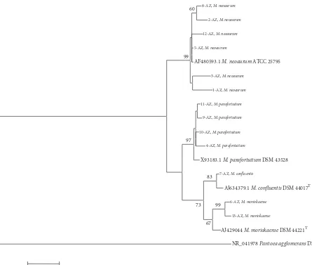
2.8. Phylogenetic Analysis. Sequences of the 16S rRNA gene were obtained for the following mycobacterial species from the American Type Culture Collection (ATCC) and the Ger-man Collection of Microorganisms and Cell Cultures (DSM): M. neoaurum ATCC25795, M. parafortuitum DSM43528, M. moriokaenseDSM44221T, andM. conluentisDSM44017T. he sequences of the collection strains and those of the strains isolated in the present investigation were aligned with the BioEdit program [22]. Phylogenetic analysis was performed using the maximum parsimony method in MEGA sotware version 4 [25]. To form the root of the cladogram, the sequence ofPantoea agglomeransDSM 3493 was used.
3. Results
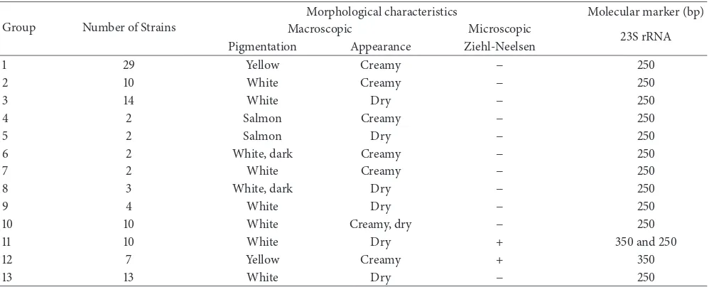
he 108 strains isolated from the 103 collected samples were distributed in 13 groups according to their macroscopic and microscopic morphological characteristics (Table 2). Groups 11 and 12, particularly, were composed of acid-fast strains.
For identiication at the species level, 39 strains were cho-sen: 10 of them belonged to group 11 and 7 to group 12. Two strains from each one of the remaining 11 groups were selected to complete the 39 strains. he 100 bp molecular marker was found in the 33% (13/39) of the selected strains. For them, the 16S rRNA gene was ampliied for sequencing and iden-tiication at the species level.
he overall prevalence of NTM on the collected samples was 12.6% (13/103) considering both milk and nasal exudate samples. However, the speciic prevalence for nasal exudate samples was 19.1% (13/68).
Table 2: Isolated strains are grouped according to their morphological characteristics and the presence of the molecular marker (100 bp) on the 23S rRNA gene.
Group Number of Strains
Morphological characteristics Molecular marker (bp)
Macroscopic Microscopic
23S rRNA
Pigmentation Appearance Ziehl-Neelsen
1 29 Yellow Creamy − 250
2 10 White Creamy − 250
3 14 White Dry − 250
4 2 Salmon Creamy − 250
5 2 Salmon Dry − 250
6 2 White, dark Creamy − 250
7 2 White Creamy − 250
8 3 White, dark Dry − 250
9 4 White Dry − 250
10 10 White Creamy, dry − 250
11 10 White Dry + 350 and 250
12 7 Yellow Creamy + 350
13 13 White Dry − 250
−: absence of acid-fast bacilli; +: presence of acid-fast bacilli; Bp: base pairs.
Table 3: Comparison of 16S rRNA gene sequences of strains isolated from cattle with those documented in GenBank, using BLAST and EzTaxon.
Strain Origin of the herd Culture medium Ampliied fragment size (bp) Similarity (Blast) % Similarity (EzTaxon) %
1-AZ 2 Middlebrook 1428 M. neoaurum 98 M. neoaurum 98.3
2-AZ 2 Stonebrink 1408 M. neoaurum 99 M. neoaurum 99.1
3-AZ 2 Stonebrink 1428 M. neoaurum 98 M. neoaurum 98.2
5-AZ 3 Stonebrink 1416 M. neoaurum 99 M. neoaurum 99.2
8-AZ 4 Middlebrook 1415 M. neoaurum 99 M. neoaurum 99.0
12-AZ 2 Middlebrook 1415 M. neoaurum 99 M. neoaurum 99.4
4-AZ 4 Middlebrook-P 1420 M. parafortuitum 99 M. parafortuitum 98.2
9-AZ 3 Middlebrook 1415 M. parafortuitum 99 M. parafortuitum 98.9
10-AZ 4 Stonebrink 1411 M. parafortuitum 99 M. parafortuitum 98.4
11-AZ 3 Stonebrink 1414 M. parafortuitum 99 M. parafortuitum 98.2
6-AZ 2 Stonebrink 1455 M. moriokaense 99 M. moriokaense 98.2
13-AZ 4 Stonebrink 1417 M. moriokaense 98 M. moriokaense 98.2
7-AZ 2 Middlebrook-P 1420 M. conluentis 99 M. conluentis 99.1
2: Zacazonapan Holstein-F; 3: Zacazonapan Holstein-F; 4: Zacazonapan Holstein-F.
M. moriokaense, and, inally, 8% (1/13) had 99% similarity withM. conluentis(Table 3).
he phylogenetic tree was formed with the genus Myco-bacteriumand four of its species by which the phylogenetic relationships between the collection strains and the strains isolated in the present investigation were observed (Figure 1).
4. Discussion
he NTM species were isolated from samples of nasal exudate only, which eliminated the samples from one of the local farms of this study (Table 1). We found that the speciic prevalence was 19.1% in herds of the south region of the State of Mexico. Similar studies in the United States, South Africa, Tanzania, and Brazil reported NTM prevalence values of
3.4%, 24.5%, 7%, and 7.8%, respectively; therefore, the preva-lence value found in this study lies within the range reported previously [26–29]. In this study, 13 of the 39 analyzed strains were identiied as the NTM speciesM. neoaurum, M. moriokaense, M. conluentis, andM. parafortuitum.
99
67 60
83
73 97 99
20
8-AZ, M. neoaurum
2-AZ, M. neoaurum
12-AZ, M. neoaurum
5-AZ, M. neoaurum
AF480593.1 M. neoaurum ATCC 25795
3-AZ, M. neoaurum
1-AZ, M. neoaurum
11-AZ, M. parafortuitum
X93183.1 M. parafortuitum DSM 43528
9-AZ, M. parafortuitum
10-AZ, M. parafortuitum
4-AZ, M. parafortuitum
7-AZ, M. confluentis
6-AZ, M. moriokaense
13-AZ, M. moriokaense
NR_041978 Pantoea agglomerans DSM 3493
AJ634379.1 M. confluentis DSM 44017T
AJ429044 M. moriokaense DSM 44221T
Figure 1: Phylogenetic tree constructed by comparing the 16S rRNA gene sequences from the isolated and reference strains.
patients with urinary infections [37], meningoencephalitis and alterations in the central nervous system [38], bacteremia and endocarditis [39], and pulmonary infection [40, 41]. Although it has been mainly isolated from clinical cases, there are also reports about its isolation from milk and cattle [28, 42, 43].
M. moriokaensewas isolated from sputum sample [44]. Although it is considered nonpathogenic for humans, it has been associated with pulmonary diseases [45].M. conluentis was isolated from sputum samples as well [46], and, along withM. parafortuitum, both are considered nonpathogenic species. M. conluentis, M. moriokaense, andM. neoaurum have been isolated from diferent bovine and wildlife tissues with tuberculous lesions, whereasM. parafortuitumhas only been isolated from bovine milk [26, 28, 47–49]. However, in our work,M. parafortuitumwas only isolated from nasal exudate samples.
he nutritional requirements of mycobacteria difer among various species, which was the reason for using difer-ent culture media. Notably, seven of the 13 strains iddifer-entiied in this study were isolated in Stonebrink medium, including
M. neoaurum, M. parafortuitum,and M. moriokaense. his result is consistent with that described by Sep´ulveda et al. [50] who indicated that Stonebrink medium is suitable for the recovery of diferent species of the genusMycobacterium. Garc´ıa-Martos and Garc´ıa-Agudo [51] reported that Middle-brook medium is optimal for the isolation of actinomycetes, which is in accord with the present investigation considering that two species,M. neoaurumandM. parafortuitum, were isolated in this medium. Notably,M. conluentiswas isolated only in Middlebrook medium supplemented with sodium pyruvate; thus, the strategy of using diferent culture media was appropriate because it allowed the isolation of diferent species of the genusMycobacterium.
5. Conclusions
Using the methodology described in this study, four NTM species were isolated and identiied:M. conluentis, M. mor-iokaense, M. neoaurum,andM. parafortuitum. hese species were isolated for the irst time from nasal exudates of bovines from the south region of the State of Mexico. hree of the identiied species (M. neoaurum, M. moriokaense, and M. conluentis) are of public health and veterinary importance.
Disclosure
his work is derived from the thesis for the degree of Doc-torate in Health Sciences (Universidad Aut´onoma del Estado de M´exico), registered in the PNPC-CONACYT.
Conflicts of Interest
All authors declare that they do not have any conlicts of interest.
Acknowledgments
he authors would like to acknowledge the inancial assis-tance from the Secretary of Research and Advanced Studies of Universidad Aut´onoma del Estado de M´exico (UAEMex) through the following research grants: (i) “Implementation of Geographic Information Systems and Techniques of Molec-ular Biology, as tools in the detection and identiication of Mycobacteriumspp.,” SIEA-UAEM 3486/2013CHT, and (ii) the network “Microbiolog´ıa y qu´ımica en las Ciencias de la Salud,” 039/2014RIF.
References
[1] A. Aranaz, D. Cousins, A. Mateos, and L. Dom´ınguez,
“Ele-vation ofMycobacterium tuberculosissubsp. caprae Aranaz et
al. 1999 to species rank asMycobacterium capraecomb. nov.,
sp. nov,”International Journal of Systematic and Evolutionary
Microbiology, vol. 53, no. 6, pp. 1785–1789, 2003.
[2] M. H. Ho, C. K. Ho, and L. Y. Chong, “Atypical mycobacterial cutaneous infections in Hong Kong: 10-Year retrospective
study,”Hong Kong Medical Journal, vol. 12, no. 1, pp. 21–26, 2006.
[3] SAGARPA, “Norma Oicial M´exicana NOM-ZOO-031-1995
Campa˜na Nacional contra la Tuberculosis bovina(M. bovis),”
Diario Oicial de la Federaci´on, vol. 1996, pp. 12–32, 1995. [4] O. R. G. Llamazares, C. B. G. Mart´ın, D. A. Nistal, V. A. D. L. P.
Redondo, L. D. Rodr´ıguez, and E. F. R. Ferri, “Field evaluation of the single intradermal cervical tuberculin test and the
inter-feron-�assay for detection and eradication of bovine
tuberculo-sis in Spain,”Veterinary Microbiology, vol. 70, no. 1-2, pp. 55–66,
1999.
[5] SENASICA, “Informes de la Situaci´on Zoosanitaria Nacional de 2016,” 2016, http://www.gob.mx/cms/uploads/attachment/ ile/169575/SITUACION_ZOOSANITARIA_2016-11-8.pdf. [6] M. D. Cave, K. D. Eisenach, P. F. McDermott, J. H. Bates, and
J. T. Crawford, “IS6110: Conservation of sequence in the Myco-bacterium tuberculosis complex and its utilization in DNA
ingerprinting,”Molecular and Cellular Probes, vol. 5, no. 1, pp.
73–80, 1991.
[7] S. R. Acosta, C. C. Estrada, and S. F. Mili´an, “Tipiicaci´on de
cepas deMycobacterium bovis,”T´ecnica Pecuaria en M´exico, vol.
47, pp. 389–412, 2009.
[8] C. Roller, W. Ludwig, and K. H. Schleifer, “Gram-positive bac-teria with a high DNA G+C content are characterized by a
common insertion within their 23S rRNA genes,”Journal of
General Microbiology, vol. 138, no. 6, pp. 1167–1175, 1992.
[9] B. A. Zaragoza, C. M. ´A. Karam, M. L. P. Bustamante, T. ´A.
H. Sandoval, and D. N. Ram´ırez, “Marcador molecular de acti-nomicetos utilizado para detectar micobacterias en muestras de
esputo,”Revista Mexicana de Ciencias Farmac´euticas, vol. 45, pp.
35–40, 2014.
[10] J. E. Clarridge, “Impact of 16S rRNA gene sequence analysis for identiication of bacteria on clinical microbiology and
infec-tious diseases,”Clinical Microbiology Reviews, vol. 17, no. 4, pp.
840–862, 2004.
[11] D. O. Sanjay, M. D. Avani, and D. O. James, “Atypical
Mycobac-terial cutaneousinfections,”Dermatologic Clinics, vol. 27, no. 1, pp. 63–73, 2009.
[12] S. Weitzul, P. J. Eichhorn, and A. G. Pandya, “Nontuberculous
mycobacterial infections of the skin,”Dermatologic Clinics, vol.
18, no. 2, pp. 359–377, 2000.
[13] H. M. Al-Abdely, S. G. Revankar, and J. R. Graybill,
“Dissem-inatedMycobacterium simiaeinfection in patients with AIDS,”
Journal of Infection, vol. 41, no. 2, pp. 143–147, 2000.
[14] H.-S. Jang, J.-H. Jo, C.-K. Oh et al., “Successful treatment of
localized cutaneous infection caused byMycobacterium
scrofu-laceumwith clarithromycin,”Pediatric Dermatology, vol. 22, no. 5, pp. 476–479, 2005.
[15] J. J. Meyer and S. S. Gelman, “Multifocal osteomyelitis due to
Mycobacterium szulgaiin a patient with chronic lymphocytic
leukemia,”Journal of Infection, vol. 56, no. 2, pp. 151–154, 2008.
[16] D. Wagner and L. S. Young, “Nontuberculous mycobacterial
infections: a clinical review,”Journal of Infection, vol. 32, no. 5,
pp. 257–270, 2004.
[17] K. P. Redbord, D. A. Shearer, H. Gloster et al., “Atypical
Myco-bacterium furunculosisoccurring ater pedicures,”Journal of the American Academy of Dermatology, vol. 54, no. 3, pp. 520–524, 2006.
[18] P. Tang, S. Walsh, C. Murray et al., “Outbreak of
acupuncture-associated cutaneousMycobacterium abscessusinfections,”
Jour-nal of Cutaneous Medicine and Surgery, vol. 10, no. 4, pp. 166– 169, 2006.
[19] INEGI, “Censo Agr´ıcola, Ganadero y Forestal 2007,” 2007, http:// www.inegi.org.mx/est/contenidos/proyectos/Agro/ca2007/Re-sultados_Agricola/default.aspx.
[20] B. A. Zaragoza, M. L. P. Bustamante, T. ´A. H. Sandoval, and D.
N. Ram´ırez, “Spatial analysis of bovine tuberculosis in the State
of Mexico, Mexico,”Veterinaria Italiana, vol. 53, no. 1, pp. 29–37,
2017.
[21] OPS, “Organizaci´on Panamericana de la Salud para el diagn´os-tico bacteriol´ogico de la tuberculosis, Normas y gu´ıa t´ecnica,” Parte I Basiloscopia, 2008.
[22] T. A. Hall, “BioEdit: a user. friendly biologycal sequence align-ment editor ana analysis program for Windows 95/98/NT,”
Nucleic Acids Symposium Series, vol. 41, no. 41, pp. 95–98, 1999. [23] S. F. Altschul, T. L. Madden, J. Zhang, Z. Zhang, W. Miller, and D. J. Lipman, “Gapped BLAST and PSI-BLAST: a new
generation of protein database search programs,”Nucleic Acids
[24] J. Chun, J.-H. Lee, Y. Jung et al., “EzTaxon: a web-based tool for the identiication of prokaryotes based on 16S ribosomal
RNA gene sequences,”International Journal of Systematic and
Evolutionary Microbiology, vol. 57, no. 10, pp. 2259–2261, 2007. [25] K. Tamura, J. Dudley, M. Nei, and S. Kumar, “MEGA4:
molec-ular evolutionary genetics analysis (MEGA) sotware version
4.0,”Molecular Biology and Evolution, vol. 24, no. 8, pp. 1596–
1599, 2007.
[26] T. C. hacker, S. Robbe-Austerman, B. Harris, M. V. Palmer, and W. R. Waters, “Isolation of mycobacteria from clinical samples
collected in the United States from 2004 to 2011,”BMC
Veteri-nary Research, vol. 9, pp. 100–110, 2013.
[27] N. Gcebe, V. Rutten, N. C. Gey van Pittius, and A. Michel, “Prevalence and distribution of non-tuberculous mycobacteria (NTM) in cattle, African bufaloes (syncerus cafer) and their
environments in South Africa,”Transboundary and Emerging
Diseases, vol. 60, supplement 1, pp. 74–84, 2013.
[28] B. Z. Katale, E. V. Mbugi, L. Botha et al., “Species diversity of non-tuberculous mycobacteria isolated from humans, livestock
and wildlife in the Serengeti ecosystem, Tanzania,”BMC
Infec-tious Diseases, vol. 14, no. 1, pp. 1–8, 616, 2014.
[29] M. M. J. Franco, A. C. Paes, M. G. Ribeiro et al., “Occurrence of mycobacteria in bovine milk samples from both individual and collective bulk tanks at farms and informal markets in the
southeast region of Sao Paulo, Brazil,”BMC Veterinary Research,
vol. 9, pp. 1–8, 2013.
[30] L. L. Washer, J. Riddell, J. Rider, and C. E. Chenoweth, “
Myco-bacterium neoaurumbloodstream infection: report of 4 cases
and review of the literature,”Clinical Infectious Diseases, vol. 45,
no. 2, pp. e10–e13, 2007.
[31] H. Awadh, M. Mansour, and M. Shorman, “Bacteremia with an
unusual pathogen:Mycobacterium neoaurum,”Case Reports in
Infectious Diseases, vol. 2016, Article ID 5167874, 3 pages, 2016. [32] M. B. Davison, J. G. McCormack, Z. M. Blacklock, D. J. Dawson,
M. H. Tilse, and F. B. Crimmins, “Bacteremia caused by
Myco-bacterium neoaurum,”Journal of Clinical Microbiology, vol. 26, pp. 762–764, 1988.
[33] C.-C. Lai, C.-K. Tan, C.-C. Chen, and P.-R. Hsueh, “
Mycobac-terium neoaurum infection in a patient with renal failure,”
International Journal of Infectious Diseases, vol. 13, no. 5, pp. e276–e278, 2009.
[34] M. L. Becker, A. A. Suchak, J. N. Wolfe, R. Zarychanski, A.
Kabani, and L. E. Nicolle, “Mycobacterium neoaurum
bactere-mia in a hemodialysis patient,”Canadian Journal of Infectious
Diseases, vol. 14, no. 1, pp. 45–48, 2003.
[35] E. J. Hayton, O. Koch, M. Scarborough, N. Sabharwal, F. Drob-niewski, and I. C. Bowler, “Rapidly growing mycobacteria as emerging pathogens in bloodstream and device-related
infec-tion: a case of pacemaker infection withMycobacterium
neoau-rum,”JMM Case Reports, vol. 2, no. 1–3, 2015.
[36] A. Kumar, G. S. Pazhayattil, A. Das, and H. A. Conte, “
Mycobac-terium neoaurumcausing prosthetic valve endocarditis: a case
report and review of the literature,”Brazilian Journal of
Infec-tious Diseases, vol. 18, no. 2, pp. 235–237, 2014.
[37] S. Zanetti, R. Faedda, G. Fadda et al., “Isolation and
identiica-tion ofMycobacterium neoaurumfrom a patient with urinary
infection,”New Microbiologica, vol. 24, no. 2, pp. 189–192, 2001.
[38] G. A. Heckman, C. Hawkins, A. Morris, L. L. Burrows, and C.
Bergeron, “Rapidly progressive dementia due toMycobacterium
neoaurummeningoencephalitis,”Emerging Infectious Diseases, vol. 10, no. 5, pp. 924–927, 2004.
[39] B. A. Brown-Elliott, R. J. Wallace Jr., C. A. Petti et al., “
Mycobac-terium neoaurumandMycobacterium bacteremicumsp. nov. as
causes of mycobacteremia,”Journal of Clinical Microbiology, vol.
48, no. 12, pp. 4377–4385, 2010.
[40] Y. Morimoto, E. D. Chan, L. Heifets, and J. M. Routes,
“Pul-monary infection withMycobacterium neoaurumidentiied by
16S ribosomal DNA sequence,”Journal of Infection, vol. 54, no.
4, pp. e227–e231, 2007.
[41] C.-K. Kim, S. I. Choi, B. R. Jeon, Y.-W. Lee, Y. K. Lee, and H. B. Shin, “Pulmonary infection caused by mycobacterium
neoau-rum: the irst case in Korea,”Annals of Laboratory Medicine, vol.
34, no. 3, pp. 243–246, 2014.
[42] S. A. Sgarioni, R. D. C. Hirata, M. Hiroyuki Hirata et al.,
“Occur-rence ofMycobacterium bovisand non-tuberculous
mycobac-teria (NTM) in raw and pasteurized milk in the northwestern
region of Paran´a, Brazil,”Brazilian Journal of Microbiology, vol.
45, no. 2, pp. 707–711, 2014.
[43] L. Padya, N. Chin’Ombe, M. Magwenzi, J. Mbanga, V. Ruhanya,
and P. Nziramasanga, “Molecular identiication of
mycobac-teriumspecies of public health importance in cattle in zimbabwe
by 16S rRNA gene sequencing,”Open Microbiology Journal, vol.
9, pp. 38–42, 2015.
[44] M. Tsukamura, I. Yano, and T. Imaeda, “Mycobacterium
mor-iokaense sp. nov., a rapidly growing, nonphotochromogenic
Mycobacterium,”International Journal of Systematic
Bacteriol-ogy, vol. 36, no. 2, pp. 333–338, 1986.
[45] A. Somoskovi and M. Salinger, “Nontuberculous mycobacteria in respiratory infections: advances in diagnosis and
identiica-tion,”Clinics in Laboratory Medicine, vol. 34, no. 2, pp. 271–295,
2014.
[46] P. Kirschner, A. Teske, K.-H. Schroder, R. M. Kroppenstedt, J.
Wolters, and E. C. Bottger, “Mycobacterium conluentissp. nov,”
International Journal of Systematic Bacteriology, vol. 42, no. 2, pp. 257–262, 1992.
[47] M. Pate, U. Zajc, D. Kuˇsar et al., “Mycobacteriumspp. in wild
game in Slovenia,”Veterinary Journal, vol. 208, pp. 93–95, 2016.
[48] L. Botha, N. C. Gey van Pittius, and P. D. van Helden,
“Myco-bacteria and disease in Southern Africa,”Transboundary and
Emerging Diseases, vol. 60, no. 26, pp. 147–156, 2013.
[49] J. J. Camarena Mi˜nana and R. Gonz´alez Pellicer, “Micobacterias at´ıpicas y su implicaci´on en patolog´ıa infecciosa pulmonar,”
Enfermedades Infecciosas y Microbiolog´ıa Cl´ınica, vol. 29, no. 5, pp. 66–75, 2011.
[50] A. Sep´ulveda, P. M. Garc´ıa, M. J. Rodr´ıguez, A. M´arquez, and J. L. Puerto, “Evaluaci´on del medio de Stonebrink para la
recu-peraci´on de micobacterias,”Revista de Diagn´ostico Biol´ogico,
vol. 50, pp. 189–192, 2001.
[51] P. Garc´ıa-Martos and L. Garc´ıa-Agudo, “Infecciones por
mico-bacterias de crecimiento r´apido,” Enfermedades Infecciosas y
Submit your manuscripts at
https://www.hindawi.com
Stem Cells
International
Hindawi Publishing Corporationhttp://www.hindawi.com Volume 2014
Hindawi Publishing Corporation
http://www.hindawi.com Volume 2014
INFLAMMATION
Hindawi Publishing Corporation
http://www.hindawi.com Volume 2014
Behavioural
Neurology
Endocrinology
International Journal ofHindawi Publishing Corporation
http://www.hindawi.com Volume 2014
Hindawi Publishing Corporation
http://www.hindawi.com Volume 2014
Disease Markers
Hindawi Publishing Corporation
http://www.hindawi.com Volume 2014
BioMed
Research International
Oncology
Journal of Hindawi Publishing Corporationhttp://www.hindawi.com Volume 2014
Hindawi Publishing Corporation
http://www.hindawi.com Volume 2014
Oxidative Medicine and Cellular Longevity
Hindawi Publishing Corporation
http://www.hindawi.com Volume 2014
PPAR Research
The Scientiic
World Journal
Hindawi Publishing Corporationhttp://www.hindawi.com Volume 2014
Immunology Research
Hindawi Publishing Corporation
http://www.hindawi.com Volume 2014
Journal of
Obesity
Journal ofHindawi Publishing Corporation
http://www.hindawi.com Volume 2014
Hindawi Publishing Corporation
http://www.hindawi.com Volume 2014
Computational and Mathematical Methods in Medicine
Ophthalmology
Journal of Hindawi Publishing Corporationhttp://www.hindawi.com Volume 2014
Diabetes Research
Journal of Hindawi Publishing Corporationhttp://www.hindawi.com Volume 2014
Hindawi Publishing Corporation
http://www.hindawi.com Volume 2014
Research and Treatment
AIDS
Hindawi Publishing Corporation
http://www.hindawi.com Volume 2014
Gastroenterology Research and Practice
Hindawi Publishing Corporation
http://www.hindawi.com Volume 2014
Parkinson’s
Disease
Evidence-Based Complementary and Alternative Medicine
Volume 2014