* corresponding author: [email protected]
Multiple mini punch grafts for extensive
ulcer: a case report
Yohanes Widodo Wirohadidjojo*
Department of Dermato-Venereology, Faculty of Medicine, Gadjah Mada University/ Dr. Sardjito General Hospital, Yogyakarta, Indonesia
ABSTRACT
Multiple mini punch grafts is the placing of mini size of full thickness skins on to ulcer bed. They consist of epidermal and dermal component composed with hair follicles and other skin appendices where epidermal stem cells are located. The epidermal stem cells are the best source of epidermal cells in reconstruction of skin equivalent that is usually used for replacing classic split thickness skin graft in recovering extensive ulcer. In this article, the application of multiple mini punch grafts onto extensive ulcer is reported. A case of extensive ulcer was suffered by a 6-year-old boy whose left foot is injured in a traffic accident. His toes had already been amputated by surgeon but a classic skin graft failed to recover the ulcer. Multiple mini punch grafts had been harvested from his inguinal and buttock skin and they were placed onto his ulcer. Pre and post mini punch grafting photographs were reviewed. After eight weeks, placed multiple mini punch tissues onto large ulcer reveals lateral extensions and more than 90% of epithelialization. Multiple mini punch grafts can be used as a method to cover large ulcer.
ABSTRAK
Punch graftmini jamak merupakan penananaman potongan kulit berukuran kecil dengan ketebalan penuh pada dasar ulkus. Potongan kulit seperti ini tersusun oleh epidermis, dermis yang mengandung folikel rambut dan apendik kulit lainnya tempat sel punca epidermal berada. Sel punca epidermal merupakan sumber terbaik sel-sel epidermal untuk menyusun kulit ekivalen yang dewasa ini sering dipakai sebagai pengganti split thickness skin graft untuk menutup ulkus yang luas. Dalam artikel ini dilaporkan penggunaan punch graft mini jamak pada kasus ulkus luas yang diderita oleh anak laki-laki berusia 6 tahun akibat kecelakaan lalu lintas pada kaki kirinya. Jari-jari kaki telah diamputasi sebelumnya, tetapi skin graftklasik gagal menyembuhkan ulkus luas pada kaki tersebut. Punch graftmini jamak yang diambil dari kulit lipat paha dan kulit bokong selanjutnya di tanamkan pada dasar ulkus tersebut. Setelah 8 minggu, kulit yang ditanam-kan tumbuh melebar kesamping dan menutup lebih dari 90 % permukaan ulkus. Disimpulditanam-kan bahwa teknik punch graft mini jamak dapat digunakan untuk menutup ulkus yang luas.
Key words: mini punch grafts-large ulcer-epithelialization-epidermal-stem cells
INTRODUCTION
In the traffic accident, full thickness of skin may be extensively loose. The split thickness skin graft may be failed to recover the loss tissues because of limitation of skin donors and
organ as shown in its responding to physical injury in a staged and specific pattern of cell migration, re-epithelialization, and cytokine expression.1The best source of epidermal cells
for this purpose is epidermal stem cells that are located along basal layers, sebaceous glands and around the hair follicles.2-5 Several
problems in skin reconstructive transplantation are the handling of the fragile sheets, variable ‘‘take rates’’,6blister formation due to ulcer bed
secretion, scarring and contracture of the graft.7
Other disadvantages are: high cost and the fact that it can only be reconstructed by clinicians who are familiar or have a relation with human cells culture technique researchers.
Multiple mini punch grafts are grafting of multiple mini size full thickness skins with multipurpose. It is most popular in hair transplantation for preparing follicular unit transplantation,8-9but it is also used in acne scar
correction,10-11and re-pigmentation of vitiligo. 12-13 They consist of epidermal and dermal
component rich with hair follicles and other skin appendixes where epidermal stem cells are located. Placing mini punch tissues onto ulcer bed maybe similar with cultivation of stem cells in hypoxia condition, a method for propagating stem cells to release growth factors.14,15 This
presumption is based on a report by Kirsneret al.16Punch graft acts as a pharmacologic agent
in stimulating granulation of the venous ulcer
bed as well as migration of previously dormant wound edges.16
Application of mini punch grafts onto large ulcer has never been published. It has been reported in the treatment limited sixe of chronic leg ulcers.15In this case report, it was reported
the application of multiple mini punch grafts onto extensive traffic accident ulcer.
CASE REPORT
A six-year-old boy who had a traffic accident was referenced because of epithelialization failures. Eight months before, he had the first operation performed by orthopedic surgeon for amputating the toes, fixing fragmented bones, and classic skin grafting. Unfortunately, the skin grafting was not taking and skin grafted wound become an extensive chronic ulcer.
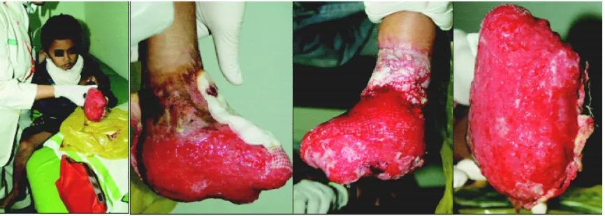
At the first visit, an extensive chronic ulcer on both dorsal and plantar left foot was observed. The ulcer showed an easy bleeding of granulation tissue with profuse fluid secretion covered by normal saline immersed gauges (FIGURE 1). Based on this condition, it was decided to take care of the ulcer by Sofratule®,
0.1% gentamycine ointment in occlusive dressing technique to prevent new bleeding in the replacing next new dressing. Cefixime 30 mg twice a day and 150 mg of tranexamic acid twice a day are also given for a week.
After a week of therapy, bleeding in replaced dressing as well as fluid secretion was not observed anymore but epithelialization was still poor. The treatment has been continued but oral therapy was stopped. After a month of
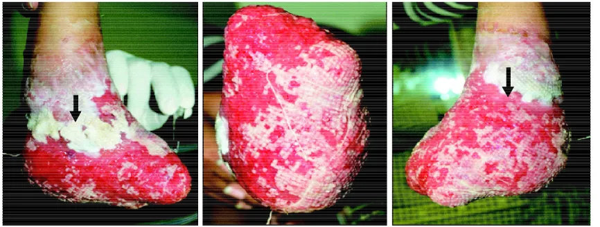
treatment, epithelialization was just limited in a small area in the ankle as shown by black arrows on FIGURE 2. Based on this condition, it was decided to perform multiple mini-punch grafts.
FIGURE 2. Ulcer condition after a month of occlusive treatment.
Surgical Procedures
Multiple mini-punch grafts (MMPGs) were performed in three phases. First, testing of MMPGs taken from left inguinal skin was placed onto dorsal and inner side of feet. Second, MMGs taken from right inguinal skin was placed onto lateral side of feet. Third, MMPGs taken from buttock skin was placed onto plantar side of feet. The third operation has been performed after previous MMPGs of dorsal feet showed confluence in skin sheet as a space for placing adhesive tape for fixing MMPGs onto plantar feet against gravitation force.
Skin donor harvesting and grafting
After disinfecting skin with povidone iodine 10 % and placing sterile linen onto operation area, the skin was injected with
tumescent anesthetic (1: 300.000 epinephrine plus 0.2% lidocaine in sterile normal saline). Skin harvesting was performed by 0.2 mm disposable punch device. MMPGs were then harvested by cutting a piece of skin in dermal-fat level and placed them onto gauges immersed with sterile normal saline (to prevent skin dehydration) before they were placed onto ulcer bed.
Before grafting, ulcer bed was washed and immersed with sterile normal saline. Placing of MMPGs onto ulcer bed was performed piece by piece with blepharoplasty skin’s forceps. All of procedure was performed without any anesthetic drugs (FIGURE 3). The ulcer and MMPGs were then covered by netting gauze (Sofratule®) followed by an occlusive dressing
for a week. In second week, topical silver ointment (Burnazin®) was then applied to
FIGURE 3. Harvesting (left) and placing MMPGS (right). Black arrow shows the lateral punched tissues expansion of previously grafting.
RESULTS
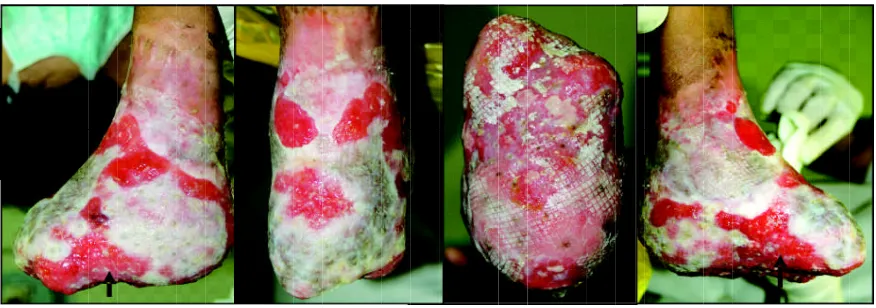
After a week of grafting, punched tissue islands showed that they were tightly attached onto ulcer bed. They were proved by resistance attachment in dry removing of sofratule. The beginning of their lateral extension was
observed in week two. At the same time, ulcer edge tissue migration to the central area was also observed. Confluences of punched tissue to perform epithelial sheet were observed in week three (FIGURE 4).
FIGURE 4. Ulcer Edge Tissue Migration and Confluences Of MMPGs. Black arrow shows new MMPGs.
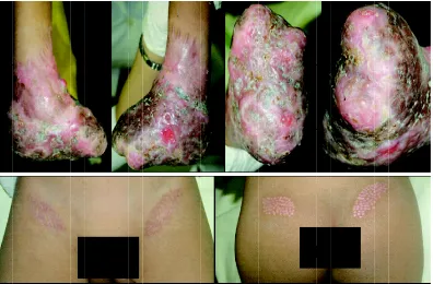
The final result can be observed in FIGURE 5. Eight weeks after the last operation, more than 90% of epithelialization was observed on
FIGURE 5. Eight weeks after the last operation
DISCUSSION
In case of extensive ulcer when enough source of skin donor is not available, such as in case of extensive burn, most of modern surgeons may perform grafting of reconstructive skin as skin substitute.17,18This material is made from
a small piece of patient’s own skin by in-vitro cultivating fibroblasts from dermal side and keratinocytes from epidermal side. Fibroblasts monolayer is then cultivated in the collagen matrixes to perform dermal equivalent and in media-air interphase, keratinocytes are then dropped onto dermal equivalent surface to perform a large reconstructive skin.19
Donor can be taken from any site of skin. Epidermal cells are composed of three population cells, namely: epidermal stem cells (SCs), transient amplifying cells, and non proliferative basal cells. Epidermal construction from transient amplifying cells formed faster than the others; however, the
epidermis from the SCs population shows continuity in growing and expressing the reporter gene for long time period.20In a case of limited
source of epidermal cells, autologous of epidermal SCs can be generated from follicular outer root sheath cells as previously published by Limat and Hunziker.21
A controversy of this technique is arisen due to use of recombinant epidermal growth factor (EGF) for keratinocytes and fetal bovine serum for fibroblast cultivation. The future effect of both materials in patient is not fully predicted, especially in a case of bovine viral infection. In another site, handling of reconstructive skin grafting among surgeons is not easy. It is fragile sheet and easy to be broken.6Clinical studies show that grafting takes
rates are variable, blister formations are sometimes found, scarring and contractures are also often observed.7 In addition, except for
Those problems lead researches to develop a technique namely cells spray for epithelialization. The cells can be directly harvested from a small piece (1: 100 of wound size) of patient’s skin, or cultivated in vitro to achieve numbers of viable cells. In this technique, donor skin is cut and enzymatic digestion with 2.5 unit/mL dispase followed by 0.05% trypsin-EDTA in Ringer lactate solution is used to harvest epidermal cells. Mixed epidermal cells are then sprayed onto wound bed. They reported that this technique shows a successful result of various depth of burn victims.22,23Compared to the classic graft, cells
spray showed similar functional and esthetic outcome.24 In order to mask scar formation of
donor area, scalp skin can also be used.25
Different with reconstructive skin which needs a month for cultivation, the cells spray technique may only need time less than an hour if direct cells isolation is chosen.
Mini punched tissues consist of full thickness skin where epidermal stem cells are located. Epidermal stem cells have high plasticity to proliferate and difference to mature cells that are needed in damage tissues repair.26
They are also resistant against aging process agents that commonly exist in chronic ulcer.27
Placing stem cells in hypoxia condition, as commonly found on ulcer bed surface, can propagate them to release various growth factors that are needed for healing process, with anti aging properties that are needed for aged cells in wound edge to proliferate and migrate to central area of ulcer. All of those theories are proved in our case. Placing MMPGs is not only capable to proliferate and perform skin sheet but it also shows evidence of stimulating ulcer edge epthelialization and migration (FIGURE 4).
In limited facility of hospital such as ours, skin reconstructive and cells spray may be practiced in the few next years. Unfortunately, limited available skin donor in extensive ulcer patient, such as in this case, cannot wait for application of those techniques. Other more simple techniques should be defined. In this
case, MMPGs have a proof to solve the problem. According to Nordström and Hansson28, MMPGs are very simple techniques
and they can be performed even by nurses in primary health program
CONCLUSION
A case of extensive ulcer suffered by a 6– year-old boy that fails to be treated by classic skin graft has been presented. Due to limited source of skin donor, another skin grafting was impossible to be performed. This case can be handled by an easy technique of multi mini-punch grafts that was performed in two months of a three-step surgery
AKNOWLEDGMENT
Author would like thank Dr. Nurhidayah, Dr Meliana, Dr Sari and all of Nurses from Dr. Sardjito General Hospital, Yogyakarta for assisting the graftsand and also Kris Lantoro for taking the pictures.
REFERENCES
1. Falanga V, Isaacs C, Paquette D, Downing G, Kouttab N, Butmarc J, Badiavas E, Hardin-Young J. Wounding of bioengineered skin: cellular and molecular aspect after injury. J Invest Dermatol 2002; 119:653-60.
2. Fuchs E. Skin stem cells: rising to the surface. J Cell Biol 2008; 180: 273–84.
3. Webb A, Li A, Kaur P. Location and phenotype of human adult keratinocyte stem cells of the skin. Differentiation 2004; 72:387–95.
4. Kamstrup M, Faurschou A, Gniadecki R, Wulf HC. Epidermal stem cells – role in normal, wounded and pathological psoriatic and cancer skin. Curr Stem Cell Res Ther 2008; 3(2): 146–50. 5. Pianigiani E, Ierardi F, Mazzanti B, Saccardi R,
Cuciti C, Fimiani M. Human de-epidermized dermis as a stem cell carrier. Transplant Proc 2010; 42(6): 2244-6.
7. Paddle-Ledinek JE, Cruickshank DG, Masterton JP. Skin replacement by cultured keratinocyte grafts: an Australian experience. Burns 1997; 23(3):204–11.
8. Harris JA. Follicular unit transplantation: dissecting and planting techniques. Facial Plast Surg Clin North Am 2004;12(2):225-32. 9. Martinick JH. The latest developments in surgical
hair restoration. Facial Plast Surg Clin North Am 2004;12(2):249-52.
10. Jacob CI, Dover JS, Kaminer MS. Acne scarring: a classification system and review of treatment options. J Am Acad Dermatol 2001;45(1):109-17.
11. Basta-Juzbašiæ A. Current therapeutic approach to acne scars. Acta Dermatovenerol Croat 2010;18(3):171-5.
12. Malakar S, Lahiri K. Punch grafting for lip leucoderma.. Dermatology 2004; 208(2):125-8. 13. Barman KD, Khaitan BK, Verma KK. A comparative study of punch grafting followed by topical corticosteroid versus punch grafting followed by PUVA therapy in stable vitiligo. Dermatol Surg 2004; 30(1):49-53.
14. Lee EY, Xia Y, Kim WS, Kim MH, Kim TH, Kim KJ, Park BS, Sung JH. Hypoxia-enhanced wound-healing function of adipose-derived stem cells: increase in stem cell proliferation and up-regulation of VEGF and bFGF. Wound Repair Regen 2009; 17 (4):540-7.
15. Chung HM, Won CH, Sung JH. Responses of adipose-derived stem cells during hypoxia: enhanced skin-regenerative potential. Expert Opin Biol Ther 2009; 9(12):1499-508.
16. Kirsner RS, FalangaV, Kerdel FA, Katz MH, Eaglstein WH. Skin grafts as pharmacological agents: pre-wounding of the donor site. Brit J Dermatol 1996; 135:292-6.
17. Waymack P, Duff RG, Sabolinski M. The effect of a tissue engineered bilayered living skin analog, over meshed split-thickness autografts on the healing of excised burn wounds. Burns 2000; 26:609-19.
18. Loss M, Wedler V, Künzi W, Meuli-Simmen C, Meyer VE. Artificial skin, split-thickness autograft and cultured autologous keratinocytes combined to treat a severe burn injury of 93% of
TBSA. Burns 2000; 26:644-52.
19. Bottcher-Haberzeth S, Biedermann T, Reichmann E. Tissue engineering of skin. Burn 2010; 36: 450-60.
20. Dunnwald M, Chalkley AT, Alexandrunas D, Fishbaugh J, Bickenbach JR. Isolating a pure population of epidermal stem cells for use in tissue engineering. Exp Dermatol 2001; 10(1): 45-54. 21. Limat A, Hunziker T. Use of epidermal equivalents
generated from follicular outer root sheath cells in vitro and for autologous grafting of chronic wounds. Cells Tissues Organs 2002;172:79-85. 22. Hartmann B, Ekkernkamp A, Johnen C, Gerlach
JC, Belfekroun C, Kuntscher MV. Sprayed cultured epithelial autografts for deep dermal burns of the face and neck. Ann Plast Surg 2007; 58(1):70–3.
23. Gerlach JC, Johnen C, McCoy E, Brautigam K, Plettig J, Corcos A. Autologous skin cell spray-transplantation for a deep dermal burn patient in an ambulant treatment room setting. Burns 2011; 37:e19-23.
24. Gravante G, Di Fede MC, Araco A, Grimaldi M, De Angelis B, Arpino A, Cervelli V, Montone A. A randomized trial comparing ReCellW system of epidermal cells delivery versus classic skin grafts for the treatment of deep partial thickness burns. Burns 2007; 33: 966-72.
25. Schlabe J, Johnen C, Schwartlander R, Moser V, Hartmann B, Gerlach JC, Ku¨ ntscher MV. Isolation and culture of different epidermal and dermal cell types from human scalp suitable for the development of a therapeutical cell spray. Burns 2008; 34:376-84.
26. David PJ, Hunt DPJ, Morris PN, Sterling J, Anderson JA, Joannides A, Jahoda C, Compston A, Chandran S. A highly enriched niche of precursor cells with neuronal and glial potential within the hair follicle dermal papilla of adult skin. Stem Cells 2008; 26(1):163-72.
27. Matthew M, Stern MM, Bickenbach JR. Epidermal stem cells are resistant to cellular aging. Aging Cell 2007; 6(4):439-52.