www.elsevier.nlrlocateraqua-online
Toxicity of acidified chitosan for cultured rainbow
ž
/
trout Oncorhynchus mykiss
Graham Bullock
a,), Vicki Blazer
b, Scott Tsukuda
a,
Steve Summerfelt
aa
Freshwater Institute, P.O. Box 1746, Shepherdstown, WV 25443, USA
b
National Fish Health Research Laboratory, U.S. Geological SurÕey, Biological Resources DiÕision, 1700 Leetown Road, KearneysÕille, WV 25430, USA
Accepted 7 November 1999
Abstract
Chitosan is a deacetylation product of chitin. It is used as a flocculent for sewage and brewery wastes, and as a chelator of heavy metals. In aquaculture, chitosan has been used as an immunostimulant for protection against bacterial diseases in fish, for controlled release of vaccines, and as a diet supplement. Chitosan has generally been considered to be nontoxic to animals, but when it was dissolved in acetic acid and added to a culture system at 1.0 ppm to
Ž .
remove organic solids, we found acute toxicity to rainbow trout Oncorhynchus mykiss . In
controlled experiments to determine the extent of toxicity, we found that trout died after several hours exposure to 0.75 ppm and died in 24 h after exposure to 0.075 ppm. Exposure to 0.038 ppm resulted in mortality after 6 days exposure, while exposure to 0.019 ppm resulted in no mortality after 14 days exposure. Histological examination of gills, skin, muscle, and internal organs indicated significant and consistent pathological changes only in gills. Lifting of lamellar epithelium, hypertrophy and hyperplasia of lamellar epithelial cells occurred in trout exposed to 0.019 and 0.038 ppm. In trout exposed to 0.75 or 0.075 ppm chitosan, large areas of lamellar fusion were observed. These results show that soluble acidified chitosan is highly toxic to rainbow trout even at low concentrations.q2000 Elsevier Science B.V. All rights reserved.
Keywords: Chitosan; Rainbow trout; Toxicity; Pathology
)Corresponding author. Tel.:q1-304-870-2200; fax:q1-304-870-2208.
0044-8486r00r$ - see front matterq2000 Elsevier Science B.V. All rights reserved. Ž .
1. Introduction
Chitin is second only to cellulose as the most plentiful natural polymer. Along with its deacetylation product chitosan, chitin is manufactured commercially from the outer shell of crustaceans, particularly crabs and shrimp. The vast quantities of available shellfish wastes easily supply the chitosan needed for many applications. Most chitosan is used as a nontoxic cationic flocculent in treatment of wastes from sewage, sludge,
Ž .
breweries, etc., and as a chelator of heavy and radioactive metals Sandford, 1989 . Chitosan has also been found to be useful as a flocculent for several species of algae ŽNigam et al., 1980; Morales et al., 1985; Lubian, 1989 . It is being evaluated in human. medicine for wound dressings, hemostatic agents, drug delivery systems, and as a cholesterol reducing agent. Agricultural applications include coatings for seeds, fruit preservation, as a fungistat and as a flocculent for recovering proteinaceous wastes ŽSandford, 1989; Elson, 1996 . In aquaculture, chitosan has been used as an immunos-.
Ž
timulant to enhance protection of salmonids against bacterial disease Anderson and .
Siwicki, 1994; Siwicki et al., 1994 and, combined with alginate, for controlled release
Ž . Ž
of proteins such as bacterins Polk et al., 1994 , and as a diet supplement Kono et al., .
1987 .
When incorporated as a food additive, free chitosan has been reported to be nontoxic
Ž .
to mice fed at 10 grkg of body weight Arai et al., 1968 . However, when fed at 20 grkg as chitosan acetate, 40% of test mice died while no mortality occurred in mice fed
Ž . Ž .
the same level of free chitosan or chitosan formate Arai et al., 1968 . Kono et al. 1987 reported that diets supplemented with 10% free chitosan did not stimulate growth rates in three species of marine fish nor were there any toxic effects. When 5% chitosan was
Ž .
added to the diet of rainbow trout Oncorhynchus mykiss as an immunostimulant there
Ž .
were no reports of toxic effects Siwicki et al., 1994 .
We tested chitosan as an aid in removing solids from a rainbow trout recirculation Ž .
system previously described by Bullock et al. 1993 . Chitosan was dissolved in 1% acetic acid and added to the system at 1.0 ppm. A severe toxic effect resulting in death was noted within several hours after addition of chitosan. Examination of affected trout showed gills were pale with excess mucus and hemorrhages. Internal organs appeared normal. Because of the previous reports of its nontoxic nature, we decided to test the toxicity of chitosan for rainbow trout under controlled conditions.
2. Materials and methods
2.1. Toxicity testing
WA and a 1.0% solution was prepared in 1.0% acetic acid. A masterflex peristaltic
Ž .
pump Cole Palmer Instrument, Burling, IL set at 0.22 mlrmin was used to deliver a calculated 1 ppm chitosan to the experimental tanks. Test levels below 1 ppm were obtained by diluting the 1% chitosan solution and keeping the pump delivery constant. In order to obtain actual chitosan levels rather than calculated levels, we attempted to quantify the 1 ppm chitosan in springwater delivered by the peristaltic pump. When the pH of springwater was raised to precipitate chitosan, the calcium and magnesium in the springwater also precipitated. However, incoming chitosan concentration was quantified by turning on the peristaltic pump for 1 min, which was calculated to deliver 2-mg chitosan, and collecting the chitosan sample in 50-ml deionized water. Chitosan was precipitated from deionized water by raising the pH of the sample to 10 using 1.0 normal NaOH. After stirring for 4 h, the sample was filtered onto an 8-mm filter ŽMillipore, Redford, MA previously dried at 608C to a constant weight. The filtered. sample was dried at 608C and weighed to a constant weight. This procedure was repeated three times. The chitosan levels selected for testing were 1.0, 0.1, 0.05, and 0.025 ppm. However, as will be discussed later, the quantification studies showed that only 75% of the calculated level of chitosan were delivered by the peristaltic pump. Therefore, actual test levels were 0.75, 0.075, 0.038, and 0.019 ppm.
Rainbow trout with an average weight of 120.5 g were used in all tests. In preliminary trials using 0.75, 0.075, 0.038, and 0.019 ppm chitosan plus a 1% acetic acid control, and a nontreated rainbow trout control, a single 57-l tank containing 15
Ž .
rainbow trout was used for each test level and controls total of six tanks . In subsequent tests with 0.038 and 0.019 ppm chitosan, triplicate tanks, each containing 15 trout, were used for each concentration. Preliminary trials were carried out up to 7 days while later trials with the 0.038 and 0.019 ppm levels were carried out for 14 days.
2.2. Histopathology
In all tests, three to five rainbow trout were taken for histological examination from each exposure level, acetic acid, and nontreated controls. In preliminary trials, surviving fish were sampled at 24 h from the 0.75 ppm treatment tank and day 4 from the 0.075 ppm, because of acute toxicity. Samples from the 1.0 ppm acetic acid control and nontreated control were taken on day 7. In trials with the 0.038 and 0.019 ppm chitosan treatments, samples were taken on day 14. Pieces of gill, muscle, heart, spleen, liver, kidney, and intestine were removed from each trout, and fixed in 10% formalin or Deitrich’s fixative. They were processed routinely for histology, sectioned at 5mm and
Ž . stained with hematoxylin and eosin H & E .
3. Results
3.1. Toxicity tests
precipitation of chitosan was found to be 1.5 ppm, which is 75% of predicted value. The actual concentration exposure levels to trout were 0.75, 0.075, 0.038, and 0.019 ppm. In the initial 7-day exposure, chitosan was acutely toxic at 0.75 and 0.075 ppm concentrations. Twelve of 15 trout died within 24 h at 0.75 ppm, and six trout died within 24 h at 0.075 ppm, and 11 died within 3 days. During the 7-day trial, only one trout died on day 7 at the 0.038 ppm concentration, and none died at the 0.019 ppm concentration. No toxicity was noted in trout exposed to 1.0% acetic acid or in nontreated control trout. During the 14-day experiment, trout exposed to 0.038 ppm began dying on day 6 in one tank and on day 8 in the other two tanks. Mortality continued during the 14-day study period with a total of nine, seven, and five trouts dying in the three tanks. No trout died in the three tanks exposed to 0.019 ppm.
3.2. Histopathology
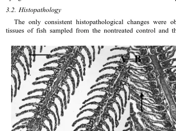
The only consistent histopathological changes were observed in gill tissue. Gill tissues of fish sampled from the nontreated control and the acetic acid control were
Ž .
Fig. 1. Histological sections of gill tissues of rainbow trout. A Control gill tissue, with filaments consisting of
Ž .
thin capillaries covered by flattened epithelial cells. B Trout exposed to 0.019 ppm chitosan. The epithelium
Ž . Ž .
is lifted off the basement membrane. Edematous fluid is often evident arrows . C Trout exposed to 0.038
Ž .
ppm chitosan. Epithelial lifting long arrow in some areas. Thickening of the lamellae due to hypertrophy of
Ž . Ž .
epithelial cells short arrows and proliferation of mucous cells clear cells within lamellar epithelium are
Ž . Ž
present. D Trout exposed to 0.075 ppm chitosan. Large areas of adjacent lamellae are fused single-headed
. Ž .
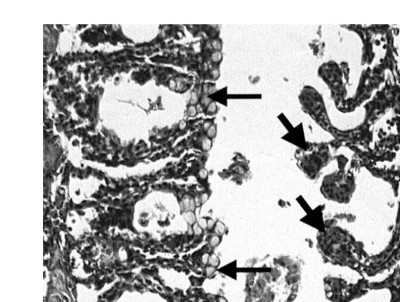
Fig. 2. Section of gill tissue from rainbow trout exposed to 0.75 ppm chitosan. Adjacent lamellae are often
Ž .
fused by proliferation of mucous cells thin arrows . Tips of the lamellae are engorged with red blood cells
Žthick arrows . H&E stain, scale bar equals 100. mm.
normal with blood spaces of the lamellae obvious and uniform in size. They were
Ž .
covered by the lamellar epithelium, a single layer of flattened cells Fig. 1A . Although no mortality was observed in fish exposed to 0.019 ppm chitosan, epithelial lifting, often with edematous fluid between the basement membrane and epithelium, was observed in
Ž .
all fish sampled at the end of the 14-day exposure period Fig. 1B . In a fish exposed to 0.038 ppm chitosan and surviving for 14 days, epithelial lifting, as well as hypertrophy of epithelial cells were noted, giving the lamellae a thickened appearance. Hyperplasia of epithelial cells, particularly mucous cells, contributed to the thickened appearance ŽFig. 1C . These changes were present in all fish sampled, although severity differed. among individual fish. Trout exposed to 0.75 ppm for 24 h and 0.075 ppm for 4 days had severe gill lesions. Large areas of secondary lamellae were fused. A common
Ž .
observation was fusion of the tips of lamellae lamellar synechiae leading to cyst-like
Ž .
formations throughout the gill Fig. 1D . In the group exposed to 0.75 ppm for 24 h, mucous cell proliferation andror hypertrophy was obvious with the mucous cells forming a bridge between adjacent lamellae. Tips of lamellae were often rounded and
Ž .
filled with red blood cells Fig. 2 . Again, all fishes, which survived to these periods, had similar lesions, although severity differed among individuals.
4. Discussion
Ž .
Aeromonas salmonicida for 14 days Anderson and Siwicki, 1994 . Microencapsulation of proteins such as bovine serum albumin or Vibrio bacteria in chitosan-alginate microcapsules facilitated controlled release of these proteins and provides a simple
Ž .
inexpensive method for oral delivery of vaccines Polk et al., 1994 . Incorporation of 10% chitosan into diets fed to three species of marine fish did not enhance growth, but
Ž .
produced no toxic effect to the fish Kono et al., 1987 .
However, as a water additive to facilitate solids removal in recirculating culture systems, chitosan exhibited dose-dependent toxic effects. Death of rainbow trout seems to be related to the gill lesions described and severity was dose-dependent. The toxicity was due to the acidified chitosan and not the acetic acid solvent because trout exposed to 1.0% acetic acid showed no gill abnormalities. Although we found chitosan acidified in
Ž
acetic acid to be highly toxic to rainbow trout, toxicity data Technical Data Sheet Sea .
Klear Chitosan Toxicity Data 11r8r96, provided by Vanson, Redmond, WA showed that chitosan dissolved in malic acid was virtually nontoxic to fathead minnows ŽPimephales promelas . The median lethal concentration was 590 ppm and the no effect. concentration was 250 ppm. These differences in toxicity may be explained by species differences in tolerance or form of chitosan used for testing.
Most gill lesions observed at the light microscopic level are considered nonspecific ŽEller, 1975; Mallatt, 1985 . It is recognized that higher resolution transmission electron.
Ž .
microscopy is required to recognize specific changes i.e., mechanism of toxicity within
Ž .
gill epithelial cells Mallatt et al., 1995 . Hence, we cannot determine the exact mechanism of chitosan toxicity. However, the lesions are similar to previously reported irritantrtoxicant-responses and some hypotheses about mechanisms can be made. The lifting of the branchial epithelium off the basement membrane, accompanied by edema, as we saw in chronic sublethal exposures, was the most commonly reported lesion in a
Ž .
statistical review of gill changes induced by toxicants and other irritants Mallatt, 1985 . It has been reported as a chronic sublethal response to a variety of waterborne
Ž .
compounds, including suspended wood debris Magor, 1988 , and as a sublethal or
Ž .
lethal response to a variety of contaminants Mallatt, 1985 . The other more chronic and sublethal response was the hypertrophy of epithelial cells leading to the thickening of the lamellar epithelium. This was also commonly reported after both lethal and sublethal
Ž .
exposures to irritantsrtoxicants Mallatt, 1985 . More recent works have indicated that these disturbances in gill structure are invariably accompanied by a drop in blood
Ž
electrolyte levels due to increased permeability of the gills to ions Wendelaar Bonga .
and Lock, 1992 .
The higher concentrations of chitosan were acutely toxic with most fish dying within 24 h when exposed to 0.75 ppm and within 72 h when exposed to 0.075 ppm. In these fishes, neither the epithelial lifting nor the cellular hypertrophy was obvious. Rather, there was severe fusion of the lamellae, particularly at the tips, and a proliferationr hy-pertrophy of mucous cells. In addition, blood flow appeared to be affected as the tips of lamellae were often swollen with red blood cells, often outside of the capillary walls. These lesions have been reported as acute responses to heavy metals such as cadmium,
Ž .
mercury, and copper Ferguson, 1989 . It is believed that the toxicantrirritant alters the glycoprotein in the mucous covering, affecting a negative charge of the epithelium,
Ž .
The lesions we observed are similar to those observed after exposure to mercury. Ž
Hypertrophy or swelling of epithelial cells Paulose, 1989; Mallatt et al., 1995; Jagoe et
. Ž .
al., 1996 , lifting of lamellar epithelium and edema Daoust et al., 1984 , and fusion of
Ž .
lamellae Paulose, 1989 have all been reported after acute or chronic exposures to mercury compounds. Mercury compounds, which have a high affinity for sulfhydryl
Ž
groups, bind to cell proteins, and in this way, disrupt many cell processes Rothstein, .
1970 . The mechanism for the hypertrophy of epithelial cells, edema and fusion of Ž
lamellae by mercury is proposed to be altered ion flux due to this binding Mallatt et al., .
1995; Jagoe et al., 1996 . Chitosan readily adheres to mucopolysaccharides and proteins,
Ž .
such as those found in skin and hair Sandford, 1989 . Perhaps, its toxic mechanism is similar to mercury in that binding to the gill epithelium leads to ionic imbalances and potentially interferes with oxygen uptake. Further research is necessary to determine the exact toxic mechanism of solubilized chitosan.
Acknowledgements
The authors thank Cliff Starliper, Julie Bebak, Scott Tsukuda, and Mike Schwartz for critical review of the manuscript, and Darlene Bowling and Kathy Spring for preparation of histological slides.
References
Anderson, D.P., Siwicki, A.K., 1994. Duration of protection against Aeromonas salmonicida immunostimu-lated with glucan or chitosan by injection or immersion. Prog. Fish-Cult. 56, 258–261.
Arai, K., Kinumaki, T., Fujita, T., 1968. Toxicity of chitosan. Bull. Tokai Reg. Fish. Res. Lab. 56, 89–94. Bullock, G.L., Hankins, J., Heinen, J., Starliper, C., Teska, J., 1993. Qualitative and quantitative bacteriologi-cal studies on a fluidized sand biofilter used in a semiclosed trout culture system. U.S. Fish and Wildlife Service Biological Report 17.
Daoust, P.Y., Wobeser, G., Newstead, J.D., 1984. Acute pathological effects of inorganic mercury and copper in gills of rainbow trout. Vet. Pathol. 21, 93–101.
Ž .
Eller, L.L., 1975. Gill lesions in freshwater teleosts. In: Ribelin, W.E., Migaki, G. Eds. , The Pathology of Fishes. University of Wisconsin Press, Madison, pp. 305–330.
Elson, C.M., 1996. Agricultural and medical applications of N,O-carboxymethylchitosan, a derivative of shrimp processing waste. Bull. Aquacult. Assoc. Can. 96, 39–44.
Ferguson, H.W., 1989. Systemic Pathology of Fish. Iowa State University Press, Ames, 263 pp.
Jagoe, C.H., Faivre, A., Newman, M.C., 1996. Morphological and morphometric changes in the gills of
Ž . Ž .
mosquitofish Gambusia holbrooki after exposure to mercury II . Aquat. Toxicol. 34, 163–183. Kono, M., Matsui, T., Shimizu, C., 1987. Effect of chitin, chitosan, and cellulose as diet supplements on the
growth of cultured fish. Nippon Suisan Gakkaishi 53, 125–129.
Lubian, L.M., 1989. Concentrating cultured marine microalgae with chitosan. Aquacult. Eng. 8, 257–265. Magor, B.G., 1988. Gill histopathology of juvenile Oncorhynchus kisutch exposed to suspended wood debris.
Can. J. Zool. 66, 2164–2169.
Mallatt, J., 1985. Fish gill structural changes induced by toxicants and other irritants: a statistical review. Can. J. Fish. Aquat. Sci. 42, 630–648.
Morales, J., de la Noue, J., Picard, G., 1985. Harvesting microalgae species by chitosan flocculation. Aquacult. Eng. 4, 257–270.
Nigam, B.P., Ramanathan, P.K., Venkataraman, L.V., 1980. Application of chitosan as a flocculent for the cultures of green algae: Scenedesmus acutus. Arch. Hydrobiol. 88, 378–387.
Paulose, P.V., 1989. Histological changes in relation to accumulation and elimination of inorganic and methyl mercury in gills of Labeo rohita Hamilton. Indian J. Exp. Biol. 27, 146–150.
Polk, A., Amsden, B., Scarratt, D., Gonzal, A., Okhamafe, O., Goosen, M., 1994. Oral delivery in aquaculture: controlled release of proteins from chitosan-alginate microcapsules. Aquacult. Eng. 13, 311–323. Rothstein, A., 1970. Sulfhydral groups in membrane structure and function. In: Bronner, F., Kleinzeller, A.
ŽEds. , Current Topics in Membranes and Transport. Academic Press, New York, pp. 135–176..
Sandford, P.A., 1989. Chitosan: commercial uses and potential applications. In: Skjak-braek, G., Anthonsen,
Ž .
T., Sanford, P. Eds. , Chitin and Chitosan: Sources, Properties, and Applications. Elsevier, London, pp. 51–69.
Siwicki, A.K., Anderson, D.P., Rumsey, G.L., 1994. Dietary intake of immunostimulants by rainbow trout affects nonspecific immunity and protection against furunculosis. Vet. Immunol. Immunopathol. 41, 125–139.