Unipolar Major Depression: The Role of Stress and
Medical Comorbidity
Yvette I. Sheline
Increasing evidence has accumulated for structural brain changes associated with unipolar recurrent major depres-sion. Studies of neuroanatomic structure in early-onset recurrent depression have only recently found evidence for depression-associated structural change. Studies using high-resolution three-dimensional magnetic resonance imaging (MRI) are now available to examine smaller brain structures with precision. Brain changes associated with early-onset major depression have been reported in the hippocampus, amygdala, caudate nucleus, putamen, and frontal cortex, structures that are extensively inter-connected. They comprise a neuroanatomic circuit that has been termed the limbic– cortical–striatal–pallidal– thalamic tract. Of these structures, volume loss in the hippocampus is the only consistently observed change to persist past the resolution of the depression. Possible mechanisms for tissue loss include neuronal loss through exposure to repeated episodes of hypercortisolemia; glial cell loss, resulting in increased vulnerability to glutamate neurotoxicity; stress-induced reduction in neurotrophic factors; and stress-induced reduction in neurogenesis. Many depressed patients, particularly those with late-onset depression, have comorbid physical illnesses pro-ducing a high rate of hyperintensities in deep white matter and subcortical gray matter and brain damage to key structures involved in the modulation of emotion. Combin-ing MRI studies with functional studies has the potential to localize abnormalities in blood flow, metabolism, and neurotransmitter receptors and provide a better integrated model of depression. Biol Psychiatry 2000;48: 791– 800 ©2000 Society of Biological Psychiatry
Key Words:Depression, MRI, atrophy, limbic– cortical– striatal–pallidal–thalamic (LCSPT) circuit, hippocampus, stress
Introduction
U
ntil recently, the major psychiatric illnesses, includ-ing major depression, have been described as “func-tional,” unassociated with structural brain pathology. In the last two decades with the development of new imaging tools, increasing evidence has accumulated that challenges this assumption. Studies have found both generalized and localized structural brain changes in major depression. In this review, brain changes associated with early-onset recurrent depression (EORD) and potential etiologic mechanisms are described, with emphasis on the role of stress and the hypothalamic–pituitary–adrenal (HPA) axis. Brain changes associated with late-onset depression and potential causal factors, primarily medical comorbidity, are also described, and a neuroanatomic circuit associated with depression is discussed. For the past decade there have been a number of studies that revealed brain changes in late-onset depression including diffuse cortical atrophy, loss in regional volumes, and increases in white matter hyperintensities. Late-onset depression typically occurs in the setting of age-related illnesses, such as Parkinson’s disease, Alzheimer’s disease, poststroke syndromes, and myocardial infarction (see below for a discussion of late-life depression).Early-Onset Recurrent Depression
There is now emerging evidence for brain changes asso-ciated with EORD as well. Differences in three-dimen-sional magnetic resonance imaging (MRI) volumes have been identified in the frontal cortex (Coffey et al 1993; Drevets et al 1997; Krishnan et al 1992), caudate nucleus (Krishnan et al 1992), putamen (Husain et al 1991), pituitary gland (Axelson et al 1992), hippocampus (Brem-ner et al 2000; Shah et al 1998; Sheline et al 1996, 1999), and the core nuclei of the amygdala (Sheline et al 1998). In addition, some studies have reported negative findings for the amygdala/hippocampus complex (Ashtari et al 1999; Axelson et al 1993; Pantel et al 1997; Swayze et al 1992) and for the caudate nucleus, putamen, and
lentic-From the Departments of Psychiatry, Radiology, and Neurology and the Mallinck-rodt Institute of Radiology, Washington University School of Medicine, St. Louis, Missouri.
Address reprint requests to Yvette I. Sheline, M.D., Washington University School of Medicine, Departments of Psychiatry, Radiology, and Neurology, The Mallinckrodt Institute of Radiology, 4940 Children’s Place, Box 8134, St. Louis MO 63110.
Received March 28, 2000; revised July 10, 2000; accepted July 19, 2000.
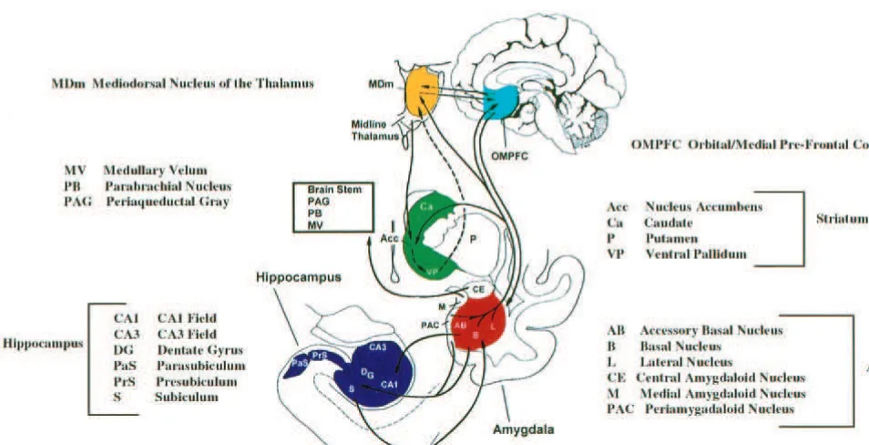
ular nucleus (Dupont et al 1995; Lenze and Sheline 1999). The studies reporting negative findings typically had lower resolution, ranging from 3 to 10 mm (Ashtari et al 1999; Axelson et al 1993; Dupont et al 1995; Swayze et al 1992), compared with 0.5–2 mm (Bremner et al 2000; Drevets et al 1997; Shah et al 1998; Sheline et al 1996, 1998) for studies reporting significant differences in major depression in these same structures, although one study reporting negative findings in the caudate nucleus and putamen (Lenze and Sheline 1999) also had high resolu-tion. In addition, a study reporting negative findings (Dupont et al 1995) measured the amygdala/hippocampus complex in bipolar subjects with major depression rather than in subjects with unipolar depression. Many of the reported changes occur in structures comprising a neuro-anatomic circuit that has been called the limbic– cortical– striatal–pallidal–thalamic (LCSPT) tract (Swerdlow and Koob 1987; Figure 1). Depression appears to involve abnormalities in specific components of this brain circuit. There is extensive interconnectivity between these struc-tures, including the prefrontal cortex, amygdala, hip-pocampus, basal ganglia, thalamus, and the connecting white matter tracts (Price et al 1987).
In postmortem studies of the prefrontal cortex in major depression (Rajkowska et al 1999), depressed subjects differed significantly from control subjects in several prefrontal cortical areas. They had decreases in cortical thickness, neuronal size decrease, and loss of glial cells in
layers II–IV of the rostral orbitofrontal cortex. Caudal orbitofrontal cortex findings were reductions in glial cells in layers V and VI and decreases in neuronal sizes. In the dorsolateral prefrontal cortex depressed subjects had re-ductions in glial and neuronal cells throughout all layers as well as reduction in cell size. Ongur et al (1998) have also reported glial cell loss in the subgenual region of the prefrontal cortex in major depression. These neuropatho-logic changes may account for MRI volumetric findings in the frontal cortex. Substantial volume reduction of 39 – 48% in the subgenual prefrontal cortex has been reported (Drevets et al 1997), as well as a much smaller 7% overall reduction in frontal lobe volume in major depression (Coffey et al 1992). The prefrontal cortex is a particularly important component of the LCSPT tract as a target of monoamine projections, and there is substantial evidence for disturbances in monoamine receptors, transporters, and second messenger systems (Arango et al 1995; Duman 1998; Mintun et al 2000; Price 1999). In addition, it is possible to speculate that overactivation in one part of this interconnected neuroanatomic circuit may lead to overex-citation in the other components, resulting in excitotoxic damage. The orbitomedial prefrontal cortex has high concentrations of glucocorticoid (GC) receptors, poten-tially rendering it vulnerable to stress-mediated damage (see below). Mechanisms involving stress and elevated GC concentrations may be more relevant in EORD than in late-onset depression.
Hippocampal Volume Loss
Some recent articles have utilized high-resolution MRI technology to examine hippocampal volumes in individu-als whose depressions were in remission, thus avoiding studying brain changes potentially due to hypercortisol-emia of depression and revealing changes that persisted beyond the acute depression. The first study (Sheline et al 1996) involved volumetric MRIs from 10 women with histories of severe, recurrent depression but in current remission for at least 6 months and a mean of 82.8 months. Case– control matching and exclusion of other physical illness or any current or past drug or alcohol abuse were important aspects of the study design. Subjects were matched within 2 years for age and education, and all were female and right-handed; the groups were matched for height. The study found reductions of 15% in left hip-pocampal volume and 12% in right hiphip-pocampal volume. Exdepressives also showed low signal foci throughout the hippocampus. The extent of left hippocampal atrophy and numbers of foci correlated with depression duration, with a similar trend for right hippocampal volume. Differences in hippocampal volume were still demonstrated after controlling for depression severity and for a history of electroconvulsive therapy (ECT). Subjects did not differ from control subjects in basal cortisol concentrations or cortisol response to dexamethasone, and there was no difference in overall cerebral volume.
A follow-up study of 24 women with histories of severe depression and remission for a minimum of 4 months (Sheline et al 1999) employed an identical case– control design. The study reported 10% and 8% reductions in left and right hippocampal volumes, respectively, with no change in total cerebral volumes. Exdepressives also had smaller volumes of the core nuclei of the amygdala, which correlated with the extent of hippocampal atrophy. In this study also, longer total duration of depression predicted greater atrophy. Post hoc analyses showed that hippocam-pal atrophy remained after controlling for a history of ECT, for postmenopausal status, and for history of estro-gen replacement therapy, and the mean duration of remis-sion was 51.7 months. Exdepressive subjects also had deficits on neuropsychologic tests of verbal memory, which are dependent on hippocampal function. There was no relationship between age and hippocampal volume reduction in either exdepressives or in control subjects, differing from several prior reports. Since the study had carefully ruled out any depressed or control subjects with medical problems, it was speculated that the subjects constituted “supernormals.”
Bremner et al (2000) examined 10 men and 6 women with severe, recurrent depressive episodes who had been in remission for an average of 7 months. Control subjects
were matched for age, gender, handedness, education, and history of alcohol abuse. Magnetic resonance imaging scan measurement revealed an average of a significant 19% volume loss in the left hippocampus and a nonsig-nificant 12% loss in the right. Of note, the method used by Bremner measured a portion of the hippocampus that includes most but not all of the structure (Bremner et al 1995). There was no change in overall brain volume or in left amygdala, caudate nucleus, or frontal or temporal lobe volumes. Exdepressives exhibited a surprising increase in volume in the right amygdala. Amygdala volumes are difficult to compare between studies because the cortical amygdala blends in with surrounding gray matter and anatomic boundaries may differ from one study to the next. Hippocampal atrophy in the Bremner study was not related to number of depressive episodes, duration of remission, hospitalizations, age, or severity of alcohol abuse. Finally, a study (Shah et al 1998) that examined brain volumes in three groups— chronic depression, remit-ted depression, and control subjects—found hippocampal atrophy in patients with chronic depression but no evi-dence of hippocampal atrophy in patients with remitted depression. Clinical characteristics of depression were not described in the remitted group, however, making com-parison with other studies difficult. Two studies (Axelson et al 1993; Swayze et al 1992) did not find hippocampal volume loss in depression but used less sensitive MRI methodology that could not differentiate the hippocampus from the amygdala. In summary, in studies that assessed depression severity and used high-resolution MRI tech-niques, depression was associated with bilateral hip-pocampal atrophy, ranging from 8% to 19%. The volume loss appears to have functional significance with an association between acute depression and abnormalities of declarative memory (Burt et al 1995) as well as an association between severe depression in remission and verbal memory (Sheline et al 1999).
Stress and Depression
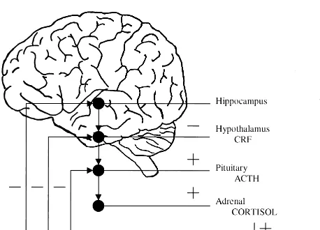
pro-cesses that can affect the hippocampus. Under normal conditions the HPA axis carries out an appropriate acute response to stress (Figure 2); there is an endocrine cascade starting with the brain, continuing to the pituitary, and ending with secretion of GCs by the adrenal gland. Negative feedback loops operate at each of these levels to restore the system to normal homeostasis; however, during conditions of chronic stress, such as occurs in depression, alterations occur in the system so that the feedback mechanisms do not operate normally and there is damage to hippocampal neuronal cells (Gold et al 1984; Holsboer et al 1987; Sapolsky et al 1991; Young et al 1991).
Animal Models of Stress and Hippocampal
Damage
A substantial body of data in animal systems indicates that recurrent episodes of stress are associated with damage to hippocampal neurons. It has been demonstrated that re-peated episodes of stress or elevated GC levels, also characteristic of depression, can produce neurotoxic dam-age to hippocampal pyramidal cells. Even 21 days of restraint stress in rats resulted in atrophy of apical den-drites of CA3 pyramidal neurons (Watanabe et al 1992a). Similarly, chronic multiple stressors (e.g., shaking in addition to restraint) produced dendritic atrophy of CA3 neurons. Multiple stressors produced a more robust in-crease in corticosterone, implicating a permissive role of another factor (excitatory amino acids) in producing dam-age (Magarinos and McEwen 1995). After repeated stres-sor episodes, ultrastructural changes (McEwen and
Mar-garinos, 1997) occur in mossy fiber projections from the granule cells in the dentate gyrus, the major excitatory input to CA3 pyramidal neurons. It is important to note, however, that these changes in ultrastructure are reversible (Conrad et al 1999), and hence by themselves cannot explain the volume loss occurring in repeated major depression. A more severe social stress (Uno et al 1989) or long-term GC treatment produced hippocampal neuronal damage in primates. At autopsy, monkeys that died after exposure to severe stress were found to have multiple gastric ulcers and hypertrophy of the adrenal cortex, indicating ongoing GC release. Furthermore, the CA3 subfield of the hippocampus was found to be damaged, and follow-up studies indicated that this damage involved hippocampal exposure to GCs (Sapolsky et al 1990). In other studies, however (Leverenz et al 1999), primates exposed to GCs in the absence of stress did not exhibit hippocampal cell loss, indicating that, in the absence of stress, chronically elevated GC levels may not produce hippocampal neurotoxicity. A recent article by Starkman et al (1999) also found that the hippocampal atrophy induced by high levels of GCs in Cushing’s disease patients was partially reversible with treatment of Cush-ing’s disease and reversal of elevated GC levels.
The mechanisms leading to GC-induced hippocampal cell death are not fully delineated, but enhanced vulnera-bility to excitotoxicity may be a critical factor (Armanini et al 1990; for reviews, see McEwen 1992; Reagan and McEwen 1997; Sapolsky et al 1986). Glucocorticoid- or psychosocial stress–induced atrophy of hippocampal py-ramidal neurons is attenuated by N-methyl-D-aspartate
receptor blockers and by phenytoin, a sodium and T-type calcium channel blocker (Magarinos et al 1996). Collec-tively, these results support a hypothesis that an interaction between GCs and glutamate is involved in stress-induced neuronal atrophy.
HPA Axis and Depression
The relevance to depression of studies demonstrating that chronically elevated GCs damage hippocampal neurons de-pends on the assumption that depression is associated with dysregulation of the GC system, and also on the assumption that exdepressives with hippocampal volume loss had ele-vated GCs. Dexamethasone nonsuppression occurs in ap-proximately half of individuals with major depression (Arana and Mossman 1988). Since baseline cortisol levels and dexamethasone suppression tests are not routinely obtained in clinical practice during acute depressive episodes, it is diffi-cult to establish a history of hypercortisolemia. None of the volumetric studies in humans, which were all retrospective, reported cortisol data as well as hippocampal volume data, making a causal link with GC levels impossible. Subjects in
these studies had long histories of recurrent, severe depres-sion, often involving hospitalization and ECT. Since more severe depression frequently involves hypercortisolism (Whiteford et al 1987), it has been hypothesized that, despite the lack of cortisol data, many subjects in these studies were likely to have been hypercortisolemic when depressed (She-line et al 1999). There have been many studies indicating that depression is accompanied by dysregulation of the HPA axis resulting in elevated cortisol levels. The first report of humans with depression secreting excessive quantities of cortisol and exhibiting insensitivity to GC feedback inhibi-tion was in 1962 (Gibbons and McHugh 1962).
Hypercortisolism and insensitivity to feedback suppression during depression have been extensively investigated. These studies determined the contributions to HPA dysfunction of adrenal hypersensitivity to adrenocorticotropic hormone (ACTH; Amsterdam et al 1989), pituitary resistance to GC feedback (Holsboer et al 1987), abnormalities in pituitary response to corticotropin-releasing factor (CRF) and other hormones (Gold et al 1984), and resistance to negative feedback at the hippocampus (Sapolsky et al 1991; Young et al 1991). The hypercortisolemia of depression involves hy-persecretion of CRF, a compensatory decrease in sensitivity of the pituitary, and increased sensitivity to ACTH by the adrenal. Gold et al (1986) hypothesized that during chronic depression with time the pituitary becomes less sensitive to CRF and the adrenal becomes hyperplastic, resulting in increased sensitivity to ACTH. Thus, HPA axis dysregulation in depression can produce repeated episodes of hypercorti-solemia, which may result in hippocampal neurotoxicity. It is important to note that the role of cortisol may vary across diagnostic/biological groups and may be more critical in early-onset depression, particularly in cases in which early life trauma occurred, than in late-life mood disorders.
Mechanisms for Volume Loss in
Early-Onset Recurrent Depression
Although follow-up studies of exdepressive subjects have not been conducted, the reported hippocampal volume loss
appears to be persistent over years, and therefore any hypothesis proposed to explain hippocampal atrophy in exdepressives must account for its seeming irreversibility. Several different mechanisms could potentially explain these findings. A narrow interpretation of the endanger-ment model (McEwen 1992; Sapolsky et al 1986) of excess GC levels combined with another insult, such as hypoxia or ischemia, appears unlikely because there is no evidence for such an accompanying insult in depression. Applied more broadly, an endangerment model might encompass damage from excitatory amino acids or sup-pression of neurogenesis during episodes of stress. Atro-phy of dendritic processes induced by excess GC levels (Watanabe et al 1992a) is unlikely as a sole etiology because of the demonstrated reversibility of dendritic atrophy (Conrad et al 1999); however, it is not known which cases of hippocampal atrophy are reversible versus irreversible, and over what period of time.
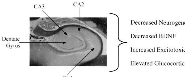
At least three potential mechanisms exist that could account for the findings to date (Figure 3). One is that the atrophy may reflect neurotoxicity (Bremner et al 2000; Sheline et al 1996, 1999), with repeated hypercortisolemic episodes of depressions giving rise to an irreversible atrophy. In the case of hippocampal volume loss, the inverse correlations between the total amount of time patients have been depressed and hippocampal volume provide evidence for recurrent depressive episodes having a causal relationship. Further, a recent study (Lupien et al 1998) has shown that in normal human aging higher cortisol levels correlated longitudinally with greater hip-pocampal volume loss. Another potential mechanism is glial cell loss either directly or indirectly producing volume loss. Several studies report gray matter atrophy in the prefrontal cortex, one in an area ventral to the genu of the corpus callosum, indicated by MRI findings (Drevets et al 1997), which were shown in postmortem studies to be due to glial cell loss (Ongur et al 1998). Another study found glial cell loss in postmortem studies of depressed subjects in two different areas of the prefrontal cortex (Rajkowska et al 1999). In addition, glial cell loss has been
reported in postmortem studies in the amygdala and in the entorhinal cortex of the hippocampus (Bowley et al, in press).
It is possible, however, that through excitatory connec-tions between the amygdala and hippocampus (White and Price 1993) damage in one structure could produce dam-age in the connected structure. Likewise, the interconnec-tions between the prefrontal cortex and the hippocampus (Carmichael and Price, 1995) could produce excitotoxic damage. Glial cells sequester glutamate, maintain meta-bolic and ionic homeostasis, and produce trophic factors, including brain-derived neurotrophic factor (BDNF; Ran-som and Sontheimer 1992; Szatkowski and Attwell 1994). Loss of glial cells could therefore increase vulnerability to neurotoxic damage and supports the idea that glutamate neurotoxicity may be involved in the volume loss in the limbic– cortical–striatal–pallidal circuit. In animal studies even brief kindled seizures may induce selective hip-pocampal volume loss (Cavazos et al 1994).
Finally, stress-induced inhibition of neurogenesis seems an attractive hypothesis, although high rates of baseline neurogenesis would be needed to produce atrophy of the scale required by hippocampal volume loss in depression. Gould et al (1997) have shown that in the tree shrew psychosocial stress suppressed neurogenesis. Likewise, corticosterone treatment in adult rats also produced sup-pression of neurogenesis, which was reversed by removal of the adrenal gland (Cameron and Gould 1994). Although neither suppression of neurogenesis nor dendritic remod-eling appears to account for the seeming irreversibility of hippocampal volume loss, there could be a combination of irreversible damage and reversible atrophy that increas-ingly converted to damage with time.
Depression and Comorbid Illness
It is important to note factors that potentially confound or contribute to anatomic changes due to depression, partic-ularly comorbid illness. This applies much more to late-life depression than to early-onset depression, due largely to the increased prevalence of comorbid illness with age; however, although comorbidity may contribute to a higher proportion of late-life depressions than in younger pa-tients, there are other important factors as well. Several computed tomography and MRI studies have shown dif-fuse cortical and subcortical atrophy and ventricular en-largement in late-life depression (Pantel et al 1997; Rabins et al 1991; Rothschild et al 1989; Soares and Mann 1997). A likely explanation for some findings of generalized brain atrophy in some studies is the comorbidity of major depression with other illnesses, especially in the case of patients with late-onset depression. Clinically significant depressive symptoms are detectable in approximately
12–36% of patients with another nonpsychiatric general medical condition, compared with approximately 5% in the general population (U.S. Department of Health and Human Services 1993). Conversely, patients with depres-sion have a significantly higher rate of other medical illnesses. Specific illnesses that have been determined to cause brain atrophy include hypertension (Kobayashi et al 1991), diabetes (Aronson 1973), Cushing’s disease (Stark-man et al 1992), and alcohol abuse (Charness 1993); however, any condition that produces neuronal ischemia or neurotoxicity is a potential candidate for producing brain atrophy.
Late-Onset Depression
Late age– onset depression frequently occurs in patients with medical and neurologic disorders. It is characterized by greater medical morbidity and mortality than early-onset depression (Jacoby et al 1981); higher rates of neuroradiologic abnormalities, particularly white matter hyperintensities (Coffey et al 1988; Figiel et al 1991); lower frequency of affective disorders in families of patients (Baron et al 1981); and, in some studies, higher rates of treatment refractoriness (Alexopoulos et al 1996). It is well established that late-onset depression may be precipitated by damage to key brain structures caused by age-associated medical or neurologic disorders (Alexo-poulos et al 1988). A number of neurologic illnesses associated with both cortical and subcortical atrophy are associated with unusually high rates of depression, includ-ing dementia of the Alzheimer’s type (Burns et al 1990), poststroke syndromes (Starkstein and Robinson 1989), Parkinson’s disease (Cummings 1992), and Huntington’s disease (Folstein et al 1983). These findings suggest that late-onset depression often is associated with age-related illnesses, which can produce damage to key brain struc-tures. In illnesses with high rates of depression, the same brain structures that have been implicated in more classic or early-onset major depression are involved—namely, the frontal cortex, hippocampus, caudate nucleus, thalamus, and basal ganglia. Not all studies find evidence for generalized atrophy in addition to volume loss in struc-tures of the LCSPT circuit. For example, Kumar et al (1998) have found loss in prefrontal lobe volume in late-onset depression in the absence of generalized atro-phy, suggesting that, as in early-onset depression, subjects with late-onset depression may also have focal loss in volume. Whether this focal volume loss involves the same etiologic mechanisms is not known.
The finding of increased numbers of hyperintensites seen on T2-weighted scans (T2H) in elderly subject groups
1991; Rabins et al 1991; Zubenko et al 1990). This result has also been reported in studies that included younger subjects (Coffey et al 1993; Hickie et al 1995), though negative findings with younger groups have been reported as well (Dupont et al 1995; Guze and Szuba 1992); however, T2H are also noted to occur at rates of up to 60% in healthy elderly patients (Fazekas et al 1991), in whom their significance is unknown (Mirsen et al 1991). In late-life depression clinical correlates of MRI-defined T2H have included older age, vascular risk factors, and late-onset depressive illness (Coffey et al 1993; Krishnan et al 1988). Fujikawa et al (1993, 1994) found a higher rate of “silent” cerebral infarctions (T2H) in late-onset major depressive disorder than in early onset. A subtype of “vascular depression” with increased cardiovascular dis-ease risk factors and incrdis-eased T2H has been proposed (Alexopoulos et al 1996; Krishnan et al 1997).
Future Directions
Given the fairly small differences in brain structure vol-umes between depressed and control subjects, it is impor-tant to enhance the ability to detect these differences. Recent advances in MRI technology have allowed much finer resolution; it is currently possible to obtain a resolu-tion of 0.5 mm, compared with the 3- to 5-mm resoluresolu-tion of many previous studies. At this resolution, for example, it is possible to distinguish the thin white matter layer separating the hippocampus from the amygdala, and to measure the hippocampal gray matter volume alone rather than the hippocampus/amygdala complex. The specific nature of the association between recurrent depression and volume reduction in regions of the LCSPT circuit is not known. The possibility cannot be excluded that these volume decreases precede the onset of depression or that the volume decrease is simply a marker of some other brain abnormality that predisposes to depression. Prospec-tive studies are needed to examine this question more fully. Studies in high-risk populations, such as first-degree relatives of affected individuals, will assist in determining whether focal atrophy changes are genetic/neurodevelop-mental or acquired and whether they predate or follow the development of depression. Additional postmortem stud-ies are also needed. Studstud-ies are needed of individuals at the time of their initial diagnosis of depression, to test the idea of atrophy as preceding and predisposing toward repeated depression. In addition, it will be useful to test explicitly whether only depressives with baseline hyper-cortisolemia show hippocampal atrophy. Furthermore, it will be important to combine structural studies with functional studies to determine the functional significance of brain structure changes. Combining MRI and functional studies such as positron emission tomography, single
photon emission computed tomography, and functional MRI has the potential to more precisely localize abnor-malities in blood flow, metabolism, and neurotransmitter receptors. This integrated perspective will allow further development of a structural–functional model of depres-sion. Neuroprotective strategies aimed at preventing the damage associated with depression are likely to be an important future direction for research. Preclinical studies provide preliminary strategies for preventing stress-in-duced hippocampal damage. These include, for example, prevention of stress-induced decreases in BDNF with antidepressants (Nibuya et al 1995, 1996; Vaidya and Duman 1999), prevention of stress-induced excitotoxic injury with phenytoin (Dilantin; Watanabe et al 1992b), prevention of stress-induced decreases in neurogenesis with antidepressants (Duman and Malberg 1998; Jacobs et al 1998), and increase in dendritic branching with seroto-nin reuptake inhibitors (Duman et al 1997).
Supported in part by Grants Nos. MH01370 and MH5844.
Aspects of this work were presented at the conference “Depression in the Twenty-First Century: New Insights into Drug Development and Neurobiology,” February 21–22, 2000, Dana Point, California. The conference was sponsored by the Society of Biological Psychiatry through an unrestricted educational grant provided jointly by Pharmacia & Upjohn and Janssen Pharmaceutica.
References
Aggleton JP (1992): Amygdala: Neurobiological Aspects of Emotion, Memory, and Mental Dysfunction. New York: Wiley-Liss.
Alexopoulos GS, Meyers B, Young R, Kakuma T, Feder M, Einhorn A, Rosendahl E (1996): Recovery in geriatric de-pression.Arch Gen Psychiatry53:305–312.
Alexopoulos GS, Young RC, Meyers BS, Abrams RC (1988): Late onset depression.Psychiatr Clin North Am11:101–115. Amsterdam J, Maislin G, Berwish N, Phillips J, Winokur A (1989): Enhanced adrenocortical sensitivity to submaximal doses of cosyntropin in depressed patients.Arch Gen Psychi-atry46:550 –554.
Arana GW, Mossman D (1988): The dexamethasone suppression test and depression.Endocrinol Metab Clin North Am17:21–39. Arango V, Underwood MD, Gubbi AV, Man JJ (1995): Local-ized alterations in pre- and postsynaptic serotonin binding sites in the ventrolateral prefrontal cortex of suicide victims.
Brain Res688:121–133.
Armanini MP, Hutchins C, Stein BA, Sapolsky RM (1990): Glucocorticoid endangerment of hippocampal neurons is NMDA-receptor dependent.Brain Res532:7–12.
Aronson S (1973): Intracranial vascular lesions in patients with diabetes mellitus.J Neuropathol Exp Neurol32:183–196. Ashtari M, Greenwald BS, Kramer-Ginsberg E, Hu J, Wu H,
Patel M, et al (1999): Hippocampal/amygdala volumes in geriatric depression.Psychol Med29:629 – 638.
McDonald WM, Ritchie JC, et al (1992): In vivo assessment of pituitary volume with magnetic resonance imaging and systematic stereology: Relationship to dexamethasone sup-pression test results in patients.Psychiatry Res44:63–70. Axelson DS, Doraiswamy PM, McDonald WM, Boyko OB,
Tupler LA, Patterson LJ, et al (1993): Hypercortisolemia and hippocampal changes in depression.Psychiatry Res47:163– 173.
Baron M, Mendlewicz J, Klotz J (1981): Age-of-onset and genetic transmission in affective disorders. Acta Psychiatr Scand64:373–380.
Bjorklund A, Hokfelt T (1987):Handbook of Chemical Neuro-anatomy.Amsterdam: Elsevier.
Bowley MP, Drevets WC, Ongur D, Price JL (in press): Glial changes in the amygdala and entorhinal cortex in mood disorders.Soc Neurosci Abstr.
Bremner JD, Narayan M, Anderson ER, Staib LH, Miller HL, Charney DS (2000). Hippocampal volume reduction in major depression.Am J Psychiatry157:115–118.
Bremner JD, Randall PR, Scott TM, Bronen RA, Seibyl JP, Southwick SM, et al (1995): MRI-based measurement of hippocampal volume in posttrumatic stress disorder.Am J Psychiatry152:973–981.
Burns A, Jacoby R, Levy R (1990): Psychiatric phenomena in Alzheimer’s disease III: Disorders of mood.Br J Psychiatry
157:81– 86.
Burt DB, Zembar MJ, Niederehe G (1995): Depression and memory impairment: A meta-analysis of the association, its pattern, and specificity.Psychol Bull117:285–305. Cameron HA, Gould E (1994): Adult neurogenesis is regulated
by adrenal steroids in the dentate gyrus. Neuroscience 61: 203–209.
Carmichael ST, Price JL (1995): Limbic connections of the orbital and medial prefrontal cortex in macaque monkeys.
J Comp Neurol363:615– 641.
Cavazos JE, Das I, Sutula TP (1994): Neuronal loss induced in limbic pathways by kindling: Evidence for induction of hippocampal sclerosis by repeated brief seizures.J Neurosci
14:3106 –3121.
Charness ME (1993): Brain lesions in alcoholics.Alcohol Clin Exp Res17:2–11.
Coffey C, Figiel G, Djang W (1988): Leukoencephalopathy in elderly depressed patients referred for ECT.Biol Psychiatry
24:143–161.
Coffey CE, Figiel GS, Djang WT, Weiner RD (1990): Subcor-tical hyperintensity on magnetic imaging: A comparison of normal and depressed elderly subjects. Am J Psychiatry
147:187–189.
Coffey CE, Wilkinson WE, Parashos IA, Soady SA, Sullivan RJ, Patterson LJ, et al (1992): Quantitative cerebral anatomy of the aging human brain: A cross-sectional study using mag-netic resonance imaging.Neurology42:527–536.
Coffey CE, Wilkinson WE, Weiner RD, Parashos IA, Djang WT, Webb MC, et al (1993): Quantitative cerebral anatomy in depression: A controlled magnetic resonance imaging study.
Arch Gen Psychiatry50:7–16.
Conrad CD, LeDoux JE, Magarinos AM, McEwen BS (1999): Repeated restraint stress facilitates fear conditioning indepen-dently of causing hippocampal CA3 dendritic atrophy.Behav Neurosci113:902–913.
Cummings JL (1992): Depression and Parkinson’s disease: A review.Am J Psychiatry149:443– 454.
DeArmond SJ, Fusco MM, Dewey MM (1989):Structure of the Human Brain: A Photographic Atlas, 3rd ed. New York: Oxford University Press.
Drevets WC, Price JL, Simpson JR Jr, Todd RD, Reich T, Vannier M, Raichle ME (1997): Subgenual prefrontal cortex abnormalities in mood disorders.Nature386:824 – 827. Duman RS (1998): Novel therapeutic approaches beyond the
serotonin receptor.Biol Psychiatry44:324 –335.
Duman RS, Heninger GR, Nestler EJ (1997): A molecular and cellular theory of depression.Arch Gen Psychiatry54:597– 606.
Duman RS, Malberg JE (1998): Neural plasticity in the patho-physiology and treatment of depression.Am Coll Neuropsy-chopharmacol37:261.
Dupont RM, Jernigan TL, Heindel W, Butters N, Shafer K, Wilson T, et al (1995): Magnetic resonance imaging and mood disorders—localization of white matter and other sub-cortical abnormalities.Arch Gen Psychiatry52:747–755. Fazekas F, Kleinert R, Offenbacher H, Payer F, Schmidt R,
Kleinert G, et al (1991): The morphologic correlate of incidental punctate white matter hyperintensities on MR images.AJNR Am J Neuroradiol12:915–921.
Figiel GS, Krishnan KRR, Doraiswamy PM, Rao VP, Nemeroff CB, Boyko OB (1991): Subcortical hyperintensities on brain magnetic resonance imaging: A comparison between late age onset and early onset elderly depressed subjects.Neurobiol Aging26:245–247.
Folstein SE, Abbott MH, Chase GA, Jensen BA, Folstein MF (1983): The association of affective disorder with Hunting-ton’s disease in a case series and in families.Psychol Med
13:537–542.
Fujikawa T, Yamawaki S, Touhouda Y (1993): Incidence of silent cerebral infarction in patients with major depression.
Stroke24:1631–1634.
Fujikawa T, Yamawaki S, Touhouda Y (1994): Background factors and clinical symptoms of major depression with silent cerebral infarction.Stroke25:798 – 801.
Gibbons J, McHugh P (1962): Plasma cortisol in depressive illness.J Psychiatry Res1:162–171.
Gold P, Chrousos G, Kellner C, Post R, Roy A, Augerinos P, et al (1984): Psychiatric implications of basic and clinical studies with CRF.Am J Psychiatry141:619 – 627.
Gold P, Loriaux L, Roy A, Kling MA, Calabrese JR, Kellner CH, et al (1986): Responses to corticotropin-releasing hormone in the hypercortisolism of depression and Cushing’s disease.
N Engl J Med314:1329 –1335.
Gould E, McEwen BS, Tanapat P, Galea LAM, Fuchs E (1997): Neurogenesis in the dentate gyrus of the adult tree shrew is regulated by psychosocial stress and NMDA receptor activa-tion.J Neurosci17:2492–2498.
Guze BH, Szuba MP (1992): Leukoencephalopathy and major depression: A preliminary report.Psychiatry Res45:169 –175. Hickie I, Scott E, Mitchell P, Wilhelm K, Austin MB, Bennett
B (1995): Subcortical hyperintensities on magnetic reso-nance imaging: Clinical correlates and prognostic signifi-cance in patients with severe depression.Biol Psychiatry
37:151–160.
aldosterone and ACTH release after human CRH administra-tion in depressed patients.Am J Psychiatry144:229 –231. Howard RJ, Beats B, Forstl H, Graves P, Bingham J, Levy R
(1993): White matter changes in late onset depression: A magnetic resonance imaging study.Int J Geriatr Psychiatry
8:183–185.
Husain MM, McDonald WM, Doraiswamy PM, Figiel GS, Na C, Escalona PR, et al (1991): A magnetic resonance imaging study of putamen nuclei in major depression.Psychiatry Res
40:95–99.
Jacobs BL, Tanapat P, Reeves AJ, Gould E (1998): Serotonin stimulates the production of new hippocampal granule neu-rons via the 5-HT1A receptor in the adult rat.Soc Neurosci
24:1992.
Jacoby RJ, Levy R, Bird JM (1981): Computed tomography and the outcome of affective disorder: A follow-up study of elderly patients.Br J Psychiatry139:288 –292.
Kobayashi S, Okada K, Yamashita K (1991): Incidence of silent lacunar lesion in normal adults and its relation to cerebral blood flow and risk factors.Stroke22:1379 –1383.
Krishnan K, Hays J, Blazer D. (1997): MRI-defined vascular depression.Am J Psychiatry154:497–501.
Krishnan KR, Goli V, Ellinwood EH, France RD, Blazer DG, Nemeroff CB (1988): Leukoencephalopathy in patients diag-nosed as major depressive.Biol Psychiatry23:519 –522. Krishnan KR, McDonald W, Escalona P, Doraiswamy PM, Na
C, Husain MM, et al (1992): MRI of the caudate nuclei in depression.Arch Gen Psychiatry49:553–557.
Krishnan KR, McDonald WM, Doraiswamy PM (1993): Neuro-anatomical substrates of depression in the elderly.Eur Arch Psychiatry Clin Neurosci243:41– 46.
Kumar A, Jin Z, Bilker W, Udupa J, Gottlieb G (1998): Late-onset minor and major depression: Early evidence for common neuroanatomical substrates detected by using MRI.
Proc Natl Acad Sci U S A95:7654 –7658.
Lenze E, Sheline Y (1999): Absence of striatal volume differ-ences between healthy depressed subjects and matched com-parisons.Am J Psychiatry6:1989 –1991.
Lesser IM, Miller BL, Boone KB, Hill-Gutierrez E, Mehringer CM, Wong K, Mena I (1991): Brain injury and cognitive function in late-onset psychotic depression.J Neuropsychia-try Clin Neurosci3:33– 40.
Leverenz JB, Wilkinson CW, Wamble M, Corbin S, Grabber JE, Raskind MA, Peskind ER (1999): Effect of chronic high-dose exogenous cortisol on hippocampal neuronal number in aged nonhuman primates.J Neurosci19:2356 –2361.
Lupien SJ, de Leon M, deSanti S, Convit A, Tarshish C, Nair NP, et al (1998): Cortisol levels during human aging predict hippocampal atrophy and memory deficits. Nat Neurosci
1:69 –73.
Magarinos AM, McEwen BS (1995): Stress induced atrophy of apical dendrites of hippocampal CA3C neurons: Involvement of GC secretion and excitatory amino acid receptors. Neuro-science69:89 –98.
Magarinos AM, McEwen BS, Flugge G, Fuchs E (1996): Chronic psychosocial stress causes apical dendritic atrophy of hippocampal CA3 pyramidal neurons in subordinate tree shrews.J Neurosci16:3534 –3540.
Mai JK, Assheuer J, Paxinos G (1995): Atlas of the Human Brain.Orlando: Academic Press.
McEwen BS (1992): Re-examination of the glucocorticoid hy-pothesis of stress and aging. In: Swaab D, Hofman M, Mirmiran M, Ravid R, van Leeuwen F, editors.Progress in Brain Research.New York: Elsevier, 356 –383.
McEwen BS, Magarinos AM (1997): Stress effects on morphol-ogy and function of the hippocampus. Ann N Y Acad Sci
821:271–284.
Mintun MA, Sheline YI, Moerlein SM, Snyder AZ (2000): Regional [18F]altanserin binding in the treatment of major depression.Neuroimage11:S83.
Mirsen TR, Lee DH, Wong CJ, Diaz JF, Fox AJ, Hachinski VC, Merskey H (1991): Clinical correlates of white-matter changes on magnetic resonance imaging scans of the brain.
Arch Neurol48:1015–21.
Nibuya M, Morinobu S, Duman RS (1995): Regulation of BDNF and trkB mRNA in rat brain by chronic electoconvulsive seizure and antidepressant drug treatments. J Neurosci15: 7539 –7547.
Nibuya M, Nestler EJ, Duman RS (1996): Chronic antidepres-sant administration increases the expression of cAMP re-sponse element binding protein (CREB) in rat hippocampus.
J Neurosci16:2365–2372.
Ongur D, Drevets WC, Price JL (1998): Glial reduction in the subgenual prefrontal cortex in mood disorders. Proc Natl Acad Sci U S A95:13290 –13295.
Pantel J, Schroder J, Essig M, Popp D, Dech H, Knopp MV, et al (1997): Quantitative magnetic resonance imaging in geri-atric depression and primary degenerative dementia.J Affect Disord42:69 – 83.
Price J, Russchen F, Amaral D (1987): The limbic region. II: The amygdaloid complex. In: Bjorkland A, Hokfelt T, Swanson L, editors.Handbook of Chemical Neuroanatomy,Vol 5. New York: Elsevier, 279 –388.
Price JL (1999): Prefrontal cortical networks related to visceral function and mood.Ann N Y Acad Sci877:383–396. Rabins PV, Pearlson GD, Aylward E, Kumar AJ, Dowell K
(1991): Cortical magnetic resonance imaging changes in elderly inpatients with major depression. Am J Psychiatry
148:617– 620.
Rajkowska G, Miguel-Hidalgo JJ, Wei J, Dilley G, Pittman SD, Meltzer HY, et al (1999): Morphometric evidence for neuro-nal and glial prefrontal cell pathology in major depression.
Biol Psychiatry45:1085–1098.
Ransom BR, Sontheimer H (1992): The neurophysiology of glial cells.J Clin Neurophysiol9:224 –251.
Reagan LP, McEwen BS (1997): Controversies surrounding glucocorticoid-mediated cell death in the hippocampus.
J Chem Neuroanat13:149 –167.
Rothschild AJ, Benes F, Hebben N, Woods B, Luciana M, Bakanas E, et al (1989): Relationships between brain CT scan findings and cortisol in psychotic and nonpsychotic depressed patients.Biol Psychiatry26:565–575.
Sapolsky RM, Krey LC, McEwen BS (1986): The neuroendo-crinology of stress and aging: The glucocorticoid cascade hypothesis.Endocrinol Rev7:284 –301.
Sapolsky RM, Uno H, Rebert CS, Finch CE (1990): Hippocam-pal damage associated with prolonged glucocorticoid expo-sure in primates.J Neurosci10:2897–2902.
glucocorticoid secretion by the hippocampal formation in the primate.J Neurosci11:3695–3704.
Selye H, Tuchweber B (1976): Stress in relation to aging and disease. In: Everitt A, Burgess J, editors. Hypothalamus, Pituitary and Aging.Springfield, IL: C.C. Thomas, 557. Shah PJ, Ebmeier KP, Glabus MF, Goodwin GM (1998):
Cortical grey matter reductions associated with treatment resistant chronic unipolar depression. Controlled magnetic resonance imaging study.Br J Psychiatry172:527–532. Sheline Y, Sanghavi M, Mintun M, Gado M (1999): Depression
duration but not age predicts hippocampal volume loss in women with recurrent major depression. J Neurosci 19: 5034 –5043.
Sheline Y, Wang P, Gado M, Csernansky J, Vannier M (1996): Hippocampal atrophy in recurrent major depression. Proc Natl Acad Sci U S A93:3908 –3913.
Sheline YI, Gado MH, Price JL (1998): Amygdala core nuclei volumes are decreased in recurrent major depression. Neuro-report9:2023–2028.
Soares JC, Mann JJ (1997): The anatomy of mood disorders— review of structural neuroimaging studies.Biol Psychiatry41: 86 –106.
Starkman MN, Gebarski SS, Berent S, Schteingart DE (1992): Hippocampal formation volume, memory dysfunction, and cortisol levels in patients with Cushing’s syndrome. Biol Psychiatry32:756 –765.
Starkman MN, Giordani B, Gebarski SS, Berent S, Schork MA, Schteingart DE (1999): Decrease in cortisol reverses human hippocampal atrophy following treatment of Cushing’s dis-ease.Biol Psychiatry46:1595–1602.
Starkstein SE, Robinson RG (1989): Affective disorders and cerebral vascular disease.Br J Psychiatry154:170 –182. Swayze VW, Andreasen NC, Alliger RJ, Yuh WTC, Ehrhard JC
(1992): Subcortical and temporal structures in affective dis-order and schizophrenia: A magnetic resonance imaging study.Biol Psychiatry31:221–240.
Swerdlow NR, Koob GF (1987): Dopamine, schizophrenia, mania
and depression: Toward a unified hypothesis of cortico-striato-pallido-thalamic function.Behav Brain Sci10:197–245. Szatkowski M, Attwell D (1994): Triggering and execution of
neuronal death in brain ischemia: Two phases of glutamate release by different mechanisms. Trends Neurosci 17:359 – 365.
Uno H, Tarara R, Else JG, Suleman MA, Sapolsky RM (1989): Hippocampal damage associated with prolonged and fatal stress in primates.J Neurosci1705–1711.
U.S. Department of Health and Human Services (1993):Clinical Practice Guideline Number 5, Depression in Primary Care: Volume 1. Detection and Diagnosis.Washington, DC: U.S. Department of Health and Human Services.
Vaidya VA, Duman RS (1999): Role of 5-HT2A receptors in down-regulation of BDNF by stress. Neurosci Lett 287: 1– 4.
Watanabe Y, Gould E, McEwen BS (1992a): Stress induces atrophy of apical dendrites of hippocampal CA3 pyriamidal neurons.Brain Res588:341–345.
Watanabe Y, Gould H, Cameron D, Daniels D, McEwen BS (1992b): Phenytoin prevents stress and corticosterone in-duced atrophy of CA3 pyramidal neurons. Hippocampus
2:431– 436.
White LE, Price JL (1993): The functional anatomy of limbic status epilepticus in the rat. I. Patterns of [14C]2-deoxyglu-cose uptake and Fos immunocytochemistry. J Neurosci13: 4787– 4809.
Whiteford H, Peabody C, Csernansky J, Warner M, Berger P (1987): Elevated baseline and postdexamethasone cortisol levels. A reflection of severity or endogeneity? J Affect Disord12:199 –202.
Young EA, Haskett RF, Murphy-WeinbergV, Watson SJ, Akil H (1991): Loss of glucocorticoids fast feedback in depression.
Arch Gen Psychiatry48:693– 698.
Zubenko GS, Sullivan P, Nelson JP, Belle SH, Wolf G (1990): Brain imaging abnormalities in mental disorders of late life.