www.elsevier.com / locate / bres
Research report
Protractive effects of chronic treatment with an acutely sub-toxic
regimen of diisopropylflurophosphate on the expression of cholinergic
receptor densities in rats
a a,b c d
J. Derek Stone , Alvin V. Terry Jr. , James R. Pauly , Mark A. Prendergast ,
a ,
*
Jerry J. Buccafusco
aAlzheimer’s Research Center, Department of Pharmacology and Toxicology, Medical College of Georgia, and the Veterans Affairs Medical Center, Augusta, GA 30912-2300, USA
b
College of Pharmacy, University of Georgia, Augusta, GA, USA
c
College of Pharmacy, University of Kentucky, Lexington, KY, USA
d
Tobacco and Health Research Institute, University of Kentucky, Lexington, KY, USA
Accepted 5 July 2000
Abstract
Individuals chronically exposed to low levels of organophosphate insecticides may present with subtle impairments in cognition. In addition, low level diisopropylflurophosphate (DFP) exposure (0.25 mg / kg per day for 2 weeks) in rats resulted in protracted working memory impairment [29]. The current studies attempt to show a temporal relationship between the DFP-induced impairment in performance of a spatial memory task and the protracted decrease in the expression of cholinergic receptors and acetylcholinesterase in
3 3
specific brain regions. Cholinergic receptors labeled with the ligands [ H]epibatidine and [ H]AFDX-384 were affected to a much greater 3 extent and for a longer period of time than were both acetylcholinesterase activities and cholinergic receptors labeled with [ H]QNB. Pre-testing administration of nicotine was shown to completely reverse this DFP-induced impairment in memory-related task performance. Additionally, prophylaxis with pyridostigmine bromide (PB) caused DFP-treated animals to exhibit near normal levels of memory-related task performance. These results are consistent with the development of a protracted phase of learning impairment to sub-acute DFP exposure, which may involve the loss of hippocampal nicotinic receptors, and may be prevented or reversed by PB or nicotine, respectively. 2000 Elsevier Science B.V. All rights reserved.
Theme: Disorders of the nervous system
Topic: Neurotoxicity
Keywords: DFP; Organophosphate; Acetylcholinesterase; Cholinergic receptor; Nicotine; Pyridostigmine bromide; Spatial learning; Memory
1. Introduction death may occur following acute exposure to high levels of
OP insecticides; effects attributed largely to postsynaptic
Acute exposure to organophosphorus (OP) compounds cholinergic receptor overstimulation. During chronic
expo-such as diisopropylfluorophosphate (DFP) increases neural sure, animals and humans may become tolerant to the
activity in CNS regions and peripheral organs innervated acutely toxic effects of OP agents, such as OP-induced
by acetylcholine-containing neurons. Severe toxicity and locomotor abnormalities or gastrointestinal disturbances
[4,10,32]. This behavioral and autonomic tolerance gener-ally is considered to reflect the down regulation or adaptation of cholinergic muscarinic [5,36,37,48] and
*Corresponding author. Tel.: 11-706-721-6355; fax: 1
1-706-721-nicotinic [37] receptors in various brain regions. Although
9861.
E-mail address: [email protected] (J.J. Buccafusco). significant tolerance to OP agents has been documented,
10 J.D. Stone et al. / Brain Research 882 (2000) 9 –18
tolerance develops to some but not all of the behavioral 2. Materials and methods
effects of DFP, and this tolerance may develop at different
rates [33,44]. 2.1. Subjects
Despite the onset of tolerance in these situations, the
adverse effects of OP compounds on higher brain func- Male Wistar rats (Harlan Sprague–Dawley),
approxi-tions, such as learning and memory may persist for quite mately 4 months old (weighing 350–400 g) were used in
some time after termination of toxicant exposure. The these studies. Each rat was housed individually in a
results from several studies have demonstrated the pres- stainless steel mesh cage in a temperature controlled room
ence of OP-induced learning impairments several days (258C) with free access to food (NIH-07 formula) and
after the behavioral signs of DFP, disulfoton, or soman water, and maintained on a 12-h light / dark cycle (lights on
toxicity have subsided [1,6,8,27]. Additionally, workers at 18:00 h). All animal protocols were previously approved
chronically exposed to OP agents present with a variety of by the institutional Committee on Animal Use for
Re-psychiatric sequelae, including depression, apathy, ir- search and Education.
ritability, and schizophreniform illness. One predominate
set of symptoms includes loss of concentration, difficulty 2.2. Drug administration
in thinking, and memory impairment [13,18,26]. Memory
impairments induced by chronic OP administration appear DFP (250 mg / kg; Sigma, St. Louis, MO) dissolved in
to be most evident on novel learning tasks (i.e., those saline was administered (s.c.) daily for 14 consecutive
which require the greatest reliance on working memory) days. This is referred to in the present manuscript as the
[16,42] and may persist for extended periods of time after standard DFP regimen. All injections were given in a
DFP withdrawal. For example, we reported that spatial volume of 1 ml / kg body weight between 09:00 and 11:00
learning in rats is impaired for up to 21 days after h.
withdrawal from a 14-day treatment regimen with DFP
(250 mg / kg per day) [29]. This impaired learning was not 2.3. Receptor autoradiography
temporally associated with DFP-induced reductions in
brain acetylcholinesterase activity. However, a comparable Separate groups of rats (n55–6 / group) were examined
DFP regimen did not impair performance of a well-learned at 1, 7, and 21 days after OP discontinuation. These tissues
delayed matching to sample task (in monkeys) or a were derived from rats that had participated in earlier
previously experienced spatial navigation task (in rats), behavioral studies [29,30]. Immediately on completion of
indicating that tasks dependent on reference memory were the behavioral studies, the brains were removed and flash
not significantly affected by DFP exposure [30]. These frozen in dry ice / isopentane. The frozen tissues were
results support the possibility that chronic exposure to OP stored at2708C until use. Each frozen brain was sectioned
agents can result in specific long-term cognitive deficits coronally from the frontal pole through the level of the
3
even when overt symptoms of excessive cholinergic cerebellar peduncles. [ H]QNB, a non-selective muscarinic
3
activity are not present. receptor ligand, and [ H]AFDX-384, a partially selective
Although the neuropathological basis for this protracted M2 muscarinic receptor ligand, were used to determine the
cognitive impairment is unknown, it is not likely that the numbers of muscarinic receptors in various brain regions.
insult represents a severe pathological event as may be Brain sections were preincubated in Tris–phosphate buffer
observed in idiopathic neurodegenerative disorders such as for 30 min at 258C. The sections were then transferred to
3
Alzheimer’s disease. Rather, it is more likely that the fresh Tris–phosphate buffer containing either [ H]QNB
3
behavioral changes, which are observed after accidental (800 pM) or [ H]AFDX-384 (5 nM) and incubated for 120
exposure to an OP agent, result from more subtle neuro- min at 258C. Similarly, the number of nicotinic receptors
3
chemical alterations. Therefore, the purpose of the present was estimated using [ H]epibatidine as the radioactive
study was to temporally relate the specific alterations in ligand. These brain sections were preincubated in Tris–
cholinergic neurochemical markers in specific brain areas Hepes buffer for 30 min at 48C, then incubated for an
with the time-course of behavioral changes observed additional 120 min in fresh Tris–Hepes buffer containing
3
previously [29] in the impairment of a spatial memory [ H]epibatidine (200 pM) at 48C. All sections were washed
task. We also sought to determine whether nicotine (a three times in their respective incubation buffers (minus
memory enhancing agent) could reverse the deficits in radioactive ligands) for 4 min each at 48C. Additionally,
cognitive function commonly observed after DFP expo- the sections were washed for 10 s in deionized water at
sure. Because of the modern use of the reversible cholines- 48C. Unlabeled atropine or nicotine (10mM) was added to
terase inhibitor pyridostigmine bromide (PB) as a the incubation buffer to determine non-specific binding for
prophylactic protecting agent against nerve gas poisoning, muscarinic and nicotinic receptors, respectively. After
we examined whether co-administration of PB with DFP incubations and washings, all sections were air dried and
could mitigate the behavioral or cognitive changes caused stored in a dessicator at room temperature overnight. The
Hyperfilm for 2–6 weeks. Receptor binding in specific traveled), to a video tracking system (Poly-Track, San
brain nuclei was quantified from autoradiographs using Diego Instruments, San Diego, CA).
NIH Image software. In all cases, non-specific ligand
binding was equal to background and receptor quantifica- 2.6. Hidden platform test
tion was only performed for those brain regions that
expressed a signal greater than background. Molar quan- Each rat was given four trials per day (session) for four
tities of ligand bound were determined using values consecutive days. On days 1–4, a trial began by placing
3
interpolated from the optical density versus a tissue ( H the rat in the water facing the pool wall in one of the four
3
brain paste standards) radioactivity standard curve. H quadrants. Rats were not allowed to acclimate to the pool
paste standards were applied to each autoradiographic film, prior to testing at any point during days 1–4. The daily
and each structure was measured bilaterally in at least four order of entry into individual quadrants was randomized
sections for each animal. Binding analyses for each ligand such that all four quadrants were used once every day. For
were performed in consecutive sections from the same each trial, the rat was allowed to swim a maximum of 90 s
brain sample. in order to find the hidden platform. When successful, the
rat was allowed a 30-s rest period on the platform. If unsuccessful within the allotted time period, the rat was 2.4. Brain acetylcholinesterase assay
assigned a score of 90 s, and was physically placed on the platform and allowed the 30-s rest period. In either case, Brain regions (frontal cortex and hippocampus) were
the rat was given the next trial (intertrial interval, 30 s) isolated and assayed spectrophotometrically in phosphate
after the rest period. buffer at pH 7.9 according to previously published
meth-ods [14]. Briefly, brain tissues were dissected and
homogenized (25%, w / v, in buffer) for 1 min using a 2.7. Administration of nicotine in DFP-treated rats
Bellco glass homogenizer with Teflon pestle. The
homoge-nate was then centrifuged at 40 0003g for 30 min at 48C. In this experimental series, the standard 14-day saline
The supernatant (100ml) was subsequently introduced into (one group) or DFP (three groups) regimen was used. Two
a cuvette containing the reaction mixture (7.5 mM weeks after completion of the regimen all four groups
acetylthiocholine iodide–substrate and 10.0 mM dithiobis- (n510–12 rats / group) were initiated in the standard water
nitrobenzoic acid, DTNB). Absorbance at 412 nm was maze task (as described above). The saline regimen group
recorded at 258C for 4 min. Protein concentrations were received a subcutaneous injection of saline 15 min before
spectrophotometrically measured using the Bio-Rad Pro- the first trial of the first day’s session. Likewise, one of the
tein Assay (Richmond, CA, USA) system with bovine groups that had received the DFP regimen was injected
serum albumin as the standard. Enzyme velocities were with saline before each day’s session. For the other two
expressed as mM of substrate hydrolyzed / min per mg DFP groups, one group received 0.5 mg / kg of nicotine
protein and the percentage of inhibition of enzyme activity (s.c.), and the other received 1.0 mg / kg of nicotine (s.c.)
for each OP dose (relative to control levels) was de- before each day’s session.
termined.
2.8. Evaluation of olfactory behavior 2.5. Water maze testing
Olfactory behavior (rearing and sniffing frequency) was measured immediately following daily administration of Maze testing was performed in a circular pool (diameter,
PB and / or DFP (see below) to assess for signs of overt 180 cm; height, 76 cm) made of plastic (Bonar Plastics,
cholinergic toxicity. Animals were placed in clear poly-Noonan, GA) with the inner surface painted black. The
propylene containers (25345325 cm) for 35 min. After a
pool was filled to a depth of 35 cm of water (non-opaque,
5-min acclimation period, rearing and sniffing behavior
maintained at 258C) which covered a black 10-cm square
was recorded for 30 min. Following the 30-min observa-platform. The platform was submerged one cm below the
tion period, animals were returned to their home cages. surface of the water and placed in the center of the
northeast quadrant on all trials. The pool was located in a
large room with a number of visual extra-maze cues 2.9. Co-administration of pyridostigmine bromide (PB)
including highly reflective geometric images (squares, and DFP
triangles, circles, etc.) mounted on the wall. Diffuse
lighting and black curtains were used to hide the ex- Rats (n510–12 / group) were administered either saline
perimenter and the awaiting rats. Swimming activity of (0.9%; controls), PB (0.40 mg / kg, p.o., t.i.d.), DFP (250
each rat was monitored via a ccTV camera mounted mg / kg, s.c., once daily), or a combination of PB and DFP
overhead which relayed information, including the latency for 7 days. Immediately after treatment with the drugs, the
12 J.D. Stone et al. / Brain Research 882 (2000) 9 –18
above) and the data recorded. Standard water maze testing shown in Fig. 1. Even at 1 day after DFP withdrawal, there
3
was initiated 1 week after exposure. is clear evidence of loss of [ H]QNB binding sites within
the parietal cortex. This loss in binding sites was
observ-2.10. Statistics able in all three sections representing rostral through
caudal levels of the brain (Fig. 1). During maze acquisition, data were collapsed across
trials for each day and averaged to obtain a mean latency 3.1.2. Muscarinic (M2) receptor binding
for each animal. A two-way analysis of variance with post The relative levels of muscarinic receptors labeled with
hoc Newman–Keuls comparisons were used to compare AFDX-384 (primarily the M2 subtype) were also estimated
daily group latencies and swim speeds during days 1–4 of in sections from the same control and DFP-treated rats. M2
3
water maze testing. A one-way ANOVA and paired receptor distribution did not always parallel [ H]QNB
3
Student’s t-test (post hoc) were used to determine differ- binding sites, suggesting that [ H]QNB labeled a different
3
ences in receptor numbers between each experimental population of receptors than did [ H]AFDX-384. For
group and its respective control (saline) group. Significant example, M2 receptors were not as highly concentrated in
differences were determined at both the P,0.05 and P, cortical and hippocampal regions (except for the
0.01 levels. subiculum), rather there were high levels found in the basal
ganglia and the superior colliculus (Fig. 1). In contrast to
3
the results with [ H]QNB, there was a more generalized
3
3. Results decrease in the expression of [ H]AFDX-384 binding sites
in the regions examined (Table 2). In sections taken from
3.1. Effect of the DFP regimen on brain cholinergic brains harvested 1 day after the last the DFP exposure, the
receptor subtype densities number of binding sites was significantly reduced in the
striatum, dentate gyrus, CA1 hippocampal region, parietal
3.1.1. Muscarinic (total) receptor binding cortex, and subiculum as compared with saline infused
Table 1 presents the autoradiographic data derived from control rats (Table 2). As with the effect on nicotinic
DFP-treated and control rats for which brain sections were receptor density (see below), the maximal reduction in M2
labeled with the non-subtype selective muscarinic antago- receptor expression occurred on withdrawal day 7. By this
3
nist [ H]QNB. Forebrain regions of relatively high levels time only the decreased receptor expression in the
(reproducibly greater than background) of binding are striatum, subiculum, and nucleus accumbens was evident.
listed. Relatively high levels of expression of binding sites In fact, by withdrawal day 21 the receptor numbers in the
were located in cortical, striatal, and hippocampal regions. striatum and subiculum continued to be significantly
3
Although a non-subtype selective ligand was used, the reduced. The most dramatic reduction in [ H]AFDX-384
expression pattern observed reflects primarily the M1 binding site density was measured in the subiculum where
subtype of muscarinic receptor [46]. Despite the robust levels declined on day 7 by 64%, with little recovery by
level of receptor expression in these areas, there was very withdrawal day 21 (Table 2).
3
little change in the number of [ H]QNB binding sites in
rats treated with DFP. The primary exception occurred in 3.1.3. Nicotinic (neuronal) receptor binding
layers three and four of the parietal cortex, which exhibited Table 3 presents autoradiographic data derived from
a time-dependent loss in binding sites up to 7 days after DFP-treated and control rats for which brain sections were
3 3
DFP withdrawal (Table 1). This loss in [ H]QNB binding labeled with [ H]epibatidine, a ligand that preferentially
sites, which amounted to 23% change from control levels, labels high affinity (neuronal) nicotinic receptors. Unlike
3
had returned almost to control levels within 21 days. The the distribution of muscarinic receptors, [ H]epibatidine
results from a typical autoradiographic experiment are preferentially labeled sites within thalamic regions,
Table 1
3
[ H]QNB binding to brain muscarinic cholinergic receptors at varying time intervals after withdrawal from chronic low-level DFP treatment
Region Control 1 day WD 7 days WD 21 days WD
CA1 hippocampus 10062.5 94.063.7 90.464.7 97.964.7
Superior colliculus 10067.3 100.264.7 85.065.9 110.669.1
.
Stone
et
al
.
/
Brain
Research
882
(2000
)
9
–
18
13
3 3
Fig. 1. Left panel: color-enhanced autoradiographs of [ H]QNB binding (primarily M1 subtype) and [ H]AFDX-384 binding (M2 subtype) to muscarinic cholinergic receptors in different levels of the brain from rostral through caudal (left to right) sections in saline (vehicle)-treated rats or in rats that received the standard DFP regimen. Brains were harvested 1 day after completion of the regimens. Relative levels of binding are depicted from high through low according to the color scheme shown. Note the lower level of QNB binding in the parietal cortex for the DFP-treated rat and the lower
3
level of [ H]AFDX-384 binding in many regions like striatum, hippocampus and parietal cortex of the DFP-treated rats when compared with those of the saline-treated rats. Right panel:
3
14 J.D. Stone et al. / Brain Research 882 (2000) 9 –18
Table 2
3
[ H]AFDX-384 binding to brain muscarinic cholinergic receptors at varying time intervals after withdrawal from chronic low-level DFP treatment
Region Control 1 day WD 7 days WD 21 days WD
CA1 hippocampus 10067.2 57.068.3** 74.3613.5 94.6610.8
Dentate gyrus 100610.6 69.9610.1* 87.9612.9 69.2610.6
Nucleus accumbens 10067.9 62.069.5** 63.969.7* 105.0612.4
Parietal cortex (layers 1–2) 10069.3 83.167.9 75.3610.1 90.165.4
Parietal cortex (layers 3–4) 100612.4 73.069.9* 71.0613.1 85.565.8
Parietal cortex (layers 5–6) 10069.8 76.068.9* 69.9610.7 82.7612.0
Striatum 100610.1 56.867.7** 61.1611.0* 77.1611.3*
Subiculum 100614.6 49.6614.6** 36.067.37** 42.362.8**
Superior colliculus 10064.1 92.568.2 96.164.3 92.066.4
P,0.05; **P,0.01 compared with control means (saline-infused rats); n56 / group. Data are normalized with regard to their respective controls.
striatum, subiculum, dentate gyrus of the hippocampus, carinic M2-subtype and nicotinic receptors were affected
medial habenula, interpeduncular nucleus, and superior to a greater extent by the DFP treatment than were the
colliculus (Fig. 1). Again, these results replicate earlier muscarinic M1-subtype receptors and acetylcholinesterase.
reports for high affinity nicotine binding in the rat brain
3
[12]. As with the results from the [ H]QNB study, there 3.2. Effect of pre-training injection of nicotine in
DFP-3
was a very selective loss of [ H]epibatidine binding sites, treated rats
in this case, the greatest effect was observed within the
dentate gyrus of the hippocampus (Table 3). The loss of The DFP regimen resulted in impaired performance in
nicotinic receptor sites differed from that observed for the the spatial memory task as compared with saline controls
3
[ H]QNB-labeled sites, and it was similar to that for (Fig. 3). Even after 4 days of water maze sessions, the
3
DFP-treated rats did not approach the proficiency of [ H]AFDX-384-labeled sites. The decrease in dentate
controls in terms of swim latencies. For those rats that nicotinic receptor expression (by almost 50% of control
3
received pre-session injections of nicotine, task acquisition levels) was similar to that of [ H]AFDX-384, whereas
3
was similar to saline controls and task performance was [ H]QNB binding was essentially unaltered in this brain
improved compared with the DFP group that received region. Also, 21 days after DFP withdrawal, the density of
3
pre-session injections of saline (Fig. 3). The same relation-[ H]AFDX-384 binding sites was unchanged, and there
ship between the experimental groups was obtained when was only a partial recovery of dentate nicotinic receptors
swimming distance was measured (Fig. 3). There was a (the receptor levels were still significantly reduced by
significant main ‘drug’ effect by ANOVA, F(3,227)54.25,
about 30% at this timepoint). This reduction in dentate
P50.006. The DFP/ saline group was significantly
differ-nicotinic receptor expression 21 days after the DFP
ent from control and nicotine-treated groups. Additionally, regimen is evident in the typical autoradiograph shown in
swim speed (the ratio of swim latency / swim distance) Fig. 1.
remained constant among all experimental groups. In Fig. 2, the DFP-induced changes in cholinergic
receptor binding sites and acetylcholinesterase levels in
3.3. Effect of co-administration of pyridostigmine cortical and hippocampal regions from 1 to 21 days after
bromide on DFP-induced impaired spatial memory
DFP withdrawal are compared. One readily apparent feature of the profile of changes in these cholinergic
Pyridostigmine bromide (PB) was evaluated for its receptors is that hippocampal (dentate and subiculum)
ability to mitigate the acute as well as the long-term changes were often greater in magnitude and reduced in
(cognitive) effects of chronic DFP exposure. The data recovery compared with cortical changes. Also, the
mus-Table 3
3
[ H]Epibatidine binding to brain nicotinic cholinergic receptors at varying time intervals after withdrawal from chronic low-level DFP treatment
Region Control 1 day WD 7 days WD 21 days WD
Dentate gyrus 10069.4 69.268.6** 50.964.2** 70.363.2**
Medial geniculate nucleus 10065.2 99.7610.7 82.464.8 94.268.7
Parietal cortex (layers 4–6) 10064.4 77.8614.2 68.9618.3 105.365.4
Parietal cortex (layer 3) 10063.2 77.9610.1* 82.9610.5 105.265.4
Striatum 10062.9 80.168.1 81.7610.2* 109.168.4
Subiculum 10065.0 89.967.9 88.267.0 90.766.5
Superior colliculus 10063.9 103.269.2 90.864.7 97.863.9
Fig. 3. Effect of pre-session injection of nicotine (Nic, 0.5 or 1.0 mg / kg, s.c.) on water maze performance (as measured by swim latency and swim distance) of rats previously treated with either the standard low-level regimen of DFP (DFP/ Sal; 0.25 mg / kg per day for 14 days) or control saline (Sal / Sal) regimen. Water maze testing was initiated 2 weeks after completion of the DFP or saline regimen. Both doses of nicotine significantly reversed the DFP-induced impairment in task acquisition as measured by swimming latencies (time required to locate the hidden platform) and swimming distance (distance traveled to locate the hidden platform). There was a significant treatment effect by ANOVA, F(3,227)54.25, P50.006. The DFP/ SAL group was significantly differ-ent from the other three groups; P,0.05 (Student–Newman–Keuls multiple comparison test).
group. The animals that received the combined PB / DFP regimen exhibited near saline levels of maze performance.
Fig. 2. Summary of the time-course for the changes in brain cerebral
The group that received PB alone exhibited a similar level
cortical or hippocampal levels of acetylcholinesterase (AChE) activity,
3 of task performance except for the anomalously high mean
neuronal nicotinic cholinergic ([ H]epibatidine) receptors, and muscarinic
3 3
cholinergic ([ H]QNB and [ H]AFDX) receptors after completion of the latency recorded during the third session (day) of testing.
standard low-level regimen of DFP (0.25 mg / kg per day for 14 days). There was a delay in the recovery of hippocampal AChE levels relative to cortical levels. Also, nicotinic receptor expression in the hippocampus
4. Discussion
was significantly reduced for up to 3 weeks after DFP exposure. Changes
3
in muscarinic receptors as measured by [ H]QNB binding were of smaller
magnitude than those for nicotinic receptors, and there was less of a Chronic, low-level DFP exposure has been shown to
3
regional difference between the time courses. However, [ H]AFDX-384 produce memory deficits in rats in the absence of overt binding, selective for M2 receptors, revealed a significant reduction in
signs of cholinergic toxicity [29,30]. These earlier results
this subtype up to 3 weeks after DFP exposure similar to that of the
demonstrated that memory impairments were still evident
epibatidine binding. The data for AChE activity was summarized from an
21 days after DFP withdrawal; although, decreases in
earlier study [29] for comparison purposes. Each point represents the
mean from five to six experiments and data are represented as percent acetylcholinesterase activity by DFP, which were initially
control (rats that received 14 consecutive saline injections). See Tables observed, had returned to control levels within this time 1–3 for statistical comparisons.
period [29]. Therefore, reduced cholinesterase activity per se did not appear to be associated with the memory deficits
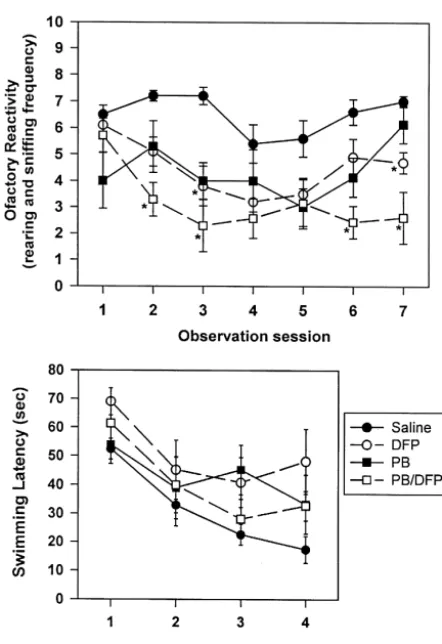
presented in Fig. 4 illustrate that each DFP regimen, observed after DFP withdrawal. Similarly, the results from
particularly the combined PB / DFP regimen, produced previous studies also have indicated that the degree of
reductions in rearing after 1–3 days of administration. DFP-induced acetylcholinesterase inhibition does not
cor-Rearing in animals receiving PB or DFP alone began to relate with the level of cognitive impairment demonstrated
recover to near-control levels after 5–6 days of exposure, in OP-exposed rats [8] or monkeys [17].
whereas those receiving the combined PB / DFP regimen In the current study, we sought to determine whether a
continued to display this motor abnormality throughout the relationship exists between specific alterations in the
observation period. The animals were subjected to water number of cholinergic receptor proteins in specific brain
maze testing 1 week after the completion of each drug areas and the pattern of behavioral changes observed upon
regimen. As demonstrated in Fig. 4, DFP-treated rats were DFP exposure. First, the present data demonstrated that
significantly impaired in water maze performance even on hippocampal cholinergic proteins were more sensitive to
16 J.D. Stone et al. / Brain Research 882 (2000) 9 –18
the current data also demonstrated that all cholinergic proteins measured (M1, M2, and nicotinic receptors, as well as acetylcholinesterase) were decreased following DFP exposure. However, the data showed that AChE and M1 receptors were either least affected (M1 receptors) or had more rapidly returned to control levels (AChE) than were the nicotinic and M2 receptors. One difference between the neuro-architecture of M1 muscarinic receptors located in the cerebral cortex and nicotinic receptors in the hippocampus is that M1 receptors are considered to exist primarily postsynaptically (located on cell bodies and dendrites), whereas nicotinic receptors [47] and M2 mus-carinic receptors [20] are largely presynaptic (located on axon terminals).
One possible explanation for the different effects of DFP on these receptor subtypes involves their specific neuronal locations. Recently, it has been demonstrated that repeated
low doses of DFP (250 mg / kg per day for 14 days)
produced significant reductions in anterograde and retro-grade fast axonal transport in peripheral nerve axons of rats at both 1 and 24 h after the last toxicant exposure [41]. These data demonstrate that a dosing regimen of DFP, which has been shown to produce protracted working memory impairment [29], as well as alterations in the numbers of cholinergic receptor subtypes in rat brain, also produces bi-directional changes in axonal transport. Al-though these transport studies were performed in peripher-al nervous system axons, no evidence exists to support the
Fig. 4. The effect of chronic administration of DFP alone or in
combina-notion that transport compromise would be different in
tion with pyridostigmine bromide (PB) on olfactory reactivity and water
maze testing. Four experimental groups of rats received either saline central nervous system axons. In fact, other neurotoxicants
vehicle (saline), PB (0.40 mg / kg, p.o., t.i.d.), DFP (250mg / kg, s.c., once have produced very similar reductions in fast transport in daily), or a combination of both PB and DFP (PB / DFP) for 7 days.
both PNS and CNS axons [39,40]. Thus, a compromise of
DFP-containing regimens, and in particular the combined PB / DFP
receptor transport would produce more significant changes
regimen reduced olfactory reactivity (rearing behavior) after 1–3 days of
in receptor numbers in areas furthest from the site of
exposure. This behavior in animals that received PB or DFP alone began
to recover to near-control levels by 5–6 days after exposure. Water maze receptor production in the cell body. For example, OP
testing was initiated 1 week after exposure. Only the DFP group was agents may directly or indirectly inactivate fast axonal significantly different from the saline group in water maze task
per-transport of receptor proteins from cell bodies in the
formance. *P,0.05 compared with SAL group.
medial septum to sites within the hippocampus. Thus, interruptions in fast transport would produce greater
of selectivity for the effects of the DFP regimen on reductions in the numbers of receptors located primarily on
hippocampal neurons as compared with cortical neurons axon terminals (nicotinic and M2 receptors) than would be
(and compared with other brain regions not illustrated observed in receptors located on or near the cell body (M1
here) is not yet apparent; however, protracted deficits in receptors). Further, receptor levels in these more distal
the hippocampus could be due to a higher percentage of locations would remain reduced after termination of
toxic-cholinergic receptors located on axon terminals (as com- ant exposure because these distal receptors could not be as
pared to dendrites and cell bodies) in the hippocampus easily or rapidly replaced as those located nearer to the site
compared to the cortex. This selectivity fits well with the of receptor synthesis. Therefore, changes in cholinergic
deficits observed in water maze performance [29,30], a receptor subtypes observed in the present experiments
task known to require intact hippocampal processing. In correspond closely to the changes that would be expected
fact, it would have been surprising if the neurochemical by an interruption in fast axonal transport.
effects to the OP treatment were not anatomically selective Another potential explanation for the decrease in
nico-in view of the fact that rats were not impaired nico-in all types tinic receptor density after DFP withdrawal is that the drug
of memory tasks [30]. caused a form of receptor down-regulation. Previous
Previous studies have demonstrated decreases in studies have reported that OP exposure causes down
3 3
[ H]QNB [43] and [ H]nicotine binding [11,38,43] in rats regulation of rat brain nicotinic receptor binding
presumably due to an increase in synaptic acetylcholine. the mechanism for the drug’s beneficial action on water
However, it has been demonstrated that increased levels of maze performance is unclear. It seems unlikely that the PB
the nicotinic agonists nicotine [25,37,48], anabasine, (1)- would directly alter the effect of DFP on brain
cholinester-anatoxin-a, cytisine [9,31,38], and the acetylcholine analog ase inhibition or on brain cholinergic receptor expression
methylcarbamylcholine [48], produced over-stimulation of unless the PB indirectly reduced the amount of DFP
nicotinic receptors generally resulting in receptor up-regu- entering the CNS. Alternatively, the ability of PB to reduce
lation. Additionally, receptor levels in the hippocampus the overall peripherally mediated toxicity of OP agents
and cortex were observed to decrease after DFP withdraw- may have provided for a better overall post-DFP treatment
al, before initiating a return towards normal levels. There- outcome. Nevertheless, under the conditions of these
fore, acetylcholine-induced receptor downregulation does experiments, prophylactic administration of PB appeared
not appear to be the cause of the reduction in nicotinic not only to provide protection against nerve agent exposure
receptor numbers observed in the present experiments. acutely, but it may also help mitigate some of the cognitive
Central nicotinic receptors (shown here to be signifi- impairment observed after toxicant exposure.
cantly affected by DFP exposure) have been studied as potential pharmacological targets to improve learning and
memory in experimental animals [2,3,7,15,21–24] and Acknowledgements
humans [28,34,35,45]. DFP-treated rats (standard DFP
regimen) exhibited a significantly reduced rate of learning This work was partly supported by
DAMD17-95-1-compared with control rats; however, the rate of learning 5036. The content of the information of this study does not
was similar to that for controls in the DFP groups that necessarily reflect the position or the policy of the
govern-received one of two doses of nicotine each day prior to ment, and no official endorsement should be inferred. This
maze testing. Whereas nicotine is used clinically only for work also was partly supported by the Office of Research
smoking cessation, several pharmaceutical companies are and Development, Medical Research Service, Department
currently evaluating novel analogs that are purported to of Veterans Affairs.
exhibit a reduced side-effect profile relative to nicotine. These compounds soon may be available for treating
cognitive deficits associated with long-term OP exposure. References
In the final experimental series, we sought to determine
whether concomitant administration of pyridostigmine [1] F.A. Abdulla, M.R. Calaminici, J.D. Stephenson, J.D. Sinden,
bromide could alter the responses to the standard DFP Chronic treatments with cholinoceptor drugs influence spatial
learn-ing in rats, Psychopharmacology 111 (1993) 508–511.
regimen. PB was administered concomitantly with DFP
[2] S.P. Arneric, M. Williams, Recent advances in the treatment of
because PB is used by our armed forces as a prophylactic
neurodegenerative disorders and cognitive function, Int. Acad.
measure against chemical nerve agents. These experiments
Biomed. Drug Res. 1993.
should be considered preliminary since only one dose of [3] S.P. Arneric, J.P. Sullivan, M.W. Decker, J.D. Brioni, A.W. Bannon,
PB (0.4 mg / kg, p.o.) was tested, although this regimen C.A. Briggs, D. Donnelly-Roberts, R.J. Radek, K.C. Marsh, J.
Kyncl, M. Williams, J.J. Buccafusco, Potential treatment of
Al-was constructed to mimic field doses administered during
zheimer’s disease using cholinergic channel activators (ChCAs) with
the Persian Gulf War (30 mg oral PB every 8 h for 1–7
cognitive enhancement, anxiolytic-like, and cytoprotective
prop-days) [19]. The mechanism for PB’s OP-protective action
erties, Alzheimer’s Dis. Assoc. Disord. 9 (1995) 50–61.
resides in its ability to prevent cholinesterase from perma- [4] J. Brodeur, K.P. Du Bois, Studies on the mechanism of acquired
nent inactivation by DFP. tolerance by rats to 0,0-diethyl S-[2-(ethylthio)-ethyl]
phosphor-Both PB alone, and DFP alone caused a reversible odithioate (DiSyston), Arch. Int. Pharmacodyn. 149 (1975) 560–
570.
inhibition of olfactory behavior in rats over the 1-week
[5] J.J. Buccafusco, R.S. Aronstam, Adrenergic agonists protect against
administration period. When the two agents were combined
soman, an irreversible acetylcholinesterase inhibitor, Toxicol. Lett.
there was a significant enhancement of the inhibition of 38 (1987) 67–76.
this behavior. This was perhaps not too surprising, since [6] J.J. Buccafusco, D.L. Heithold, S.H. Chon, Long-term behavioral
both compounds are cholinesterase inhibitors. Although and learning abnormalities produced by the irreversible
cholinester-ase inhibitor soman: effect of a standard pretreatment regimen and
PB does not produce irreversible inhibition of the enzyme,
clonidine, Toxicol. Lett. 52 (1990) 319–329.
the 3-day dosing schedule was most likely as effective as
[7] J.J. Buccafusco, W.J. Jackson, Beneficial effects of nicotine
adminis-DFP at maintaining cholinesterase inhibition. However, tered prior to a delayed matching-to-sample task in young and aged
despite the initial additive toxic effects, PB / DFP-treated monkeys, Neurobiol. Aging 12 (1991) 233–238.
animals were almost as efficient as saline controls in [8] P.J. Bushnell, S.S. Padilla, T. Ward, C.N. Pope, V.B. Olszyk,
Behavioral and neurochemical changes in rats dosed repeatedly with
learning the water maze task (although the PB regimen was
diisopropylfluorophosphate, J. Pharmacol. Exp. Ther. 256 (1991)
not quite as effective as was the pre-testing nicotine
741–750.
regimen in reversing the effects of DFP on maze per- [9] A.C. Collins, E. Romm, J.M. Wehner, Dissociation of the apparent
formance). Since PB’s actions are generally restricted to relationship between nicotine tolerance and up-regulation of
18 J.D. Stone et al. / Brain Research 882 (2000) 9 –18
[10] L.G. Costa, B.W. Schwab, S.D. Murphy, Differential alterations of [30] M.A. Prendergast, A.V. Terry, J.J. Buccafusco, Effects of chronic, cholinergic muscarinic receptors during chronic and acute tolerance low-level organophosphate exposure on delayed recall, discrimina-to organophosphorus insecticides, Biochem. Pharmacol. 31 (1982) tion, and spatial learning in monkeys and rats, Neurotoxicol. Teratol.
3407–3413. 20 (1998) 115–122.
3
[11] L.G. Costa, S.D. Murphy, [ H]Nicotine binding in rat brain: [31] P.P. Rowell, S. Wonnacott, Evidence for functional activity of alteration after chronic acetylcholinesterase inhibition, J. Pharmacol. up-regulated nicotine binding sites in rat striatal synaptosomes, J. Exp. Ther. 226 (1983) 392–397. Neurochem. 55 (1990) 2105–2110.
´ ´
[12] M.I. Davila-Garcıa, J.L. Musachio, D.C. Perry, Y. Xiao, A. Horti, [32] R.W. Russell, V.G. Carson, R.S. Jope, R.A. Booth, J. Macri,
125
E.D. London, R.R. Dannals, K.J. Kellar, [ I]IPH, an epibatidine Development of behavioral tolerance: a search for subcellular analog, binds with high affinity to neuronal nicotinic cholinergic mechanisms, Psychopharmacology 66 (1975) 155–158.
receptors, J. Pharmacol. Exp. Ther. 282 (1997) 445–451. [33] R.W. Russell, R.A. Booth, D.J. Smith, M. Roch, K.M. Rice, S.D. [13] F.H. Duffy, J.L. Burchfiel, P.H. Bartels, M. Gaon, V.M. Sim, Long- Lauretz, Roles of neurotransmitter receptors in behavior: recovery of term effects of an organophosphate upon the human electroence- function following decreases in muscarinic receptor density induced phalogram, Toxicol. Appl. Pharmacol. 47 (1979) 161–176. by cholinesterase inhibition, Behav. Neurosci. 103 (1989) 881–892. [14] G.L. Ellman, K.D. Courtney, V. Andres, R.M. Featherstone, A new
[34] J.M. Rusted, L. Graupner, A. Tennant, D.M. Warburton, Effortful and rapid colormetric determination of acetylcholinesterase activity,
processing is a requirement for nicotine-induced improvements in Biochem. Pharmacol. 7 (1961) 88–95.
memory, Psychopharmacology 138 (1998) 362–368. [15] K. Elrod, J.J. Buccafusco, W.J. Jackson, Nicotine enhances delayed
[35] B. Sahakian, G. Jones, R. Levy, J. Gray, D. Warburton, The effects matching-to -sample performance by primates, Life Sci. 43 (1988)
of nicotine on attention, information processing, and short-term 277–287.
memory in patients with dementia of the Alzheimer’s type, Br. J. [16] R. Gardner, R. Ray, J. Frankenheim, K. Wallace, M. Loss, R.
Psychiatry 154 (1989) 797–800. Robichaud, A possible mechanism for
diisopropylfluorophosphate-[36] G.D. Schiller, Reduced binding of [3H]-quinuclidiniyl benzilate induced memory loss in rats, Pharmacol. Biochem. Behav. 21
associated with chronically low acetylcholinesterase activity, Life (1984) 43–46.
Sci. 24 (1979) 1159–1164. [17] E.M. Gause, R.J. Hartmann, B.Z. Leal, Neurobehavioral effects of
[37] R.D. Schwartz, K.J. Kellar, Nicotinic cholinergic receptor binding repeated sublethal soman in primates, Pharmacol. Biochem. Behav.
sites in the brain: regulation in vivo, Science 220 (1983) 214–216. 23 (1985) 1003–1012.
3
[18] S. Gershon, F.H. Shaw, Psychiatric sequelae of chronic exposure to [38] R.D. Schwartz, K.J. Kellar, In vivo regulation of [ H]acetylcholine organophosphorus insecticides, Lancet i (1961) 1371–1374. recognition sites in brain by nicotinic cholinergic drugs, J. Neuro-[19] J.R. Keeler, C.G. Hurst, M.A. Dunn, Pyridostigmine used as a nerve chem. 45 (1985) 427–433.
agent pretreatment under wartime conditions, J. Am. Med. Assoc. [39] J.D. Stone, A.P. Peterson, J. Eyer, D.W. Sickles, Axonal neurofila-266 (1991) 693–695. ments are non-essential elements of toxicant-induced reductions in
¨
[20] J. Klein, K. Loffelholz, in: Cholinergic Mechanisms: From Molecu- fast axonal transport. Radiolabeling in CNS axons, Neurotoxicology lar Biology to Clinical Significance, Progress in Brain Research, Vol. (2000) (in press).
109, Elsevier, Amsterdam, 1996. [40] J.D. Stone, A.P. Peterson, J. Eyer, T.G. Oblak, D.W. Sickles, Axonal [21] E.D. Levin, Nicotinic systems and cognitive function, Psycho- neurofilaments are non-essential elements of toxicant-induced reduc-pharmacology 108 (1992) 417–531. tions in fast axonal transport. Video-enhanced differential interfer-[22] E.D. Levin, J.E. Rose, Nicotinic and muscarinic interactions and ence microscopy in PNS axons, Toxicol. Appl. Pharmacol. 161
choice accuracy in the radial-arm maze, Brain Res. Bull. 27 (1) (1999) 50–58.
(1991) 125–128. [41] J.D. Stone, T.G. Oblak, J.J. Buccafusco, D.W. Sickles, Diisopropyl [23] E.D. Levin, S. Kaplan, A. Boardman, Acute nicotine interactions phosphorofluoridate (DFP) reduces fast bi-directional axonal
trans-with nicotinic and muscarinic antagonists: working and reference port in vitro and in vivo, Soc. Neurosci. Abstr. 25 (1999) 2004. memory effects in the 16-arm radial maze, Behavioural Pharmacol. [42] M. Upchurch, J.M. Wehner, Effects of chronic diisopropylfluoro-8 (1997) 236–242. phosphate treatment on spatial learning in mice, Pharmacol. Bio-[24] E.D. Levin, B.B. Simon, Nicotinic acetylcholine involvement in chem. Behav. 27 (1987) 143–151.
cognitive function in animals, Psychopharmacology 138 (1998)
[43] J.L. Van De Kamp, A.C. Collins, Species differences in diisopropyl-217–230.
fluorophosphate-induced decreases in the number of brain nicotinic [25] M.J. Marks, J.B. Burch, A.C. Collins, Effects of chronic nicotine
receptors, Pharmacol. Biochem. Behav. 42 (1992) 131–141. infusion on tolerance development and nicotinic receptors, J.
[44] C.J. van Dongen, O.L. Wolthuis, On the development of behavioral Pharmacol. Exp. Ther. 226 (1983) 817–825.
tolerance to organophosphates. I. Behavioral and biochemical [26] D.R. Metcalf, J.H. Holmes, EEG, psychological and neurological
aspects, Pharmacol. Biochem. Behav. 34 (1989) 472–481. alterations in humans with organophosphorus exposure, Ann. NY
[45] D.M. Warburton, G. Mancuso, Evaluation of the information pro-Acad. Sci. 160 (1969) 357–365.
cessing and mood effects of a transdermal nicotine patch, Psycho-[27] B.E. McDonald, L.G. Costa, S.D. Murphy, Spatial memory
impair-pharmacology 135 (1998) 305–310. ment and central muscarinic receptor loss following prolonged
[46] J. Wei, E.A. Walton, A. Milici, J.J. Buccafusco, m1-m5 Muscarinic treatment with organophosphates, Toxicol. Lett. 40 (1988) 47–56.
receptor distribution in rat CNS by RT-PCR and HPLC, J. Neuro-[28] P.A. Newhouse, A. Potter, J. Corwin, R. Lenox, Acute nicotinic
chem. 63 (1994) 815–821. blockade produces cognitive impairment in normal humans,
Psycho-[47] S. Wonnacott, Presynaptic nicotinic ACh receptors, Trends Neurosci. pharmacology 108 (1992) 80–484.
20 (1997) 92–98. [29] M.A. Prendergast, A.V. Terry, J.J. Buccafusco, Chronic, low-level
![Table 1[ H]QNB binding to brain muscarinic cholinergic receptors at varying time intervals after withdrawal from chronic low-level DFP treatment3](https://thumb-ap.123doks.com/thumbv2/123dok/3139175.1382721/4.612.45.547.618.719/binding-muscarinic-cholinergic-receptors-varying-intervals-withdrawal-treatment.webp)
![Fig. 1. Left panel: color-enhanced autoradiographs of [ H]QNB binding (primarily M1 subtype) and [ H]AFDX-384 binding (M2 subtype) to muscarinic cholinergic receptors in different levels of the33brain from rostral through caudal (left to right) sections in](https://thumb-ap.123doks.com/thumbv2/123dok/3139175.1382721/5.792.99.710.51.454/enhanced-autoradiographs-primarily-muscarinic-cholinergic-receptors-different-sections.webp)
![Table 2[ H]AFDX-384 binding to brain muscarinic cholinergic receptors at varying time intervals after withdrawal from chronic low-level DFP treatment3](https://thumb-ap.123doks.com/thumbv2/123dok/3139175.1382721/6.612.44.548.81.191/binding-muscarinic-cholinergic-receptors-varying-intervals-withdrawal-treatment.webp)