CHAPTER II
LITERATURE REVIEW
Mental retardation (MR) is often found together with abnormality of the head circumference, microcephaly or macrocephaly, as an associated sign. Both microcephaly and macrocephaly increases the risk for mental retardation, and there is a correlation between the degree of head circumference abnormality and the severity of cognitive impairment. Microcephalic individuals are more likely to have MR, while MR individuals are at least twice more likely to have macrocephaly compared to intellectually normal peers (Field, 2007; Ashwal, 2009). There is a high likelihood that mental retardation and abnormality of head circumference is caused by the same explanation, for example, a genetic mutation that is causing both mental retardation and microcephaly or macrocephaly (Abuelo, 2007; Olney, 2007).
2.1. Microcephaly
2.1.1. Definition and etiology
Microcephaly is the reduced occipitofrontal circumference of the head smaller than -2 standard deviation. The incidence of microcephaly at birth is between 1.3 and 150 per 100,000 live births. The rate of the incidence depends on the population and the threshold in defining microcephaly (Kaindl, 2010).
Microcephaly is determined using measurement of occipito-frontal circumference (OFC) by placing a measuring tape around the cranial vault which includes the widest part of the forehead and the most prominent part of the occipital area (Abuello, 2007). The widely accepted definition of
OFC measurement includes observations within 2 standard deviations from the mean (Rollins 2010). Several standard charts are available for monitoring of head circumference, such as those from the CDC, WHO, Fells, United States Head Circumference Growth Reference Charts, and Nellhaus (Roche, 1987; Rollins, 2010).
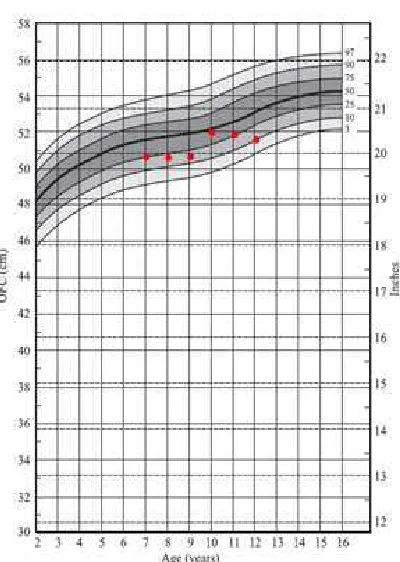
Recently, a preliminary study conducted in Semarang, Central Java (Mundhofir, unpublished) indicated that the mean value of head circumference of normal schoolchildren age 7-12 years is smaller compared to the normal range of the Nellhaus charts.
Figure 1. Head chart plotting of Semarang normal boys. The red dots show the mean OFC of boys between age 7 and 12 years old.
Figure 2. Head chart plotting of Semarang normal girls. The red dots show the mean OFC of boys between age 7 and 12 years old.
The interpretation of head circumference must be considered using the specific head charts appropriate for application in the different countries. It has also been suggested that the head circumference standard applied in each country should be reevaluated and updated periodically (Karabiber, 2001).
Table 2. Causes of microcephaly (adapted from Abuelo, 2007; Jacob, 2009; Mahmood, 2011)
Environmental and genetic causes of microcephaly
Environmental causes of microcephaly Hypoxic-ischemic encephalopathy Intrauterine infections
Teratogen (alcohol, hydantoin, radiation) Intrauterine growth retardation (IUGR) Neural tube defects (NTD)
Maternal Phenylketonuria (PKU) Poorly controlled maternal diabetes Chronic malnutrition
Genetic causes of microcephaly
Primary hereditary microcephaly (MCPH) Chromosome abnormalities (trisomy 13, trisomy 18, trisomy 21)
Deletion syndrome:
4p deletion (Wolf-Hirschhorn) syndrome 7q11.23 deletion (Williams) syndrome Rett Syndrome
Other syndromes with multiple anomalies Cornelia de Lange syndrome
2.1.2. MCPH
Autosomal recessive primary microcephaly, or also known as primary hereditary microcephaly (MCPH) is a neurodevelopmental disorder characterized by microcephaly present at birth and mental retardation. Its incidence is between 1:30,000 and 1:2,000,000 in non-consanguineous populations, in comparison with 1:10,000 in consanguineous Pakistani families (Mahmood, 2011). The current clinical definition for MCPH is as follows (Woods, 2005):
1. Congenital microcephaly
2. Mental retardation without other neurological signs such as spasticity or progressive cognitive decline. Seizures are unusual but do not exclude the diagnosis
3. For most MCPH patients, normal height, weight, appearance, chromosome analysis and brain scan are observed. In patients with MCPH1 mutations, reduction in height may be found but head circumference was significantly reduced compared to height.
Table 3. Overview of MCPH features (Adapted from Kaindl, 2010).
Main features Microcephaly (-2SD) at
birth, further relative reduction in the first years of life
Further intracranial malformations Agenesis of corpus callosum
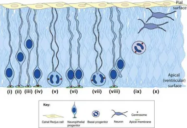
Figure 3. Development of mouse neuroepithelium. Neuroepithelial cells nuclei migrate (i,ii) to basal surface during G1 where they undergo S phase (iii). NE cells then migrate apically at G2 (iv). The centrosomes (red circles) stay at the apical membrane (green). Mitosis happens at the apical surface; the centrosomes form the spindle poles. Symmetrical division results in two identical neuroepithelial cells (v, vi) while asymmetrical division (vii) produce one neuroepithelial cell and one daughter cell which detaches and may either be a basal progenitor (ix) or neuron (x) (Adapted from Thornton, 2009).
MCPH genes affect fetal neurogenesis by affecting this shift towards asymmetrical division of neuron progenitors (Kaindl, 2010).
2.2. MCPH Genes
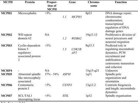
Currently, the known genetic causes of MCPH are mutations in 7 loci, with 6 identified genes: MCPH1, WDR62 (MCPH2), CDK5RAP2 (MCPH3),
ASPM (MCPH5), CENPJ (MCPH6) and STIL (MCPH7) (Table 4). As MCPH is inherited in an autosomal recessive manner, both alleles have to be mutated, either that an individual has a homozygous mutation or two compound heterozygous mutations in order to result in affected phenotype.
Table 4. MCPH genes and function (Adapted from Thornton, 2009) MCPH Protein
Propor-MCPH1 Microcephalin <5%
1.1 MCPH1
8p23 DNA damage repair; chromosome
19q13.12 Proliferative division of neural precursors and
9q33.3 Predicted role in regulating microtubule
<5% CENPJ 13q12.2 Centriole biogenesis and length; microtubule dynamics
MCPH7 SCL/TAL1 interrupting locus
<5% STIL 1p32 Spindle organisation
MCPH1
MCPH1 is located on human chromosome 8p23, encoding 835 amino acids. It has 14 exons, spanning 237 kb in the human genome (Jackson, 1998; Jackson, 2002). MCPH1 gene encodes the protein microcephalin consisted of 835 amino acids. Microcephalin mRNA is expressed in human fetal tissue especially in the brain, liver and kidney. In murine brain, microcephalin was found to be expressed during neurogenesis especially in the proximity of lateral ventricles which holds progenitor cells that produce neuron which later form the cerebral cortex (Jackson, 2002). The MCPH phenotype may be caused by defect in the cell cycle checkpoint control and DNA repair, as well as defective centrosomal function affecting the number of symmetric cell divisions of neuronal stem cells during neurogenesis. The pathologic variants include homozygous nonsense mutations, homozygous deletions and homozygous duplications (Kaindl, 2010). The p.Pro828Ser normal variant in a Chinese population was associated with variation in cranial size (Wang, 2008).
WDR62 (MCPH2)
Recently, the MCPH2 gene was identified to be WDR62, the second most common cause of MCPH. WDR62 is a gene with 32 exons and consists of 1523 amino acids. The six described mutations include four missense mutations and two duplications in 3 families. (Nicholas, 2010).
CDK5RAP2 (MCPH3)
CDK5RAP2 comprises 38 exons and spans approximately 191 kb. The human cyclin-dependent kinase 5, regulatory associated protein 2 gene encodes
CDK5RAP2 with 1893 amino acid. CDK5RAP2 is a centrosome-associated protein, and its mRNA is widely expressed in human and in embryonal mouse tissue In murine embryos, highest levels were detected in the central nervous system (Bond et al., 2005).
Studies of CDK5RAP2 homologues show that they regulate centrosome maturation, recruitment and strengthening of pericentriolar matrix at the centrioles and regulate centrosome cohesion. Loss of function causes failure of centrosome to mature and efficiently organize microtubules. This defect in humans may lead to spindle positioning abnormality that cannot be tolerated in neuroepithelial progenitors (Kaindl, 2010). Furthermore, CDK5RAP2, as well as MCPH1 was found to be essential in spindle checkpoint activation, indicating that spindle checkpoint mechanism controls the number of neurons generated by neural precursor cells (Zhang, 2009). Spindle positioning is crucial during asymmetrical division to generate daughter cells with different sizes or fates (McCarthy, 2006).
The three described mutations in the CDK5RAP2 gene predict a premature stop codon in 4 Pakistani families, suggesting in a loss of functional protein activity that causes the MCPH phenotype (Bond, 2005; Hassan, 2007).
ASPM (MCPH5)
Mutations in the ASPM gene on chromosome 1q31.3 have been considered the most common cause of MCPH and counts for half of Asian and European MCPH cases (Nicholas, 2009). The ASPM gene spans 62kb with 28 exons.
The ASPM gene encodes the abnormal spindle-like microcephaly-associated protein consisting of 3477 amino acids. The ASPM protein is highly expressed in progenitor cells and regulates the positioning and organization of spindle for proliferation. Expression is high on onset of neurogenesis at the early stage, and decreases as neurogenesis progresses. ASPM therefore determines the orientation of the cell division to be either symmetrical or asymmetrical during neurogenesis and important in maintaining the proliferative capacity of neuroepithelial cells progenitor (Thornton 2009).
The pathologic variants of the ASPM gene include translocation, deletion, insertion/ duplication or base substitution. The mutations were spread throughout the genes with no mutation hot-spot. The disease mechanism causing MCPH is thought to be nonsense mediated decay of the ASPM mRNA leading to significant reduction of the protein in neuroepithelial cells during neurogenesis (Nicholas, 2009).
CENPJ (MCPH6)
The human centromeric protein J (CENPJ) gene spans 40 kb and comprises 17 exons. CENPJ, consisting of 1338 amino acids, plays a role in the control of centrosome and spindle function during neurogenic mitosis (Bond et al 2005; Kaindl et al 2010).
STIL (MCPH7)
STIL, SCL/TAL1 interrupting locus gene is consisted of 1287 amino acids.
STIL homozygous mutations causing loss of function were very recently identified as the cause of MCPH in four of 24 consanguineous Indian familiesRecent data suggest that STIL has similarities in function with ASPM; and the phenotype of MCPH was hypothesized to be caused by impairment in spindle alignment because of STIL mutation. The described pathogenic mutations of STIL are nonsense mutation, frameshift deletion and intronic splice mutation resulting in truncated protein (Kumar, 2009).
2.3. Macrocephaly
2.3.1. Definition and aetiology
Macrocephaly refers to an abnormally large head which includes the scalp, cranial bone and intracranial contents. In macrocephaly, the measured occipitofrontal circumference (OFC) is greater than the 2 standard deviation (Williams, 2008).
Table 5. Classification of macrocephaly conditions (Williams, 2008)
Bannayan Riley Ruvulcaba syndrome/ Cowden syndrome
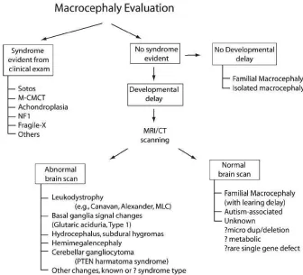
Children with macrocephaly should be examined for detailed history including family, developmental and growth history and physical examination for any other dysmorphic features or anomalies. For syndromic macrocephaly, associated manifestations may lead to diagnosis. Therefore, search for other congenital abnormalities should be conducted such as abdominal ultrasonography, echocardiogram, and ophthalmologic examination. When available, cytogenetic studies and molecular genetic studies should be performed as indicated (Olney, 2007).
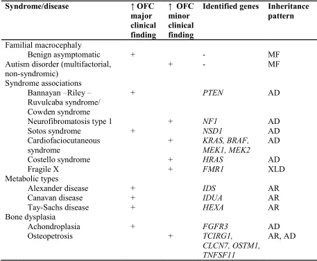
this feature may be helpful in restricting the differential diagnosis in individuals with mental retardation (Field, 2007). Table 6 shows some of the recognizable syndromic conditions and their identified genes where macrocephaly is a predominant clinical feature. However, only PTEN gene causes both mental retardation and macrocephaly (Barker, 2003).
Table 6. Macrocephaly syndromes with causative genes (Williams, 2008)
Syndrome/disease
Osteopetrosis + TCIRG1,
CLCN7, OSTM1, TNFSF11
AR, AD
MF: multifactorial disease, AD: Autosomal dominant, XLD: X-Linked, AR: Autosomal recessive
be confirmed by an appropriate and available tests. When there is no neurological dysfunction, the possibility of familial macrocephaly should be considered. However, when there are developmental concerns, a brain MRI is usually performed. When there is no informative result from MRI, tests such as chromosome study, array-CGH and fragile X molecular screening are often performed for assessment in the presence of developmental delay and/or dysmorphism together with macrocephaly. In developed countries, MRI/CT scanning is a likely option for routine diagnosis, however this may be difficult to apply in developing countries (Figure 4).
The brain enlargement found in autism patients has been speculated to occur caused by the excess of synaptic connections. However, there is increasing evidence of the association between autism and excessive brain growth (Eigsti, 2003; Frith, 2003).
The finding of PTEN mutations in autism or MR patients with macrocephaly subsequently resulted in the recommendation of PTEN testing in these patients as identification of mutation may impact on genetic counseling in recurrence risk and medical management of patients (Orrico, 2008; Varga, 2009). Moreover, PTEN testing is relevant for these patients since patients with PTEN
mutations are advocated to undergo cancer screening according to the National Comprehensive Cancer Network (NCCN) guidelines.
2.3.2. PTEN
The phosphatase and tensin homologue (PTEN) gene on 10q23.3 is a tumor suppressor gene that functions as a dual-specificity phosphatase involved with cellular growth. It functions primarily as a lipid phosphatase down-regulating the phosphoinositol 3-kinase/AKT pathway. This pathway is critical in normal cell proliferation and organ development, and its dysfunction has been associated with diseases characterised by overgrowth (Varga 2009; Barker, 2003). This phospatase is also found to affect cell migration and important for the maintenance of chromosomal integrity through association with centromere proteins (Li, 1997).
Several reports have shown germline PTEN mutations in patients with autistic behaviours or mental retardation with the presence of macrocephaly (Zori et al 1998; Goffin et al, 1998; Parisi et al, 2001; Delatycki et al, 2003; Butler et al, 2005). On the basis of these, Varga et al (2009) performed PTEN genetic testing in 114 patients referred with autism spectrum disorder; from 49 patients with MR (32 patients with both MR and macrocephaly), 6 patients (12,2%) were found to have germline PTEN mutations (Varga, 2009).
Gene targeting studies show that mutation in PTEN gene leads to increase in cell numbers, decrease in cell death, and enlargement of neuronal cell size and subsequently macrocephaly (Groszer et al, 2001; McCaffery, 2005). A study showed that there is brain overgrowth in Pten haploinsufficient mice of both sexes and that there are deficits in social approach behavior of the female mice (Page, 2009).
2.4. Analysis and interpretation of genetic variants
2.4.1. General interpretation
Molecular genetic testing by DNA sequencing will find variations within the DNA sequence. Some of the detected sequence variants can easily be recognized as pathogenic mutations, but for others the pathogenicity is still
determination of the significance of genetic variants is needed (Bell, 2007).
diagnostic laboratories, the classification of unclassified variants is usually predicted by several computer programs. Various parameters can be used to detect the structural or functional effects of amino acid substitutions, a method using mathematical calculation also known as protein prediction (Wang, 2001). The risk in the pathogenic effect of non-synonymous changes of the DNA sequence may be calculated with several bioinformatics methods, such as Grantham scoring, Align-GVGD, SIFT and Polyphen.
2.4.2. Grantham score and Align-GVGD
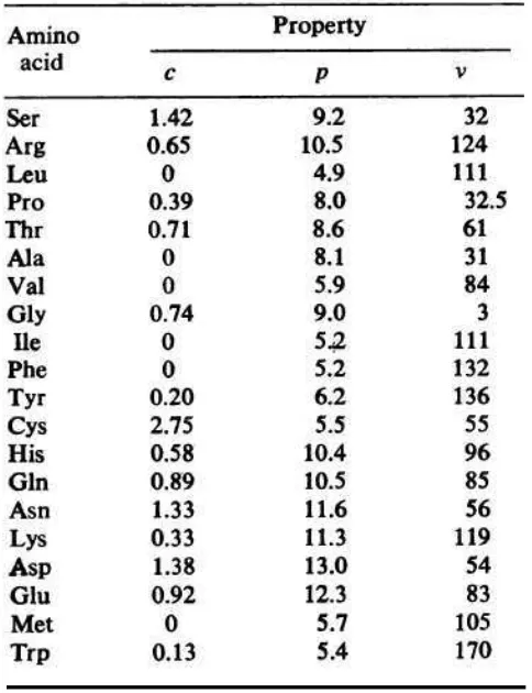
Grantham (1974) introduced the formula for calculating the difference between amino acids taking into account their composition (C), polarity (P) and molecular volume (V). This will in turn show how much difference exists between amino acids (Mathe, 2006).
The GVGD calculation is derived from the original Grantham formula (Tavtigian, 2005; Grantham, 1974):
C: Composition P: Polarity
V: Molecular volume i: Original amino acid j: Mutated amino acid
Table 7. Amino acid properties for Grantham calculations(Grantham, 1974).
C: composition index, P: polarity index, V: molecular volume.
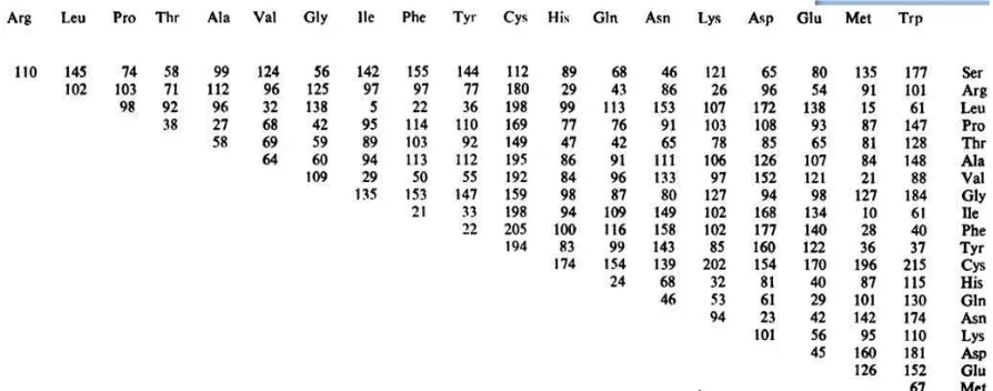
Table 8. The Grantham score table based on amino acid change (Grantham, 1974).
Arg: Arginine, Leu: Leucine, Pro: Proline, Thr: Threonine, Ala: Alanine, Val: Valine, Gly; Glysine, Ile: Isoleucine, Phe: Phenylalanine, Tyr: Tyrosine, Cys: Cysteine, His: Histidine, Gln: Glutamine, Asn: Asparagine, Lys: Lysine, Asp: Aspartat, Glu: Glutamic acid, Met: Methionine, Trp: Tryptophan
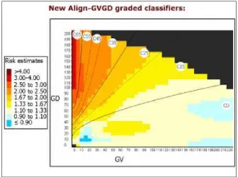
Figure 5. Align-GVGD classifiers. Using a mathematical model, GV and GD can measure the biochemical variations between the amino acids. Based on the combined GV and GD scores, missense mutations are classified between C0 to C65, interpreted as having the least to the most likelihood of interfering with function (Tavtigian, 2005).
In practice, Align-GVGD is a computational calculation that can be accessed on http://agvgd.iarc.fr/index.php.
2.4.3.
and leucine, then the SIFT assumes that this position can only have the amino acids having a hydrophobic trait. Therefore when a substitution occurs, the substitution to a hydrophobic acid was predicted as tolerated, while changes to others (for example, to a polar amino acid) in this conserved position was predicted to affect protein function (Ng, 2003).
Figure 6. The process
I: Isoleusine, R: Arginine, L: Leucine, P: Proline, M: Methionine, D: Aspartic acid (Kumar, 2009).
means that the substitution is likely to be neutral. SIFT prediction is available through http://sift.jcvi.org/.
2.4.4. PolyPhen-2
Polymorphism phenotyping (PolyPhen) is a sequence-based and structure-based method in predicting the functional effect of a mutation. Polyphen prediction relies on several strategies: (1) characterization of the substitution site, (2) calculation of position-specific independent count (PSIC), (3) mapping of the substitution to a known 3D protein structure to reveal whether the replacement has the likelihood to disturb the protein structure, or whether it tends to disrupt the
calculation of surface area, residue volume, and loss of hydrogen bond (Ramensky, 2002).
2.5. Theoretical Framework
2.6. Conceptual Framework
2.7. Hypothesis
The hypotheses of this study are the followings:
1. Pathogenic mutations which include missense, nonsense, splicing, small deletions or small insertions can be found in the ASPM, WDR62, CENPJ, CDK5RAP2, MCPH1 and STIL genes in the Indonesian mentally retarded individuals with microcephaly.
2. Pathogenic mutations which include missense, nonsense, splicing, small deletions, small insertions, or large deletions can be found in the PTEN
gene in the Indonesian mentally retarded individuals with macrocephaly.
Mental Retardation