Coronary Risk Factors and Collateral Circulation in Acute Myocardial
Infarction
Sheila Sumargo,1 Julius B. Dewanto,2 Syarief Hidayat3
1Faculty of Medicine Universitas Padjadjaran, 2Department of Biochemistry and Molecular Biology Faculty of Medicine, Universitas Padjadjaran, 3Department of Cardiology and Vascular
Medicine Faculty of Medicine Universitas Padjadjaran/Dr. Hasan Sadikin General Hospital Bandung
Abstract
Background: Coronary arterial stenosis, the major cause of acute myocardial infarction (AMI), induces shear stress to surrounding arteriolar endothelium. This stimulates changes in endothelial cells, smooth
muscle cells and fibroblast to create collaterals that can provide alternative blood flow to the jeopardized myocardial area. However, coronary collateralization is various among AMI patients. The aim of this study was to analyze the correlation between coronary risk factors and collateral sirculation in AMI patients. Methods: A retrospective cross-sectional study was carried out to 148 medical records of hospitalized AMI patients in Dr. Hasan Sadikin General Hospital Bandung, Indonesia. All patients were assessed for coronary collateral circulation which were graded as good (Rentrop score 2–3) and poor (Rentrop score 0–1). Risk factors noted in this study were age group, sex, hypertension, diabetes mellitus and diagnosis (ST-segment
Elevation Myocardial Infarction (STEMI) or Non STEMI (NSTEMI)) according to the medical record data.
Results: Hypertension was found to be associated with the presence of good coronary collateral circulation (p=0.02, PR=1.410 [95% CI 1.030-1.930]). Sex, age group, diabetes mellitus and STEMI or NSTEMI diagnosis were not statistically significant.
Conclusions: Hypertension was the only coronary risk factor associated to the presence of well-developed coronary collaterals. The increase of myocardial oxygen demand and flow of collateral feeding coronary artery in the setting of hypertension may contribute to the results. [AMJ.2015;2(4):529–33]
Keywords: Acute myocardial infarction, coronary collateral, coronary risk factors
Correspondence: Sheila Sumargo, Faculty of Medicine, Universitas Padjadjaran, Jalan Raya Bandung-Sumedang Km.21, Jatinangor, Sumedang, Indonesia, Phone: +62 81322230320 Email: [email protected]
Introduction
The majority of acute myocardial infarction
(AMI) is caused by coronary arterial occlusion which leads to the death of myocytes.1 Age, sex,
hypertension and diabetes mellitus are some
important risk factors to the development of AMI.1 The type and the nature of coronary
occlusion also determine how AMI develops, whether it is ST−segment Elevation Myocardial
Infarction (STEMI) or Non STEMI (NSTEMI).2 The presence of spontaneous coronary
collaterals may be able to limit the expansion of
infarction area, since they provide alternative
blood flow to the threatened myocardium.3
The presence of functional coronary
collaterals potentially lowers the infarct size,
the development of heart failure and mortality
rate after AMI, thus is accountable for better
prognosis.4,5,6 The development of coronary collaterals involves the arteriogenesis process,
which is the arterial defense mechanism
against the intermittent and gradual occlusion of the culprit artery.7 Sudden arterial stenosis creates larger interarterial pressure gradient
between arteries proximal and distal from
it, inducing shear stress to surrounding arteriolar endothelial cells.1,8 This will stimulate arteriolar endothelial cells, smooth
muscle cells and fibroblast leading to their
remodeling to create larger collateral arteries
that can provide alternative blood flow to the jeopardized myocardial area.9
However, the presence of coronary collateral
controversial. This phenomenon is suspected
to be the cause of the differences in AMI risk factors. This study was undertaken to assess whether coronary risk factors were associated with the presence of well-developed coronary
collateral vessels.
Methods
A retrospective cross-sectional study was
carried out in Dr. Hasan Sadikin General
Hospital Bandung Indonesia from May– November 2013 using medical records of all hospitalized patients diagnosed with AMI and underwent coronary angiography from January to December 2012. Out of 276 medical records selected, 128 medical records were excluded since data of thepatients did
not meet the inclusion criteria (history of old myocardial infarction, history of previous elective Percutaneous Coronary Intervention (PCI), history of previous Coronary Artery Bypass Graft (CABG), and missing medical
record data). One hundred and forty eight medical records were enrolled to the final study population. This investigation obtained
permission from the institutional ethics committee and all data regarding patients
were concealed.
The data of patients were classified
according to the presence of visually apparent coronary collaterals in angiogram. Coronary
angiograms of the patients were evaluated and next, collaterals were t scored based on Rentrop classification which were then further classified as good (Rentrop score 2–3) and poor (Rentrop score 0–1) coronary collaterals. The Rentrop score description is as follow; score 0: no collaterals were visible; score 1: only side branches, but no major trunk, were visualized through collaterals; score 2: partial filling of
the epicardial segment of the stenosed artery
through collaterals; score 3: complete filling
of the epicardial segment.10 The clinical risk
factors noted from the patients were sex, age, hypertension, diabetes mellitus and diagnosis of AMI (STEMI and NSTEMI) based on the medical record data. Age was then divided into two groups with the median age of patients as the cutoff point. Other risk factors were not analyzed due to incomplete medical record
data.
All data were analyzed by using the computer based Statistical Product and Service Solutions (SPSS) version 20.0. Furthermore, Chi-square test was used to compare categorical variablesand the p-value <0.05 was considered statistically significant.
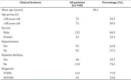
Table 1 Baseline Characteristics of Study Patients
Clinical features All patients
(n=148)
Percentage (%)
Mean age (years) 58.1
Age group (n)
≤58 years old 75 50.7
>58 years old 73 49.3
Sex (n)
Male 125 84.5
Female 23 15.5
Hypertension
Yes 93 62.8
No 55 37.2
Diabetes Mellitus
Yes 38 25.7
No 110 74.3
Diagnosis
STEMI 114 77.0
Results
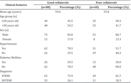
From 148 patients, there were more patients diagnosed with STEMI than NSTEMI (Table 1). Table 2 shows that the study population was divided into 2 groups based on the coronary collateralization and there were more patients with good coronary collateral vessels (59.5%). Among the 148 study patients, there were more male than female. There were more patients in the ≤58 years age group than in the >58 years age group. Over 60% patients were hypertensive, while only a quarter of study
patients were found to have diabetes mellitus. Table 3 shows that hypertension was significantly associated with the presence of better coronary collaterals (p=0.020, PR=1.410 [95% CI 1.030-1.930]), however not for the younger age (p=0.124), male sex (p=0.541) and diabetes mellitus (p=0.192).
There were more patients with
well-developed collaterals among STEMI patients
(n=65) and so were among NSTEMI (n=23) patients (Table 2). However, there was no significant relationship between the presence
of good coronary collaterals and the incidence
of STEMI or NSTEMI (p=0.551) (Table 3).
Table 2 Coronary Collaterals among Study Patients
Clinical features Good collaterals Poor collaterals
(n=88) Percentage (%) (n=60) Percentage (%)
Mean age (years) 59.6 55.8
Age group (n)
≤58 years old 40 45.5 35 58.3
>58 years old 48 54.5 25 41.7
Sex (n)
Male 73 83.0 52 86.7
Female 15 17.0 8 13.3
Hypertension
Yes 62 70.5 31 51.7
No 26 29.5 29 48.3
Diabetes Mellitus
Yes 26 29.5 12 20.0
No 62 70.5 48 80.0
Diagnosis
STEMI 65 73.9 49 81.7
NSTEMI 23 26.1 11 18.3
Table 3 Analysis of Coronary Collaterals Based on Clinical Features
Clinical features p-value PR 95% CI
Min Max
≤ 58 years old 0.124 0.779 0.570 1.066
Male 0.541 0.895 0.642 1.249
Hypertension 0.020 1.410 1.030 1.930
Diabetes Mellitus 0.891 1.214 0.925 1.593
Discussions
The study revealed that there was a significant relationship between hypertension and good collateralization. Age group, sex and diabetes mellitus were not associated with coronary
collaterals development. A previous study
corresponded with this finding.11 However, some studies pointed that a younger age,
male sex and the absence of diabetes mellitus contributed to the development of better
coronary collateral vessels.12,13,14 Additionally, age related endothelial dysfunction may
contribute to the development of poor
collaterals.12 While the ability of monocyte
migration towards growth factor stimulation in diabetes mellitus was impaired, which contributes to poor collateralization in diabetic
individuals.14
There is also no significant difference in collateralization between STEMI and NSTEMI patients, as described in a previous study by Majumder et al.15 Thus the differences
between the nature of coronary collaterals and
pathophysiological process involving STEMI
and NSTEMI may contribute to the result. This study found that hypertension was associated to the development of better coronary collaterals (p=0.020, PR=1.410 [95% CI 1.030-1.930]). This finding was in accordance with a previous study.16 The pathophysiology of hypertension involves the increase of heart
rate and peripheral resistance, which leads to the augmentation of the cardiac workload and myocardial oxygen consumption.1 Chronic
myocardial oxygen supply and demand imbalance contributes to the development of better coronary collateral circulation by increasing the expression of cytokine and growth factors needed to expand the coronary
collaterals locally.17 A previous study by Meisel et al.16 stated that the systemic blood pressure
determines the flow in the feeding coronary arteries supply collateral circulation which
generates the pressure distal to occlusion.
Thus, excessive lowering blood pressure in the setting of AMI may decrease the collateral flow and aggravate myocardial ischemia.16 This explained how hypertension can contribute to the development of better collateralization. Contrary to a previous study by Koerselman
et al.18 which showed that hypertension
is associated with poorer coronary collateralization. It showed that hypertension causes arteriolar remodeling which may lead to the obliteration of naturally occurred
collateral arterioles.19,20 Nevertheless, certain
limitations should be considered as history of
risk factors treatments was not considered in this study. Furthermore, other variables such as other AMI risk factors (lipid profile, obesity or smoking), and their treatments as well as the degree of coronary occlusion, which might actually give better association with the development of collaterals, were not analyzed in the current study. The limited number of patients may also contribute to the study
results.
This study showed that among coronary risk factors analyzed there is a significant association between hypertension and better coronary collateralization in the setting of AMI patients. The increase of myocardial oxygen demand and flow of the feeding coronary
arteries supply collateral circulation in the
setting of hypertensive state may contribute to
the results.
References
1. Bonow RO, Mann DL, Zipes DP, Libby P,
Braunwald E, editors. Braunwald’s heart disease: a textbook of cardiovascular
medicine. 9th ed. Philadelphia: Saunders
Elsevier; 2012.
2. Daga LC, Kaul U, Mansoor A. Approach to
STEMI and NSTEMI. J Assoc Physicians India. 2011;59 Suppl:19−25.
3. Ng S. Coronary collaterals: occurrence
and functions [thesis]. Utrecht: Utrecht University; 2012.
4. Plein S, Younger JF, Sparrow P, Ridgway JP, Ball SG, Greenwood JP. Cardiovascular
magnetic resonance of scar and ischemia
burden early after acute st elevation and non-st elevation myocardial infarction. J Cardiovasc Magn Reson. 2008;10:47.
5. Steg PG, Kerner A, Mancini GBJ, Reynolds
HR, Carvalho AC, Fridrich V, et al. Impact of collateral flow to the occluded
infarct-related artery on clinical outcomes in
patients with recent myocardial infarction: a report from the randomized occluded artery trial. Circulation. 2010;121(25): 2724−30.
6. Meier P, Hemingway H, Lansky AJ, Knapp G,
Pitt B, Seiler C. The impact of the coronary
collateral circulation on mortality: a meta-analysis. Eur Heart J. 2011;33(5):614−21.
7. Hershey JC, Baskin EP, Glass JD, Hartman
HA, Gilberto DB, Rogers IT, et al. Revascularization in the rabbit hindlimb: dissociation between capillary sprouting and arteriogenesis. Cardiovasc Res. 2001; 49(3):618−25.
cellular, and molecular factors on collateral
artery growth (arteriogenesis). Circ Res. 2004;95(5):449−58.
9. Seiler C. The human coronary collateral
circulation. Eur J Clin Invest. 2010;40(5): 465−76.
10. Tanboga IH, Topcu S, Nacar T, Aksakal E, Kalkan K, Kiki I, et al. Relation of coronary collateral circulation with red cell distribution width in patients with non-ST
elevation myocardial infarction. Clin Appl
Thromb Hemost. 2014;20(4):411−5.
11. Nathoe HM, Koerselman J, Buskens E,
van Dijk D, Stella PR, Plokker THW, et al. Determinants and prognostic significance
of collaterals in patients undergoing
coronary revascularization. Am J Cardiol. 2006;98(1):31−5.
12. Kurotobi T, Sato H, Kinjo K, Nakatani
D, Mizuno H, Shimizu M, et al. Reduced
collateral circulation to the infarct-related
artery in elderly patients with acute myocardial infarction. J Am Coll Cardiol. 2004;44(1):28−34.
13. Zorkun C, Akkaya E, Zorlu A, Tandogan I. Determinants of coronary collateral
circulation in patients with coronary artery disease. Anadolu Kardiyol Derg. 2013;13(2):146−51.
14. Waltenberger J. Impaired collateral vessel development in diabetes: potential cellular
mechanisms and therapeutic implications.
Cardiovasc Res. 2001;49(3):554−60.
15. Majumder A, Karim M, Rahman M, Uddin M.
Study of association of c- reactive protein
with coronary collateral development. Cardiovasc J. 2010;3(1):26−32.
16. Meisel SR, Frimerman A, Blondheim DS, Shotan A, Asif A, Shani J, et al. Relation of the systemic blood pressure to the
collateral pressure distal to an infarct-related coronary artery occlusion during
acute myocardial infarction. Am J Cardiol. 2013;111(3):319−23.
17. Schirmer SH, van Royen N, Moerland PD,
Fledderus JO, Henriques JP, van der Schaaf RJ, et al. Local cytokine concentrations and oxygen pressure are related to maturation of the collateral circulation in humans. J Am Coll Cardiol. 2009;53(23):2141−7. 18. Koerselman J, Jaegere PPTd, Verhaar MC,
Graaf Yvd, Grobbee DE. High blood pressure is inversely related with the presence and extent of coronary collaterals. J Hum Hypertens. 2005;19(10):809−17.
19. Humphrey JD. Mechanisms of arterial
remodeling in hypertension: Coupled roles of wall shear and intramural stress. Hypertension. 2008;52(2):195−200. 20. de Marchi SF, Gloekler S, Meier P, Traupe
T, Steck H, Cook S, et al. Determinants of preformed collateral vessels in the human