Relation between Preinfarction Angina and Coronary Collateral
Circulation in Patients with Acute Myocardial Infarction
Achmad Shidiq,1 Syarief Hidayat,2 Januarsih Iwan A. Rachman3
1Faculty of Medicine Universitas Padjadjaran, 2Department of Cardiology and Vascular Medicine Faculty of Medicine Universitas Padjadjaran/Dr. Hasan Sadikin General Hospital Bandung,
3Department of Anatomy and Cell Biology, Faculty of Medicine, Universitas Padjadjaran
Abstract
Background: Coronary collateral circulation conduits an alternative blood flow to the ischemic myocardium
in the setting of coronary artery occlusion which can prevent the infarction area to extend more widely. Well-developed coronary collaterals are closely related with the presence of preinfarction angina. However, the duration of preinfarction angina which can induce well-developed coronary collateralization is in controversy. The aim of this study was to evaluate the relation between duration of preinfarction angina and coronary collaterals circulation in patients with acute myocardial infarction.
Methods: This cross-sectional study was conducted from May to November 2013 in Dr. Hasan Sadikin
General Hospital, Bandung, Indonesia. Seventy three acute myocardial infarction (AMI) patients were included in the study. The patients were divided into Group 1 (<7 days) and Group 2 (≥7 days) based on their preinfarction angina history. The coronary collaterals were assesed and graded as good (Rentrop score 2−3) and poor (Rentrop score 0−1).Statistical analysis was performed using the chi-square test.
Result: The presence of a well-developed coronary collateral was not significantly different in <7days than
≥7 days duration of preinfarction angina [50.8% v 75.0%, p=0.124].
Conclusions: There is no relation between the duration of preinfarction angina and coronary collaterals
circulation in patients with acute myocardial infarction. [AMJ.2016;3(1):28–33]
Keywords: Acute myocardial infarction, coronary collaterals, preinfaction angina
Correspondence: Achmad Shidiq, Faculty of Medicine, Universitas Padjadjaran, Jalan Raya Bandung-Sumedang Km.21,
Jatinangor, Sumedang, Indonesia, Phone: +62 85794808279 Email: [email protected]
Introduction
Acute Myocardial Infarction (AMI) is the leading cause of mortality in the world. The AMI occurs by the occlusion of coronary artery consequently blood perfusion fails to meet the myocardial oxygen demand, leading to the death of the myocardial area supplied by the culprit coronary artery.1
The existence of spontaneous coronary collaterals may be able to limit the expansion of the infarction area, since they provide alternative blood flow to the threatened myocardium. Without significant coronary collaterals, the size of myocardial infarction will continually expand as long as the culprit coronary artery remains occluded.2,3 The presence of functional coronary collaterals also potentially lowers the development of heart failure4 and mortality rate5 after AMI, thus accountable for better prognosis.6
Several previous studies stated the
coronary collaterals is stimulated by ischemia, increasing shear stress in the occluded vessels, and the presence of angiogenic growth factor.7,8 On the other hand coronary collateralization is impaired in patients with hypertension and diabetes mellitus. In the case of ischemia, the episode angina pectoris as a sign of myocardial ischemia becomes the important predictor of well-developed coronary collaterals vessels.9,10 However the duration of preinfarction angina which can induce well-developed coronary collateralization is in controversy.
The current study was undertaken to evaluate whether the duration of preinfarction angina was related to the development of coronary collaterals circulation.
Methods
30 AMJ March 2016
consecutive series of 148 angiograms from patients with acute myocardial infarction in the period January to December 2012 were included for coronary collateral analysis. Then, 75 patients were excluded due to theirhistory of old myocardial infarction, history of previous elective Percutaneous Coronary Intervention (PCI), history of previous Coronary Artery Bypass Graft (CABG), and missing medical record data. Therefore, 73 patients were enrolled to the final study population. This investigation was reviewed and approved by the ethics committee and all data regarding patients were concealed.
Furthermore, the patients were divided into two groups according to the time interval between preinfarction angina and hospitalization as follows: Group 1 represent time interval <7 days and Group 2 represent ≥7 days. Preinfarction angina (a history of angina pectoris prior to AMI) was defined as a typical anginal chest pain occurring at rest or during exercise before the onset of AMI. The diagnosis of AMI was based on the typical chest pain lasting more than 30min, ST-segment elevation of at least 1 mm in 2 contiguous precordial leads, and a subsequent increase in the serum creatine kinase concentration to more than twice the upper limit of normal.11 The patients were considered to have a history of hypertension if their systolic pressure was ≥140 mmHg, and the diastolic pressure was ≥90 mmHg, or if they were currently undergoing a treatment for hypertension. A diagnosis of diabetes mellitus was established on the basis of one of the following three factors: a history of taking insulin or an oral hypoglycemic
agent, abnormal preinfarction fasting glucose levels (126 mg/dl), and positive results on a 75 g oral glucose tolerance test.
The Coronary angiograms of the patients were evaluated and collaterals were then scored based on Rentrop classification as Grade 0 (non-developed, no collaterals were visible), Grade 1(less-developed, only side branches, but no major trunk, were visualized through collaterals), Grade 2 (well-developed, partial filling of the epicardial segment of the stenosed artery through collaterals) or Grade 3 (complete filling of the epicardial segment). Then, further classified as good (Rentrop score 2−3) and poor (Rentrop score 0−1) coronary collaterals.12
Moreover, the continuous variables are presented as themean and standard deviation (SD), and thecategorical data are summarized as frequencies or percentages. TheChi-square test was used to examine the proportional differences between categorical variables. The result was considered statistically significant at p value <0.05 for 2-sided test. All data were analyzed by using the computer based Statistical Packages for Social Sciences version 20 for Windows (SPSS, Inc., Chicago, IL, USA).
Results
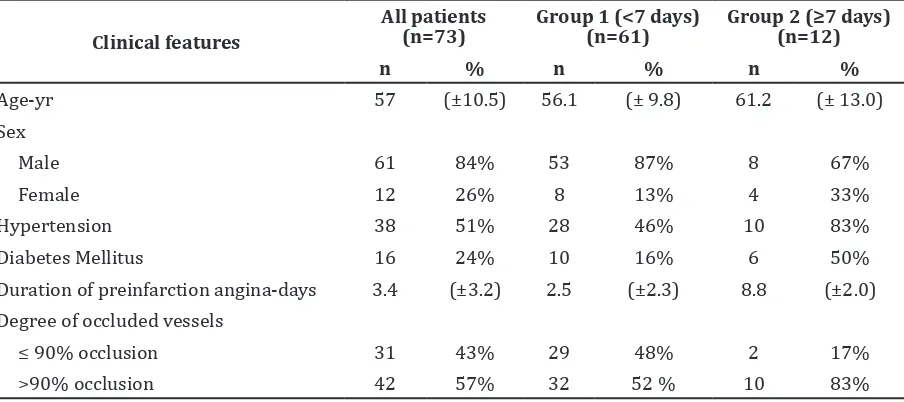
Among the 73 study patients, there was more male (n=61, 84%) than female. The overall mean age was 57 ± 10.5 with the youngest age at attack was 30 years. While hypertension, diabetes mellitus and critical occlusion were more than 90%, and were more prevalent in group 2 (Table 1).
Table 1 Baseline Characteristics of Study Patients
Clinical features
All patients (n=73)
Group 1 (<7 days) (n=61)
Group 2 (≥7 days)
(n=12)
n % n % n %
Age-yr 57 (±10.5) 56.1 (± 9.8) 61.2 (± 13.0)
Sex
Male 61 84% 53 87% 8 67%
Female 12 26% 8 13% 4 33%
Hypertension 38 51% 28 46% 10 83%
Diabetes Mellitus 16 24% 10 16% 6 50%
Duration of preinfarction angina-days 3.4 (±3.2) 2.5 (±2.3) 8.8 (±2.0)
Degree of occluded vessels
≤ 90% occlusion 31 43% 29 48% 2 17%
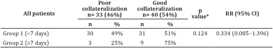
Good coronary collateral vessels were found in 40 patients (54%). We found that there were more patients with well-developed collaterals among group 2 patients (n=9, 75.0%) and were likewise among group 1 patients (n=31, 50.8%). However, it found that there was no significant difference between duration of first preinfarction angina to hospitalization and the presence of well-developed coronary collaterals (p=0.124).
Discussion
The current study revealed that there was more male than female study patients. This result is similar with many other studies which shows a higher prevalence of male among patients with acute myocardial infarction. The study also showed the proportion of well-developed collaterals was 54%. This result is in accordance with other recent data from patients with acute myocardial infarction. The documented prevalence of well-developed coronary collateral circulation in acute myocardial infarction intervention has varied from 15 to 55 %.13
On the other hand, it found longer duration of preinfarction angina particularly ≥7 days, which was not related to the presence of well-developed collateral as other studies. Herlitz et al.6 showed that the patients with chronic angina pectoris before an acute myocardial infarction had smaller infarct compared with short duration angina pectoris before the episode of MI due to the presence of well-developed coronary collaterals.14 In addition, Antoniucci et al.15 showed coronary collateral circulation were clearly visible 6 hours after myocardial ischemia. However, our finding can be explained by other experimental and clinical studies which revealed coronary collateral circulation developed perfectly 8 weeks after myocardial infarction or a period of 3 months of ischemic condition marked by preinfarction angina episode.13 These varied findings might be due to the different classification of duration preinfarction angina and the methods used to
assess coronary collaterals.
Preinfarction angina is caused by myocardial ischemia, whereas Myocardial ischemia can be a sufficient stimulus to induce coronary collateral development, possibly through biochemical preconditioning by releasing theangiogenic growth factor. Additionally, the exposure to hypoxia stimulates theaccumulation of vascular endothelial growth factor (VEGF) mRNA. Many other genes involved in angiogenesis are also upregulated in response to hypoxia including cardiac macrophage.16 However the development of collateral arteries through arteriogenesis is not dependent on ischemia. Collateral arteries develop in non hypoxic tissue, and is induced by an increase of shear stress in the setting of coronary occlusion.17
Preinfarction angina, is not only a specific marker of myocardial ishemia but is simultaneously a sign in the presence of severe coronary occlusion. The formation of coronary collateral vessels has initiated the development of an critically acute occluded coronary artery (>90%).17,18,19 An acutely reduction of the arterial diameter creates larger interarterial pressure gradient between the arterial segment before and after the stenoses, inducing shear stress to surrounding arteriolar endothelial cells.This will stimulate the arteriolar endothelial cells, smooth muscle cells and fibroblast leading to their proliferation, migration and remodeling to create larger functional muscular arteries that can provide an alternative blood flow to the jeopardized myocardial area.19,20 This explained that the pathophysiological process of preinfarction angina may lead to the development of good coronary collateral vessels through biochemical and mechanical pathways.
There were several limitations in this study. First, the use of coronary angiography, by which some collateral vessels with a diameter of <100 µm were not visualized for the evaluation of collateral circulation. Coronary collaterals may be more accurately
Table 2 Distribution of the Presence of Coronary Collaterals among StudyPatients
All patients
Group 1 (<7 days) 30 49% 31 51% 0.124 0.334 (0.085−1.396)
Group 2 (≥7 days) 3 25% 9 75%
32 AMJ March 2016
assessed by thecollateral flow index with the simultaneous measurement of aortic pressure and the distal pressure within the occluded segment of the culprit coronary artery. However, the angiographic approach to the classification of collateral flow still remain the standard of reference in the clinical setting. The second limitation was the difficulty to determine the exact origin of the symptoms of each patient since preinfarction angina is a subjective marker of myocardial ischemia. Finally, myocardial ischemia, not angina, plays an important role in the development of collateral circulation as mentioned above. Therefore, myocardial ischemia including silent ischemia that occurs before the onset of themyocardial infarction should have been evaluated. As more than half of the patients were admitted with a first symptom, it was difficult to document the presence or absence of myocardial ischemia.
In conclusion, this study shows that there is no relation between duration of preinfarction angina (<7 days or ≥7 days) and coronary collateral circulation. The development and pathophysiological process of collateralization may explain the results.
References
1. Schoen FJ, Mitchell RN. The heart. In: Kumar V, Abbas AK, Fausto N, Aster JC, editors. Robbins and Cotran: pathologic basis of disease. 8th ed. Philadelphia: WB Saunders; 2010. p. 660−71.
2. Plein S, John F Younger, Sparrow P, Ridgway JP, Ball SG, Greenwood JP. Cardiovascular magnetic resonance of scar and ischemia burden early after acute ST elevation and non-ST elevation myocardial infarction. J Cardiovasc Magn Reson. 2008;10:47−56. 3. Shen Y, Wu F, Pan C, Zhu T, Zhang Q, Zhang
R, et al. Clinical relevance of angiographic coronary collaterals during primary coronary intervention sor acute ST-elevation myocaldial infarction. Chin Med J. 2014;127(1):66−71.
4. Steg PG, Kerner A, Mancini GBJ, Reynolds HR, Carvalho AC, Fridrich V, et al. Impact of collateral flow to the occluded infarct-related artery on clinical outcomes in patients with recent myocardial infarction. A Report from the randomized occluded artery trial. Circulation. 2010;121(25):2724−30.
5. Meier P, Hemingway H, Lansky AJ, Knapp G, Pitt B, Seiler C. The impact of the coronary collateral circulation on mortality: a
meta-analysis. Eur Heart J. 2011;33(5):614−21. 6. Herlitz J, Karlson B, Richter A. Occurrence
of angina pectoris prior to acute myocardial infarction and its relation to prognosis. Eur Heart J. 1993;14:484−91.
7. Steen H, Giannitsis E, Futterer S, Merten C, Juenger C, Katus HA. Cardiac troponin T at 96 hours after acute myocardial infarction correlates with infarct size and cardiac function. J Am Coll Cardiol. 2006;48(11):2192−4.
8. Giannitsis E, Steen H, Kurz K, Ivandic B, Simon AC, Futterer S, et al. Cardiac magnetic resonance imaging study for quantification of infarct size comparing directly serial versus single time-point measurements of cardiac troponin T. J Am Coll Cardiol. 2008;51(3):307−14.
9. Traupe T, Gloekler S, Marchi SFd, Werner GS, Seiler C. Assesment of the human coronary collateral circulation. Circulation. 2010;122:1210−20.
10. Ng S, Soerianata S, Andriantoro H, Ottervanger JP, Grobbee DE. Timing of coronary collateral appearance during ST-elevation myocardial infarction. Interv Cardiol. 2012;4(1):137−43.
11. Thygesen K, Alpert JS, White HD, Jaffe AS, Katus HA, Apple FS, et al. Expert consensus document: third universal definition of myocardial infarction. J Am Coll Cardiol. 2012;60(X):2528−38.
12. Tanboga IH, Topcu S, Nacar T, Aksakal E, Kalkan K, Kiki I, et al. Relation of coronary collateral circulation with red cell distribution width in patients with non-ST elevation myocardial infarction. Clin Appl Thromb Hemost. 2012;0(0):15.
13. Schwartz H, Leiboff RH, Bren GB, Wasserman AG, Katz RJ. Temporal evolution of the human coronary collateral circulation after myocardial infarction. J Am Coll Cardiol. 1984;4(6):1088−93. 14. Lonborg J, Kelbaek H, Vejlstrup N, Botker
HE. Influence of preinfarction angina, collateral flow, and pre-procedural TIMI flow on myocardial salvage index by cardiac magnetic resonance in patients with ST-segment elevation myocardial infarction. Eur Heart J. 2012;13:433−43. 15. Antoniucci D, Valenti R, Moschi G. Relation
between preintervention angiographic evidence of coronary collateral circulation and clinical and angiographic outcomes after primary angioplasty or stenting for acute myocardial infarction. J Am Coll Cardiol. 2002;89:121−5.
Grobbee DE. Coronary collaterals: An important and underexposed aspect of coronary artery disease. Circulation. 2003;107:2507−11.
17. Lavine KJ, Kovacs A, Weinheimer C, Mann DL. Repetitive myocardial ischemia promotes coronary growth in the adult mamalian heart. J Am Heart Assoc. 2013;2(5):1−30.
18. Schaper W. Collateral circulation: past and present. Basic Res Cardiol. 2009;104(1):5– 21.
19. Pagonas N, Gross CM, Li Meijing, Bondke A, Klauss V, Buschmann E. Influence of epocardial stenosis severity and central venous pressure on the index of microcirculatory resistance in a follow-up study. Euro Intervention. 2014;9(9):1063−8.