https://doi.org/10.1007/s13577-018-0222-1
RESEARCH ARTICLE
Human amniotic fluid stem cells have a unique potential to accelerate
cutaneous wound healing with reduced fibrotic scarring like a fetus
Marie Fukutake1 · Daigo Ochiai1 · Hirotaka Masuda1 · Yushi Abe1 · Yu Sato1 · Toshimitsu Otani1 · Shigeki Sakai3 ·
Noriko Aramaki‑Hattori3 · Masayuki Shimoda2 · Tadashi Matsumoto1 · Kei Miyakoshi1 · Yae Kanai2 · Kazuo Kishi3 ·
Mamoru Tanaka1
Received: 31 July 2018 / Accepted: 8 November 2018 / Published online: 1 December 2018 © Japan Human Cell Society and Springer Japan KK, part of Springer Nature 2018
Abstract
Adult wound healing can result in fibrotic scarring (FS) characterized by excess expression of myofibroblasts and increased type I/type III collagen expression. In contrast, fetal wound healing results in complete regeneration without FS, and the mechanism remains unclear. Amniotic fluid cells could contribute to scar-free wound healing, but the effects of human amniotic fluid cells are not well characterized. Here, we determined the effect of human amniotic fluid stem cells (hAFS) on FS during wound healing. Human amniotic fluid was obtained by amniocentesis at 15–17 weeks of gestation. CD117-positive cells were isolated and defined as hAFS. hAFS (1 × 10⁶) suspended in PBS or cell-free PBS were injected around
wounds created in the dorsal region of BALB/c mice. Wound size was macroscopically measured, and re-epithelialization in the epidermis, granulation tissue area in the dermis and collagen contents in the regenerated wound were histologically analyzed. The ability of hAFS to engraft in the wound was assessed by tracking hAFS labeled with PKH-26. hAFS fulfilled the minimal criteria for mesenchymal stem cells. hAFS injection into the wound accelerated wound closure via enhancement of re-epithelialization with less FS. The process was characterized by lower numbers of myofibroblasts and higher expression of type III collagen. Finally, transplanted hAFS were clearly observed in the dermis until day 7 implying that hAFS worked in a paracrine manner. hAFS can function in a paracrine manner to accelerate cutaneous wound healing, producing less FS, a process resembling fetal wound healing.
Keywords Human amniotic fluid stem cell · Wound healing · Epithelialization · Fibrosis · Scar formation
Introduction
Cutaneous wound healing is a complex process that involves inflammation, cell proliferation, differentiation, migration, angiogenesis and remodeling of the extracellular matrix (ECM) [1]. It requires the cooperation of multiple cell types, cytokines and ECM proteins. There are phenotypic differ-ences between the collagen components in fetal and adult wounds [2–6]. In adults, it sometimes results in a non-func-tional mass of fibrotic tissue at the site of the regenerated tissue (a scar), characterized by abnormal ECM remodeling such as excessive deposition of type I collagen [7]. In con-trast, fetal wound healing results in complete regeneration without fibrotic scarring, characterized by rapid deposition of type III collagen in a fine reticular network [2–6]. The factors responsible for scarless fetal wound healing have been attributed to intrinsic factors in the developing fetal dermis secondary to gene expression associated with the
Electronic supplementary material The online version of this article (https ://doi.org/10.1007/s1357 7-018-0222-1) contains supplementary material, which is available to authorized users.
* Daigo Ochiai ochiaidaigo@keio.jp
1 Department of Obstetrics and Gynecology, Keio University
School of Medicine, 35, Shinanomachi Shinjyukuku, Tokyo 160-8582, Japan
2 Department of Pathology, Keio University School
of Medicine, 35, Shinanomachi Shinjyukuku, Tokyo 160-8582, Japan
3 Department of Plastic and Reconstructive Surgery,
Keio University School of Medicine, 35, Shinanomachi Shinjyukuku, Tokyo 160-8582, Japan
development of fetal skin [2, 3]. However, the extrinsic fac-tors responsible for scar-free fetal wound healing remain to be determined. Significantly, several studies suggested that the extrinsic factors in amniotic fluid and/or amniotic fluid cells contribute to scarless fetal wound healing [3, 8].
Mesenchymal stem cells (MSCs) are emerging as a prom-ising cell population for the promotion of wound healing and their therapeutic potential depends on the origin of the MSC [9–11]. Among the different sources of MSCs, those derived from amniotic fluid have a number of characteristics that make them attractive candidates for stem cell therapy for neonates [12, 13]. Amniotic cells can be easily collected during routine prenatal testing and could be saved for future stem cell therapy, as needed. Amniotic cells are not subject to ethical debates because the residual amniotic fluid remain-ing after genetic research is normally destroyed [12–14]. However, few studies have focused on the therapeutic effects of human amniotic fluid stem cells (hAFS) on wound heal-ing, especially on ECM remodeling.
The aim of this study was to determine the effect of hAFS on fibrotic scarring during wound healing. Towards that end, we injected hAFS around a dorsal lesion in BALB/c mice and evaluated ECM remodeling in the regenerated tissue during wound healing.
Materials and methods
Isolation and culture of CD117+ amniotic fluid cells
The study was approved by the institutional review board of Keio University School of Medicine (No. 20140285) and informed consent was obtained from all patients in writing. Patients were all adults. Five-milliliter samples of amniotic fluid were obtained from pregnant women who underwent amniocentesis at 15–17 weeks of gestation. Within 2 h, amniotic fluid samples were centrifuged at 1500 rpm for 5 min. After removal of the supernatant, the cells were cul-tured at 37 °C in a humidified incubator containing 5% CO2.
Amniotic fluid cell culture medium was composed of mini-mum essential medium (a-MEM; Gibco, Langley, OK, USA) supplemented with 20% Chang Medium (18% Chang B plus 2%Chang C; Irvine Scientific, Santa Ana, CA, USA), fetal
bovine serum (FBS) (BioWest, Miami, FL, USA), 1% glu-tamine (GIBCO) and 1% penicillin/streptomycin (Wako Pure Chemical, Osaka, Japan). For further selection of stem cell populations, growth medium was replaced every 4 days until the cell population became sub-confluent. Subsequently, CD117-positive (CD 117+) cells were isolated by a
mag-netic cell-sorting kit (Miltenyi Biotec, Auburn, CA, USA), as previously reported [12]. CD117+ cells were plated and
expanded to higher passages for further analysis.
Immunophenotypic analysis of CD117+ amniotic
fluid cells
CD117+ cells were characterized by flow cytometry for
sur-face markers of mesenchymal (CD29, CD73, and CD90) and hematopoietic (CD14, CD34, and HLA-DR) stem cells. A total of 1 × 10⁵ cells were harvested and incubated with
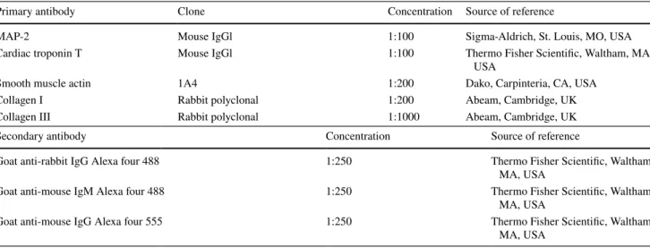
either PE, FITC or APC-conjugated antibodies against CD29, CD73, CD90, CD14, CD34, and HLA-DR mouse anti-human monoclonal antibodies and appropriate isotype controls. Stained cells were then analyzed using a MoFlo XDP flow cytometer (Beckman Coulter, Inc., Brea, CA, USA) using Cell Quest software, and data were analyzed using Summit software. Antibody information is listed in Table 1.
Analysis of the differentiation potential of CD117+
amniotic fluid cells
To investigate the differentiation capacity of hAFS, CD117+
cells were differentiated in vitro into osteogenic, adipogenic, chondrogenic, neurogenic and cardiomyogenic lineages. CD117+ cells were cultured in expansion medium until 70%
confluence was reached. The culture was then shifted to a specific induction medium at 37 °C in a humidified incuba-tor containing 5% CO2. Thus, CD117+ cells were cultured in
‘adipogenic differentiation medium’, ‘osteogenic differentia-tion medium’ (Lonza, Basel, Switzerland), ‘neurogenic dif-ferentiation medium’ (PromoCell GmbH, Heidelberg, Ger-many) or ‘cardiomyogenic differentiation medium’ (Cellular Engineering Technologies, Inc., Iowa, USA) for the appro-priate time according to the manufacturer’s recommended protocol. To induce chondrogenic differentiation, a total of
Table 1 List of antibodies used for flow cytometric analysis in this study
Antigen Clone Concentration Source of reference
CD14 TUK4 10 µl/2.5 × 105 cell Miltenyi Biotec, Auburn, CA, USA
CD29 AG89 20 µl/2.5 × 105 cell MBL CO., LTD., Nagoya, Japan
CD34 581 20 µl/2.5 × l05 cell BD Bioscience Pharmingen, San Diego, CA, USA
CD73 AD2 20 µl/2.5 × l05 cell BioLegend Inc., San Diego, CA, USA
CD90 5E10 20 µl/2.5 × 105 cell BioLegend Inc., San Diego, CA, USA
2.5 × 10⁵ cells were placed into a 15-mL conical
polypropyl-ene tube and centrifuged at 1500 rpm for 10 min. Then cell pellets (1 pellet/tube) were cultured for 21 days in ‘chondro-genic differentiation medium’ (Lonza, Basel, Switzerland). Osteogenesis was assessed by Alizarin staining (Cosmo Bio Co., Ltd. Tokyo, Japan) of the calcified extracellular matrix deposition. Oil red O staining was used for detection of intracellular lipid droplet formation to evaluate adipogen-esis. Chondrogenic differentiation was determined by Alcian Blue staining. For the evaluation of neural differentiation, we immunostained cells for the neuron-specific marker MAP-2. Differentiation into the cardiomyogenic lineage was determined by cardiac troponin T immunostaining. Antibody information is listed in Table 2.
Enzyme‑linked immunosorbent assay (ELISA) of paracrine mediators secreted from hAFS
hAFS at passages 4–6 were seeded on 35-mm culture dishes with amniotic fluid cell culture medium at a density of 1 × 10⁵ cells/well. After 72 h, the confluent cells were
washed with PBS and then incubated in amniotic fluid cell culture medium. After another 12 h, 24 h, 48 h and 72 h, the medium was collected and designated “conditioned medium derived from hAFS”. Paracrine mediator levels for vascular endothelial growth factor (VEGF), IL-10 and PGE2 were
determined using ELISA kits according to the manufac-turer’s protocol.
A dorsal excisional cutaneous wound model in BALB/c mice
BALB/c mice (8 weeks old; male; body weight, 20–23 g) were obtained from Oriental Yeast Co., Ltd. (OYC, Tokyo, Japan). To prepare skin defects, mice were anesthetized with
3% isoflurane followed by maintenance with 2% isoflurane. After hair removal from the dorsal surface, 8-mm full-thick-ness excisional skin wounds were created on each side of the midline using a sterile biopsy punch (Kai Industries Co., Ltd, Japan). We injected 240 µL PBS (containing or lacking 1 × 10⁶ hAFS) around the wound at 6 injection sites. Wounds
were then covered with an occlusive dressing (Tegaderm, Sumitomo 3M, Ltd., Tokyo, Japan) and an elastic adhesive bandage (Silkytex, ALCARE Co, Ltd., Tokyo, Japan). All procedures were performed according to the guidelines for the Care and Use of Laboratory Animals of Keio Univer-sity School of Medicine, and were approved by the Animal Study Committee of Keio University (IRB approval number 15083-(0)).
Macroscopic and histological analyses of cutaneous wounds
Mice were killed by atlantoaxial subluxation, and skin with the wound was removed. Wound size was macroscopically monitored with a microscope camera (Leica, Wetzlar, Ger-many) 0, 7, 14 and 21 days after the treatment. Wound sizes (percentage of wound area to initial wound area) were cal-culated from the photographs using ImageJ software (http:// www.rsb.info.nih.gov/ij; n = 5 per group).
Skin samples were harvested 0, 7, 14, and 21 days after the treatment. Excised specimens were fixed with 4% para-formaldehyde for paraffin embedding. Paraffin sections (4 µm) were treated with H&E, Masson’s Trichrome, Elas-tica van Gieson and Picrosirius red stains. Immunostain-ing for α-smooth muscle actin (α-SMA), type I and III col-lagen was also performed. Antibody information is listed in Table 2. Stained sections were viewed by microscopy (BZ-9000, KEYENCE, Osaka, Japan), and Picrosirius
Table 2 List of antibodies used for immunofluorescence staining in this study
Primary antibody Clone Concentration Source of reference
MAP-2 Mouse IgGl 1:100 Sigma-Aldrich, St. Louis, MO, USA
Cardiac troponin T Mouse IgGl 1:100 Thermo Fisher Scientific, Waltham, MA, USA
Smooth muscle actin 1A4 1:200 Dako, Carpinteria, CA, USA
Collagen I Rabbit polyclonal 1:200 Abeam, Cambridge, UK
Collagen III Rabbit polyclonal 1:1000 Abeam, Cambridge, UK
Secondary antibody Concentration Source of reference
Goat anti-rabbit IgG Alexa four 488 1:250 Thermo Fisher Scientific, Waltham, MA, USA
Goat anti-mouse IgM Alexa four 488 1:250 Thermo Fisher Scientific, Waltham, MA, USA
Goat anti-mouse IgG Alexa four 555 1:250 Thermo Fisher Scientific, Waltham, MA, USA
red-stained sections were observed using polarized micros-copy (BX53-P, Olympus, Tokyo, Japan).
Morphometric analysis of re‑epithelialization and formation of granulation tissue
and myofibroblasts
The width of the wound and the distance of the traversed epithelium were measured on Masson’s Trichrome-stained sections, and the percentage of the tissue that underwent re-epithelialization was calculated according to the following formula: [distance covered by epithelium]/[distance across wound bed] × 100 (n = 5) [15]. The granulation tissue area in the wounds was determined on Elastica van Gieson-stained sections according to previous methods (n = 5) [15]. Myofi-broblasts were identified by α-SMA immunostaining, a clas-sical myofibroblast-specific method.
Assessment of the ability of hAFS to engraft in wound
hAFS were labeled with PKH-26 Red fluorescent cell linker kit, as per the manufacturer’s protocol (Sigma-Aldrich, St. Louis, MO, USA). Cells (1 × 10⁶) were seeded on 35-mm
culture dishes with serum-free α-MEM. After 24 h, the cells were viewed by fluorescent microscopy (BZ-9000, KEY-ENCE). hAFS labeled with PKH-26 were injected around the wound at six injection sites. Each wound received 1 × 10⁶
cells (hAFS labeled with PKH-26) suspended in 240 µL PBS or 240 µL cell-free PBS. Skin samples were harvested 1, 4, 7, 14 and 21 days after the treatment. Cryosections were stained with Hoechst-33342. Fluorescent images were captured using a fluorescent microscopy (BZ-9000, KEYENCE).
RNA isolation and quantitative real‑time RT‑PCR Total RNA was isolated with an RNeasy mini kit (Qiagen, Hilden, Germany) according to the manufacturer’s instruc-tion. The total skin RNA underwent reverse transcription to cDNA using a Prime Script RT Master Mix (Takara Bio,
Shiga, Japan). Quantitative PCRs were performed in dupli-cate in a volume of 25 µL per reaction in a 96-well Bio-Rad CFX96 Real-time PCR System (Bio-Rad, Inc., Hercules, CA, USA). Reaction mixtures included 5 ng of genomic DNA as template, 0.4 mM each primer (Thermo Fisher Sci-entific Inc., Tokyo, Japan), SYBR Premix Ex Taq II (Tli RNaseH Plus) (Takara Bio, Shiga, Japan), and sterile H2O.
The primer sets are listed in Table 3. PCR was performed as follows: pre-denaturation at 95 °C for 30 s, 45 cycles of denaturation at 95 °C for 5 s, annealing at 60 °C for 20 s. The negative control (without reverse transcriptase) had no signal. The relative level of gene expression for each sam-ple was calculated using the 2−ΔΔCT method. Gene
expres-sion levels were normalized to that of Gapdh as an internal control.
Statistical analysis
All results are expressed as means ± SD. The quantitative variable was statistically analyzed using a one-way ANOVA followed by a Student’s t test. P < 0.05 was considered statis-tically significant. Each analysis was performed with com-mercially available software (IBM SPSS Statistics 24).
Results
Isolation, culture and immunophenotypic
characterization of CD117+ amniotic fluid cells
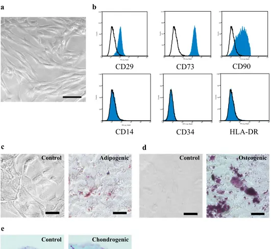
Isolated amniotic fluid cells consisted of a mixed popula-tion of adherent cells with different morphologies and sizes. Most were spindle and round-shaped cells. After immunose-lection and passage in culture, clonal spindle-shaped cells were expanded as stable lines (Fig. 1a). The cell-surface antigenic expression of CD117+ amniotic fluid cells was
determined flow cytometrically. CD117+ amniotic fluid cells
were strongly positive for mesenchymal markers, such as CD29, CD73, and CD90, whereas they were negative for hematopoietic markers such as CD14, CD34, and HLA-DR (Fig. 1b). We also determined the differentiation capability
Table 3 List of primer sequence used in this study
Gene name Amplicon
length (bp) Forward primer sequences (5′–3′) Reverse primer sequences (5′–3′) Collagen, type I. alpha 1 (Col1a1) 145 ACT GGT ACA TCA GCC CGA ACC GAC ATT AGG CGC AGG AAG GTC Collagen, type I, alpha 2 (Col1a2) 147 CAG GCC CAA CCT GTA AAC ACC CTG AGT TGC CAT TTC CTT GGAG Collagen, type III, alpha 1 (Col3a1) 170 CCA TGA CTG TCC CAC GTA AGC CCG GCT GGA AAG AAG TCT GAG α-Smooth muscle actin (Acta2) 156 CAG GCA TGG ATG GCA TCA ATCAC ACT CTA GCT GTG AAG TCA GTG TCG Glyceraldehyde-3-phosphate
Fig. 1 Isolation, culture, and immunophenotypic charac-terization of CD117+ amniotic
fluid cells. a A bright-field image shows the morphology of CD117+ amniotic fluid cells
in culture. Scale bar = 100 µm.
b CD117+ amniotic fluid cells
were stained with antibodies and analyzed by flow cytometry.
c–e Representative microscopic images of CD117+ amniotic
fluid cells. The cells were cultured with adipogenic, osteo-genic or chondroosteo-genic differ-entiation media for appropriate times, and they were assessed by Oil red O (c), Alizarin red (d) or Alcian blue (e) staining. Scale bar = 50 µm. f Cardiomy-ogenic lineage-specific signals observed after cardiac troponin T staining. g Neuron lineage-specific signals exhibited after MAP-2 staining. h ELISA analysis of paracrine media-tors secreted from CD117+
amniotic fluid cells. Images are representative of 3 independent experiments performed with cells from different donors
Troponin T
Hoechst Hoechst MAP2
Troponin T
Hoechst Hoechst MAP2 100 101 102 103 104 105 PE-Log_Height 0 553 1107 1660 2214 Co un ts 100 101 102 103 104 105 FL2-Log_Height 0 78 156 234 313 Co un ts 100 101 102 103 104 105 PE-Log_Height 0 348 696 1044 1393 Co un ts CD29 CD73 CD90 100 101 102 103 104 105 PE-Log_Height 0 273 547 821 1095 Co un ts HLA-DR CD34 100 101 102 103 104 105 PE-Log_Height 0 284 569 853 1138 Co un ts 100 101 102 103 104 105 APC-Log_Height 0 318 636 954 1273 Co un ts a b h CD14 0 2000 4000 6000 0 24 48 72 0 20 40 60 80 0 24 48 72 VEGF (pg/ ml ) Time (hours) IL -10 (pg /ml) PG E2 (pg/ml )
Time (hours) Time (hours) 0 1000 2000 3000 0 24 48 72 f c e d g
Control Adipogenic Control Osteogenic
of CD117+ amniotic fluid cells. These cells could
differ-entiate towards adipogenic, osteogenic and chondrogenic lineages, as shown by Oil red O, Alizarin red, and Alcian blue staining, respectively (Fig. 1c–e). We evaluated the
differentiation potential toward neuronal lineages and car-diomyogenic lineages. As expected, we observed the pres-ence of cells that expressed MAP-2 as a neuronal marker (Fig. 1f) and cardiac troponin T as a cardiomyogenic marker
hAFS
Day0
Day7
Day14
Day21
PBS
Time after wounding (Days)
0
20
40
60
80
100
0
7
14
21
PBS
hAFS
Relative open wound (%)
a
**
0
1
2
3
0
7
14
21
PBS
hAFS
Re-epithelialization (%)
Granulation tissue area (m
m
2
)
Time after wounding (Days)
Time after wounding (Days)
c
b
hAFS
PBS
**
0
20
40
60
80
100
0
7
14
21
PBS
hAFS
*
(Fig. 1g). Based on these results, CD117+ amniotic fluid
cells were shown to fulfill the minimal criteria of an MSC population [16] with potentials for neuronal and cardiomyo-genic differentiation, as previously reported [12, 13, 17, 18]. Paracrine mediators secreted from hAFS
To determine the molecular mediators secreted from hAFS, we used ELISA assays to examine conditioned medium derived from hAFS. ELISAs showed that VEGF levels gradually increased during the observation period, whereas IL-10 and PGE2 levels peaked at 12 h and declined to
base-line (Fig. 1h).
hAFS treatment transiently accelerated cutaneous wound closure by enhancing re‑epithelialization of the epidermis without affecting the granulation tissue area in the dermis
To investigate whether hAFS treatment affected wound healing, we injected hAFS or PBS (control) around 8-mm full-thickness excisional skin wounds created on the backs of BALB/c mice. Macroscopic measurement of wound size demonstrated that hAFS treatment significantly accelerated wound closure compared to control (Fig. 2a). Accelera-tion of wound closure in hAFS groups was observed up to day 14, but there was no obvious difference between the 2 groups at day 21 (Fig. 2a). Additionally, an analysis of re-epithelialization in the epidermis measured on Mas-son’s Trichrome-stained sections demonstrated that hAFS treatment significantly enhanced re-epithelialization after 14 days of treatment compared to control (Fig. 2b). Nev-ertheless, the granulation tissue area in the dermis meas-ured on Elastica van Gieson-stained sections did not show any difference between the 2 groups during the observation
period (Fig. 2c). These results indicated that hAFS treat-ment accelerated cutaneous wound closure by enhancing re-epithelialization in the epidermis without affecting the granulation tissue area in the dermis compared to controls. hAFS decreased α‑SMA expression in regenerated dermal tissues
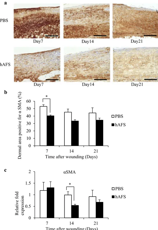
In mice, the skin contraction induced by α-SMA-positive myofibroblasts as well as skin tissue generation contributes to cutaneous wound closure [1, 19]. To investigate whether skin contraction affected wound closure, we evaluated α-SMA-positive cells in granulation tissue. We found that hAFS significantly decreased α-SMA-positive expression by myofibroblasts compared to controls (Fig. 3a, b). In addition, quantitative real-time PCR analysis also showed a significant reduction in mRNA levels of α-SMA at day 14 in the hAFS group compared to controls (Fig. 3c). These observations suggested that hAFS regulated the rate of fibroblast differen-tiation into α-SMA-positive myofibroblasts at the transcrip-tional level. Taken together, we confirmed that hAFS injec-tion accelerated cutaneous wound closure independently of wound contraction.
hAFS treatment attenuated fibrotic changes in the regenerated wound
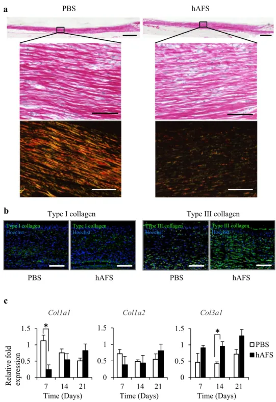
Myofibroblasts containing α-SMA protein play a central role in synthesizing fibrotic tissue containing excess type I collagen [1, 19]. Fibrotic scarring is the final consequence of wound healing and determines its quality. To investigate fibrotic scarring of the regenerated wound, we histologically examined collagen organization in regenerated wounds at day 21. Picrosirius red staining showed that type I collagen (red and yellow) bundle organization was markedly reduced and type III collagen (green) was increased in hAFS groups (Fig. 4a). As the ratio between type I collagen and type III collagen protein expression during wound healing repre-sents a valuable indicator for tissue fibrosis [2, 5], we inves-tigated type I and type III collagen expression in regenerated wounds at day 21. Although type I collagen expression was comparable between the 2 groups, type III collagen protein expression was markedly increased in hAFS groups com-pared to control (Fig. 4b). Furthermore, quantitative real-time PCR analysis showed an increase in mRNA levels for type III collagen during the observation period (Fig. 4c). There was an especially significant increase at day 14 in the hAFS group, although mRNA levels for type I collagen were comparable between the 2 groups except for a signifi-cant decrease Col1a1 at day 7 in the hAFS group (Fig. 4c). These findings suggested that hAFS treatment exerted anti-fibrotic effects resembling fetal wound healing, which were
Fig. 2 hAFS treatment enhanced cutaneous wound closure by accel-erating re-epithelialization without affecting granulation tissue. a
Representative images of full-thickness excisional wounds treated with phosphate-buffered saline (PBS), or 1.0 × 10⁶ hAFS 0, 7, 14 and
21 days after the treatment (left). Data expressed as the percentage of the initial wound size at day 0 (right). b Representative images of Masson’s Trichrome staining section 14 days after the treatment [wound margin (arrows) and the leading edge of the epithelia (arrow-heads)] (n = 5). The percentage of re-epithelialization was calculated according to the following formula: [distance covered by epithelium: distance between wound margin (arrow) − distance between the lead-ing edge of epithelia (arrowheads)]/[distance between wound margin (arrow)] × 100. Scale bar = 500 µm (upper), 100 µm (lower). c Rep-resentative images of Elastica van Gieson-stained sections 14 days after hAFS treatment. Dotted line shows the granulation tissue area. Quantification of granulation tissue areas after 0, 7, 14 and 21 days by the morphometric evaluation of granulation tissue area measured on Elastica van Gieson-stained sections (n = 5). Scale bar = 500 µm (upper), 100 µm (lower). Results are presented as mean ± SD. *P < 0.05 and **P < 0.01 compared to control
characterized by higher type III collagen expression regu-lated at the transcriptional level.
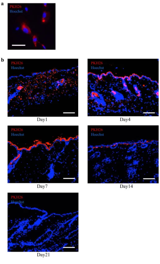
hAFS transiently engrafted in the wound
The ability of hAFS to engraft in a wound was assessed by hAFS labeled with PKH-26. We confirmed that hAFS could be labeled with 26 in vitro by mixing cells with PKH-26 on culture dishes followed by 24-h cultivation. The cells
were then assayed by fluorescent microscopy. This study clearly demonstrated that hAFS could be labeled with PKH-26 (Fig. 5a). To investigate the ability of hAFS to engraft in the wound, skin samples harvested after injection of hAFS labeled with PKH-26 were examined. At day 1 and day 4, large num-bers of hAFS were observed both in the epidermis and dermis. However, after day 7, hAFS were mainly observed in the epi-dermis but not the epi-dermis. At day 21, no labeled cells could be detected in the wound (Fig. 5b). Moreover, we also performed
Fig. 3 hAFS treatment inhibited α-SMA-positive myofibroblast formation. a Representative images of treated regener-ated wounds using antibodies against myofibroblast-specific marker α-SMA on days 7, 14 and 21. Scale bar = 500 µm. b
Morphometric analysis of the α-SMA-positive area. Results are presented as means ± SD (n = 3). *P < 0.05 compared to control. c Quantitative real-time PCR of α-SMA expression levels on days 7, 14 and 21 (n = 3). Results are presented as means ± SD. *P < 0.05 com-pared to control
a
0 0.5 1 1.5 2 7 14 21 PBS hAFSb
c
Time after wounding (Days) * 0 10 20 30 40 50 60 7 14 21 PBS hAFS
Time after wounding (Days) * Derm al area positive for α SMA (% ) hAFS PBS Day7 Day7 Day14 Day14 Day21 Day21 αSMA
immunohistochemical analysis to determine the localization of human-derived cells using human mitochondria anti-bodies (Supplemental Fig. 1). In accordance with the results of PKH26 analysis, human-derived cells could be observed at days 1, 4, 7, and 14. However, no labeled cells could be detected in the wound at day 21. These results suggested that hAFS transplanted into the epidermis might accelerate wound closure via enhancement of re-epithelialization through direct differentiation into epidermal cells (such as keratinocytes) and
through paracrine mechanisms. In contrast, hAFS in the der-mis might alter collagen organization by paracrine signaling, not by direct differentiation into dermal cells.
Fig. 4 hAFS treatment altered collagen organization in the regenerated tissues 21 days after treatment. a Representative images of regenerated tissue stained with Picrosirius red using optical (upper) and polar-ized light microscopy (lower) at day 21. The alignment of type I collagen (yellow and red) and type III collagen (green) was observed by polarized microscopy (lower). Scale bars = 1 mm (upper), 50 µm (lower). b Representative immunofluorescent stains of the regenerated tissue at day 21 using antibodies against type I (left) and type III (right) collagen, followed by Hoechst staining. Scale bars = 100 µm.
c Quantitative real-time PCR of Col1a1, Col1a2, and Col3a1 expression levels at days 7, 14 and 21 (n = 3). Images are representative of 3 independent experiments. Results are pre-sented as means ± SD. *P < 0.05 compared to control 0 0.5 1 1.5 7 14 21 PBS hAFS
a
PBS hAFSb
0 0.5 1 1.5 7 14 21 0 0.5 1 1.5 7 14 21Col1a1 Col1a2 Col3a1
䠆 䠆 Time (Days)
c
Time (Days) Time (Days)Type I collagen Type III collagen
PBS hAFS PBS hAFS
Relative fold expressio
Discussion
In the present study, we demonstrated that hAFS have a unique potential to accelerate cutaneous wound healing with less fibrotic scarring, thereby resembling fetal wound healing.
MSCs have demonstrated an ability to modulate the wound environment to accelerate wound closure [9–11]. In our study, hAFS treatment accelerated cutaneous wound closure by enhancing re-epithelialization for up to 14 days. Moreover, hAFS, which could differentiate into an ectoder-mal lineage, were engrafted in the epidermis during this
Fig. 5 Engraftment of hAFS in wound. a Representative images of hAFS labeled with PKH-26 in vitro (red). Cells were stained with Hoechst. Scale bar = 50 µm. b Immuno-fluorescent and Hoechst staining of the cutaneous tissues 1, 7, 14 and 21 days after injection of hAFS labeled with PKH-26. Scale bar = 200 µm. Images are representative of 3 independent experiments
a
Day1 Day21 Day14 Day7 Day4b
period. In the literature, there is considerable debate over the mechanisms by which MSCs promote cutaneous wound healing. Although it appears to be partially due to direct differentiation into epidermal cells and keratinocytes [20], paracrine signaling, such as the release of trophic factors that promote angiogenesis, immunomodulation, and recruitment of endogenous tissue stem cells, has been suggested as pos-sible mechanism underlying the effects of MSCs in wound healing [9, 11, 21]. Among MSCs, hAFS and their condi-tioned medium have been reported to promote wound clo-sure by direct differentiation into keratinocytes and through paracrine signaling via the TGF-β/SMAD2 pathway [20,
22]. We suggest that both direct differentiation and paracrine effects contribute to wound closure in our study [20, 22].
Murine full-thickness wounds close through re-epitheli-alization and through α-SMA-positive myofibroblast-driven contraction in granulation tissue. In granulation tissue, fibro-blasts constitute the main cell population. They are present in the dermis, proliferate rapidly, and migrate to wound sites and differentiate into myofibroblast due to stimulation by growth factors such as transforming growth factor-β in the wound. The continued presence and activation of myofibro-blasts during wound regeneration induce fibrotic scarring [1,
2, 4, 5, 7, 19, 23]. In the present study, we found that hAFS treatment suppressed α-SMA-positive myofibroblast expres-sion at the transcriptional level in the dermis, implying that hAFS improves the quality of ECM remodeling and accel-erates wound closure independent of wound contraction by regulating fibroblast differentiation into myofibroblast that is the major determinant for fibrotic scarring in the regen-erating tissue.
Few studies have focused on the anti-fibrotic effect of MSCs on ECM remodeling during wound healing. Their potential for reduced fibrotic scarring is less well estab-lished in vivo, as a result of sometimes conflicting findings [21, 24–32]. To the best of our knowledge, this is the first study focusing on the anti-fibrotic effect of hAFS treat-ment on ECM remodeling during wound healing. We found that hAFS treatment had anti-fibrotic effects in the dermis, thereby resembling fetal wound healing. Healing was char-acterized by higher expression of type III collagen at the transcriptional level, which was followed by a significant decrease of α-SMA-positive myofibroblasts. There are phe-notypic differences between the collagen contents in fetal and adult wounds. In adults, after exposure to inflamma-tory mediators in wounds, dermal fibroblasts differentiate into myofibroblasts that express excessive type I collagen, leading to scar formation. On the other hand, fetal dermal fibroblasts before 18 days of gestation in mice and 24 weeks of gestations in humans synthesize more type III collagen, leading to scar-free fetal wound healing [2–6]. Our data sug-gest that hAFS cells regulate fibroblast differentiation result-ing in improved ECM remodelresult-ing durresult-ing wound healresult-ing.
This might be achieved by converting dermal fibroblasts into fetal-like fibroblasts (not to myofibroblast of adult charac-ter), resulting in less fibrotic scarring.
MSCs possess a number of trophic functions that modu-late fibrotic scarring [29]. In the present study, paracrine factors secreted from hAFS transiently engrafted in the dermis during the inflammatory phase (i.e., at days 1 and 4) might play a pivotal role in regulating fibroblast phe-notype. Notably, IL-10 and PGE2, both of which were observed in the hAFS secretome in our study, are potent mediators that inhibit inflammatory and fibrotic responses by regulating fibroblast phenotype during wound healing [33–36]. Although further investigation is needed to eluci-date the detailed mechanisms by which the hAFS secretome regulates fibroblast differentiation and thereby enhances ECM remodeling during wound healing, our results sug-gest that hAFS regulate fibroblast differentiation and exert anti-fibrotic effects on wound healing through a paracrine mechanism.
In vivo therapeutic potential of MSCs depends on its source [37]. The limitation of the study was that we did not compare the anti-fibrotic effect of hAFS on cutaneous wound healing with other MSCs derived from bone marrow (BM) and adipose tissue. However, in the present study, we directly compared IL-10 and PGE2 secretions derived from hAFS and BM-MSCs under the same conditions in vitro (Supplemental Fig. 2). Interestingly, IL-10 concentration was comparable between hAFS group and BM-MSC group. However, PGE 2 was significantly higher in the BM-MSC group cultured over 24 h compared to hAFS. These results indicated the difference of immune responses between hAFS and BM-MSCs, suggesting that we need to compare the anti-fibrotic effect of hAFS in vivo with other MSCs. Doi et al. recently described that the therapeutic effect of MSCs derived from umbilical cord blood and Wharton’s jelly on cutaneous wound healing were comparable [38]. As to hAFS, we would like to address this question using in vivo studies in future investigations.
Amniotic fluid contains cells derived from developing fetal tissues, including fetal skin [13]. These cells might pro-mote cutaneous wound healing because the factors respon-sible for scarless fetal wound healing have been attributed to the developing fetal tissue itself [2, 3, 6]. Therefore, it is reasonable that hAFS could have significant anti-fibrotic potential for cutaneous wound healing. In other words, our findings suggest that hAFS could contribute to fetal scarless wound healing as suggested by recent studies [3, 8].
hAFS cells offer intriguing potentials for autologous stem cell treatment for a variety of complications in neonates, including congenital abnormalities and preterm birth. To prepare an adequate amount of autologous hAFS for these neonates, only a small amount of amniotic fluid cells col-lected by amniocentesis is required, with minimal invasive
risk for the patient. hAFS have an anti-fibrotic potential for treatment of intractable perinatal diseases and can target various organs, including lung [39, 40], kidney [41–44] and liver [45] as well as cutaneous wounds. Thus, anti-fibrotic treatment by hAFS could be a promising autologous stem cell therapy for intractable perinatal diseases.
Conclusion
Our study provides evidence that hAFS have a unique poten-tial to accelerate cutaneous wound healing with reduced fibrotic scarring. Anti-fibrotic treatment using hAFS could be a promising autologous stem cell therapy for intractable perinatal diseases.
Acknowledgements This work was supported by JSPS Grant-in-Aid for Scientific Research (C) Grant number JP15K09724 (https ://www.
jsps.go.jp/engli sh/e-grant s/), JSPS Grant-in-Aid for Scientific Research
(B) Grant number 17H04236 (https ://www.jsps.go.jp/engli sh/e-grant s/), JSPS Grant-in-Aid for Challenging Exploratory Research Grant number JP16K15536 (https ://www.jsps.go.jp/engli sh/e-grant s/), JAOG Ogyaa Donation Foundation (http://www.ogyaa .or.jp/), Japan Spina Bifida and Hydrocephalus Research Foundation (http://www.jikei
kai-group .or.jp/jsato shi/), Keio Gijuku Academic Development Funds
research funding (individual research) (http://www.rcp.keio.ac.jp/ora/
jukun ai/gakus hin.html#two), and Kawano Masanori Memorial Public
Interest Incorporated Foundation for Promotion of Pediatrics (https ://
kawan ozaid an.or.jp/c).
Author contributions MF, DO, HM, SS, NA, MS, YK, KK, and MT conceived and designed the experiments. MF, DO, YA, and TO per-formed the experiments. MF, DO, HM, YA, TO, SS, NA, MS, TM, KM, YK, KK, and MT analyzed the data. SS and MS contributed rea-gents/materials/analytic tools. MF, DO, HM, and MT wrote the paper. Compliance with ethical standards
Conflict of interest The authors have no conflicts of interest to declare.
References
1. Singer AJ, Clark RA. Cutaneous wound healing. N Engl J Med. 1999;341(10):738–746. https ://doi.org/10.1056/NEJM1 99909
02341 1006.
2. Kishi K, Okabe K, Shimizu R, Kubota Y. Fetal skin possesses the ability to regenerate completely: complete regeneration of skin. Keio J Med. 2012;61(4):101–8. https ://doi.org/10.2302/
kjm.2011-0002-IR.
3. Hu MS, Maan ZN, Wu JC, Rennert RC, Hong WX, Lai TS, et al. Tissue engineering and regenerative repair in wound healing. Ann Biomed Eng. 2014;42(7):1494–507. https ://doi.org/10.1007/s1043
9-014-1010-z.
4. Larson BJ, Longaker MT, Lorenz HP. Scarless fetal wound healing: a basic science review. Plast Reconstr Surg. 2010;126(4):1172–80. https ://doi.org/10.1097/PRS.0b013 e3181
eae78 1.
5. Lo DD, Zimmermann AS, Nauta A, Longaker MT, Lorenz HP. Scarless fetal skin wound healing update. Birth Defects Res
C Embryo Today. 2012;96(3):237–47. https ://doi.org/10.1002/
bdrc.21018 .
6. Leung A, Crombleholme TM, Keswani SG. Fetal wound heal-ing: implications for minimal scar formation. Curr Opin Pediatr. 2012;24(3):371–8. https ://doi.org/10.1097/MOP.0b013 e3283
53579 0.
7. Friedman SL, Sheppard D, Duffield JS, Violette S. Therapy for fibrotic diseases: nearing the starting line. Sci Transl Med. 2013;5(167):167 sr1. https ://doi.org/10.1126/scitr anslm
ed.30047 00.
8. Klein JD, Turner CG, Steigman SA, Ahmed A, Zurakowski D, Eriksson E, et al. Amniotic mesenchymal stem cells enhance normal fetal wound healing. Stem Cells Dev. 2011;20(6):969–
76. https ://doi.org/10.1089/scd.2010.0379.
9. Jackson WM, Nesti LJ, Tuan RS. Concise review: clinical trans-lation of wound healing therapies based on mesenchymal stem cells. Stem Cells Transl Med. 2012;1(1):44–50. https ://doi.
org/10.5966/sctm.2011-0024.
10. Li M, Zhao Y, Hao H, Han W, Fu X. Mesenchymal stem cell-based therapy for nonhealing wounds: today and tomor-row. Wound Repair Regen. 2015;23(4):465–82. https ://doi.
org/10.1111/wrr.12304 .
11. Lee DE, Ayoub N, Agrawal DK. Mesenchymal stem cells and cutaneous wound healing: novel methods to increase cell deliv-ery and therapeutic efficacy. Stem Cell Res Ther. 2016;7:37.
https ://doi.org/10.1186/s1328 7-016-0303-6.
12. De Coppi P, Bartsch G Jr, Siddiqui MM, Xu T, Santos CC, Perin L, et al. Isolation of amniotic stem cell lines with poten-tial for therapy. Nat Biotechnol. 2007;25(1):100–6. https ://doi.
org/10.1038/nbt12 74.
13. Loukogeorgakis SP, De Coppi P. Amniotic fluid stem cells: the known, the unknown and potential regenerative medicine appli-cations. Stem Cells. 2016. https ://doi.org/10.1002/stem.2553. 14. Roubelakis MG, Bitsika V, Zagoura D, Trohatou O, Pappa KI,
Makridakis M, et al. In vitro and in vivo properties of distinct populations of amniotic fluid mesenchymal progenitor cells. J Cell Mol Med. 2011;15(9):1896–913. https ://doi.org/10.111
1/j.1582-4934.2010.01180 .x.
15. Hattori N, Mochizuki S, Kishi K, Nakajima T, Takaishi H, D’Armiento J, et al. MMP-13 plays a role in keratinocyte migra-tion, angiogenesis, and contraction in mouse skin wound heal-ing. Am J Pathol. 2009;175(2):533–46. https ://doi.org/10.2353/
ajpat h.2009.08108 0.
16. Dominici M, Le Blanc K, Mueller I, Slaper-Cortenbach I, Marini F, Krause D, et al. Minimal criteria for defining multi-potent mesenchymal stromal cells. The International Soci-ety for Cellular Therapy position statement. Cytotherapy. 2006;8(4):315–7. https ://doi.org/10.1080/14653 24060 08559 05. 17. Perin L, Sedrakyan S, Da Sacco S, De Filippo R. Characteriza-tion of human amniotic fluid stem cells and their pluripoten-tial capability. Methods Cell Biol. 2008;86:85–99. https ://doi.
org/10.1016/s0091 -679x(08)00005 -8.
18. Yan ZJ, Hu YQ, Zhang HT, Zhang P, Xiao ZY, Sun XL, et al. Comparison of the neural differentiation potential of human mesenchymal stem cells from amniotic fluid and adult bone marrow. Cell Mol Neurobiol. 2013;33(4):465–475. https ://doi.
org/10.1007/s1057 1-013-9922-y.
19. Gurtner GC, Werner S, Barrandon Y, Longaker MT. Wound repair and regeneration. Nature. 2008;453(7193):314–321. https
://doi.org/10.1038/natur e0703 9.
20. Sun Q, Li F, Li H, Chen RH, Gu YZ, Chen Y, et al. Amniotic fluid stem cells provide considerable advantages in epidermal regeneration: B7H4 creates a moderate inflammation microen-vironment to promote wound repair. Sci Rep. 2015;5:11560.
21. Wu Y, Chen L, Scott PG, Tredget EE. Mesenchymal stem cells enhance wound healing through differentiation and angiogenesis. Stem Cells. 2007;25(10):2648–2659. https ://doi.org/10.1634/
stemc ells.2007-0226.
22. Yoon BS, Moon JH, Jun EK, Kim J, Maeng I, Kim JS, et al. Secretory profiles and wound healing effects of human amni-otic fluid-derived mesenchymal stem cells. Stem Cells Dev. 2010;19(6):887–902. https ://doi.org/10.1089/scd.2009.0138. 23. Sakai S, Sato K, Tabata Y, Kishi K. Local release of pioglitazone
(a peroxisome proliferator-activated receptor gamma agonist) accelerates proliferation and remodeling phases of wound healing. Wound Repair Regen. 2016;24(1):57–64. https ://doi.org/10.1111/
wrr.12376 .
24. Qi Y, Jiang D, Sindrilaru A, Stegemann A, Schatz S, Treiber N, et al. TSG-6 released from intradermally injected mesenchymal stem cells accelerates wound healing and reduces tissue fibro-sis in murine full-thickness skin wounds. J Invest Dermatol. 2014;134(2):526–37. https ://doi.org/10.1038/jid.2013.328. 25. Wu Y, Huang S, Enhe J, Ma K, Yang S, Sun T, et al. Bone
marrow-derived mesenchymal stem cell attenuates skin fibrosis development in mice. Int Wound J. 2014;11(6):701–10. https ://
doi.org/10.1111/iwj.12034 .
26. Huang S, Wu Y, Gao D, Fu X. Paracrine action of mesenchymal stromal cells delivered by microspheres contributes to cutaneous wound healing and prevents scar formation in mice. Cytotherapy. 2015;17(7):922–31. https ://doi.org/10.1016/j.jcyt.2015.03.690. 27. McFarlin K, Gao X, Liu YB, Dulchavsky DS, Kwon D, Arbab AS,
et al. Bone marrow-derived mesenchymal stromal cells accelerate wound healing in the rat. Wound Repair Regen. 2006;14(4):471– 478. https ://doi.org/10.1111/j.1743-6109.2006.00153 .x. 28. Chen L, Xu Y, Zhao J, Zhang Z, Yang R, Xie J, et al.
Condi-tioned medium from hypoxic bone marrow-derived mesenchy-mal stem cells enhances wound healing in mice. PLoS One. 2014;9(4):e96161. https ://doi.org/10.1371/journ al.pone.00961 61. 29. Li Q, Zhang C, Fu X. Will stem cells bring hope to pathological
skin scar treatment? Cytotherapy. 2016;18(8):943–56. https ://doi.
org/10.1016/j.jcyt.2016.05.008.
30. Kwon DS, Gao X, Liu YB, Dulchavsky DS, Danyluk AL, Bansal M, et al. Treatment with bone marrow-derived stromal cells accel-erates wound healing in diabetic rats. Int Wound J. 2008;5(3):453–
63. https ://doi.org/10.1111/j.1742-481X.2007.00408 .x.
31. Ding J, Ma Z, Shankowsky HA, Medina A, Tredget EE. Deep dermal fibroblast profibrotic characteristics are enhanced by bone marrow-derived mesenchymal stem cells. Wound Repair Regen. 2013;21(3):448–55. https ://doi.org/10.1111/wrr.12046 .
32. Fu X, Li H. Mesenchymal stem cells and skin wound repair and regeneration: possibilities and questions. Cell Tissue Res. 2009;335(2):317–21. https ://doi.org/10.1007/s0044 1-008-0724-3. 33. Seo SY, Han SI, Bae CS, Cho H, Lim SC. Effect of 15-hydroxy-prostaglandin dehydrogenase inhibitor on wound healing. Prosta-glandins Leukot Essent Fatty Acids. 2015;97:35–41. https ://doi.
org/10.1016/j.plefa .2015.03.005.
34. Kieran I, Knock A, Bush J, So K, Metcalfe A, Hobson R, et al. Interleukin-10 reduces scar formation in both animal and human cutaneous wounds: results of two preclinical and
phase II randomized control studies. Wound Repair Regen. 2013;21(3):428–36. https ://doi.org/10.1111/wrr.12043 .
35. Balaji S, Wang X, King A, Le LD, Bhattacharya SS, Moles CM, et al. Interleukin-10-mediated regenerative postnatal tissue repair is dependent on regulation of hyaluronan metabolism via fibro-blast-specific STAT3 signaling. FASEB J. 2017;31(3):868–81.
https ://doi.org/10.1096/fj.20160 0856R .
36. Sandulache VC, Parekh A, Li-Korotky HS, Dohar JE, Hebda PA. Prostaglandin E2 differentially modulates human fetal and adult dermal fibroblast migration and contraction: implication for wound healing. Wound Repair Regen. 2006;14(5):633–643. https
://doi.org/10.1111/j.1743-6109.2006.00156 .x.
37. Bortolotti F, Ukovich L, Razban V, Martinelli V, Ruozi G, Pelos B, et al. In vivo therapeutic potential of mesenchymal stromal cells depends on the source and the isolation procedure. Stem Cell Rep. 2015;4(3):332–9. https ://doi.org/10.1016/j.stemc
r.2015.01.001.
38. Doi H, Kitajima Y, Luo L, Yan C, Tateishi S, Ono Y, et al. Potency of umbilical cord blood- and Wharton’s jelly-derived mesenchy-mal stem cells for scarless wound healing. Sci Rep. 2016;6:18844.
https ://doi.org/10.1038/srep1 8844.
39. Wen ST, Chen W, Chen HL, Lai CW, Yen CC, Lee KH, et al. Amniotic fluid stem cells from EGFP transgenic mice attenuate hyperoxia-induced acute lung injury. PLoS One. 2013;8(9):e75383. https ://doi.org/10.1371/journ al.pone.00753 83. 40. Garcia O, Carraro G, Turcatel G, Hall M, Sedrakyan S, Roche
T, et al. Amniotic fluid stem cells inhibit the progression of bleomycin-induced pulmonary fibrosis via CCL2 modulation in bronchoalveolar lavage. PLoS One. 2013;8(8):e71679. https ://doi.
org/10.1371/journ al.pone.00716 79.
41. Baulier E, Favreau F, Le Corf A, Jayle C, Schneider F, Goujon JM, et al. Amniotic fluid-derived mesenchymal stem cells prevent fibrosis and preserve renal function in a preclinical porcine model of kidney transplantation. Stem Cells Transl Med. 2014;3(7):809–
20. https ://doi.org/10.5966/sctm.2013-0186.
42. Sedrakyan S, Da Sacco S, Milanesi A, Shiri L, Petrosyan A, Var-imezova R, et al. Injection of amniotic fluid stem cells delays pro-gression of renal fibrosis. J Am Soc Nephrol. 2012;23(4):661–73.
https ://doi.org/10.1681/ASN.20110 30243 .
43. Sun D, Bu L, Liu C, Yin Z, Zhou X, Li X, et al. Therapeutic effects of human amniotic fluid-derived stem cells on renal inter-stitial fibrosis in a murine model of unilateral ureteral obstruc-tion. PLoS One. 2013;8(5):e65042. https ://doi.org/10.1371/journ
al.pone.00650 42.
44. Monteiro Carvalho Mori da Cunha MG, Zia S, Oliveira Arcolino F, Carlon MS, Beckmann DV, Pippi NL, et al. Amniotic fluid derived stem cells with a renal progenitor phenotype inhibit inter-stitial fibrosis in renal ischemia and reperfusion injury in rats. PLoS One. 2015;10(8):e0136145. https ://doi.org/10.1371/journ
al.pone.01361 45.
45. Peng SY, Chou CJ, Cheng PJ, Ko IC, Kao YJ, Chen YH, et al. Therapeutic potential of amniotic-fluid-derived stem cells on liver fibrosis model in mice. Taiwan J Obstet Gynecol. 2014;53(2):151–

