Directory UMM :Data Elmu:jurnal:P:Precambrian Research:Vol103.Issue1-2.2000:
Teks penuh
Gambar


Dokumen terkait
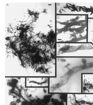
Formation of mylonites in a compressional tectonic setting have been documented in a seismic study in the Grenville orogen in Quebec (Ji et al., 1997). Consequently, the present
Three comprehensive, well-written chapters on the methods for genetic transformation cover the principles, methodological improvements and ap- plication to cereals for the three
The book brings together a series of well-writ- ten, step-by-step protocols on the most frequently used techniques in plant cell and tissue culture, covering various aspects of
Potential uses include the study of stochastic variation in endemic infection levels through time, the evaluation of heterogeneity eects in the partnership formation process, and
The inclusion of copra meal in the diet ever, as the level of copra meal in the diet increased decreased feed intake during both the finisher period in the barley
For example, inhibition of NOS specific stripes that occurs in the optic tectum of three- does not block the formation of ocular dominance columns eyed frogs [124] even though NOS
taking Takapoto lagoon Tuamotu archipelago, French Polynesia as a study site. Although the black pearl oyster was naturally one of the most abundant bivalves in the benthic fauna
scopical observations. The ratio % of normal to atretic follicles decreased with time after the irradiation in primordial follicles and in primary follicles as well. At 6 h
