Comparison Methods of Edge Detection for USG
Images
M. Khairudin
Electrical Engineering Education Dept. Faculty of Engineering, Universitas Negeri Yogyakarta
Yogyakarta, Indonesia [email protected]
Dessy Irmawati
Electronics Engineering Education Dept. Faculty of Engineering, Universitas Negeri Yogyakarta,
Yogyakarta, Indonesia [email protected]
Abstract— This study discussed on a comparison of three
techniques for edge detection of ultrasonography (USG) image. Ultrasound images are used to provide information about fetal development in the womb. The image generated by the two-dimensional ultrasound has not been able to provide complete information. Therefore, in order get the form of fetus on ultrasound image can be clearly identified with the necessary process of image analysis that can detect the boundaries of objects edges, so that it can differentiate between one object with another object on the ultrasound image. Comparison results between the edge detection methods namely Sobel, Prewitt and Canny are expected obtaining the best technique. It can be used in the ultrasound image segmentation process to obtain the best shape of the object of USG image. Based on the available data it can be concluded that the best edge detection methods is produced by Sobel method. The results show that Sobel Edge Detection has the best accuracy compared to other methods with a threshold value of 0.03. (Based on the research, Canny and Prewitt have the same value. Therefore, MSE of Sobel is lower than Canny and Prewitt. Information fetal development in the womb can be seen through a scanning process with ultrasonography technology. The resulting image of the process of two-dimensional ultrasonography has not been able to provide complete information.
Keywords—comparison, edge detection, image,
ultrasonography.
I. INTRODUCTION
Digital image processing means a processing digital computer. It uses computer algorithms to perform image processing on digital images. The investigation of ultrasound is the suplement investigation which carried out on pregnant women. Prior to ultrasound existed, a new fetal heartbeat can be heard on gestational age 16-18 weeks. Along with the development of technology, ultrasound development is also becoming more sophisticated, ultrasound shown in a variety of dimensions, including ultrasound two, three, or four dimensions. Each has advantages and disadvantages for the types of ultrasound, more higher dimensions shown on the monitor will give impact on more expensive the price of equipments. This will cause the investigations using ultrasound are also more expensive. With these problems, the type of 2-dimensional ultrasound is still widely used gynecologist, because it is still considered a representative to investigate pregnant's condition [1].
To analyse the image, it should be accurately with the noise. Noise is an undesired information that contaminates images. The desired effect for an edge detection operation is giving without responses to non-edge pixels and giving only one response to a single edge. Researchers have been explored algorithms of edge detection. Edge detections are basically image segmentation techniques, divide spatial domain, on which defining the image, til get a meaningful parts or regions. Edge detection techniques allow to observe features of an image which have a more or less abrupt change in gray level or texture that indicate the end of one part of the image [2]. Applying an edge detection for an image can significantly reduce the amount of data to be processed. Therefore filter out information that may be regarded as less relevant, while preserving the important structural properties of an image [3].
Several methods for edge detection consist of Canny, Prewitt, Sobel, Rosenfeld, Thurston, Marr-Hildreth and Laplacian methods. These methods detect edges by utilizing masks to perform the convolution on the digital image according to the sudden change of gray level pixel intensity [4]. Canny [5] modified on Sobel method. Canny finds the edge direction by inspecting the horizontal and vertical edge pixel intensity and implement non-maximum suppression to sharpen the edge. Since edges often occur at image locations representing object boundaries, edge detection is extensively used in image segmentation when images are divided into areas corresponding to different objects [6].
II. METHOD
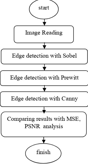
In this experiment starting from image data collection of pregnancy. Then proceed with the processing of image data. Image data processing is done with the following steps (1) conversion of the data of USG image in digital form with the age of the fetus is 20 and 28 weeks respectively, (2) continued by edge detection analysis using Sobel method. In this process also conducting analysis with Mean Square Error (MSE) and Peak Signal to Noise Ratio (PSNR), (3) the next process is edge detection analysis using Prewitt method also with MSE and PSNR analysis, (4) the last step ini this study is edge detection analysis using Canny method also with MSE and PSNR analysis.
Based on MSE and PSNR analysis from the three methods, this study has compared the three results to get the best performance of image. To analysis the images using edge detection can be found at Fig. 1:
Fig.1. Image processing
The stages in the process of fetal ultrasound image using a edge detection with techniques Sobel , Prewitt and Canny can be seen in Figure 1. The image is processed and calculated the value of MSE and PSNR by the following equations:
[
]
are image coordinate respectively.
III. RESULTS AND DISCUSSIONS
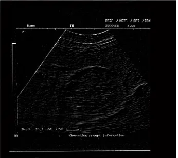
Based on the data of digital images will be used for processing the images in digital form. In this paper, the image processing performed in Fig. 2 and 3 are show the age of fetal of 20 and 28 weeks respectively below:
Fig. 2. Image with fetal age of 20 weeks
Fig. 3. Image with fetal age of 28 weeks
start
Edge detection with Sobel Image Reading
Comparing results with MSE, PSNR analysis
finish
Edge detection with Prewitt
The original data in Fig. 2 and 3 then performed with edge detection using Sobel method produced the image performances shown in Figures 4 and 5 respectively below:
Fig. 4. Sobel technique with fetal age of 20 weeks
Fig. 5. Sobel technique with fetal age of 28 weeks
The next step conducting edge detection for the same original images in Fig 2 and 3 using Prewitt method produced the image performances shown in Fig 6 and 7 respectively below:
Fig. 6. Prewitt technique with fetal age of 20 weeks
Fig. 7. Prewitt technique with fetal age of 28 weeks
The third method in this study using Canny to analysis edge detection for the same original images in Fig. 2 and 3 produced the image performances shown in Fig. 8 and 9 respectively below:
Fig. 8. Canny technique with fetal age of 20 weeks
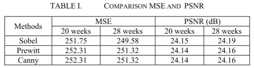
To analysis the performances of edge detection process used comparison of mean square error (MSE) and peak signal to noise ratio (PSNR) can be seen at Table 1.
TABLE I. COMPARISON MSE AND PSNR
Methods MSE PSNR (dB)
20 weeks 28 weeks 20 weeks 28 weeks Sobel 251.75 249.58 24.15 24.19 Prewitt 252.31 251.32 24.14 24.16
Canny 252.31 251.32 24.14 24.16
Based on experiments that have been carried out, the image of the edge detection with the three methods, the methods Sobel, Prewitt and Canny then compared. The results of Sobel edge detection method produces better output than the methods of Canny and Prewitt. Sobel method looks more detail, compared to the others.
Prewitt and Canny has a value of MSE is always the same, with a value greater than Sobel. Edge detection produced by the method of edge detection Sobel is more perfect compared to others, because the resulting lines of edge detection with Sobel method with morphology's are more smooth, and all the lines are connected in detail.
IV. CONCLUSSION
In this study has generated edge detection in three techniques, namely Sobel, Prewitt and Canny. Based on the available data it can be concluded that edge detection can reach the best results through methods Sobel. With Sobel method showed a morphology lines generated is more smooth and uninterrupted. In this study most of the segmentation process ultrasound images using Sobel reached the smallest MSE value.
Acknowledgment
The experiments were by grant from PIU-IDB Project Universitas Negeri Yogyakarta. The authors would like to thank the anonymous reviewers for their precious suggestions for this paper.
References
[1] Khairudin,M and Irmawati D., “Comparison Methods of Noise Elimination for Pregnancy Image Processing”, IEEE Proceeding of The 2nd ICITACEE Proceeding, Semarang-Indonesia, 16 October 2015.
[2] Rashmi, Mukesh Kumar and Rohini Saxena, “Algorithm And Technique On Various Edge Detection: A Survey”, Signal & Image Processing : An International Journal (SIPIJ) Vol.4, No.3, June 2013
[3] Kunjam Nageswara Rao, P srinivasa Rao, Allam Appa Rao, G R Sridhar, “Sobel Edge Detection Method To Identify And Quantify The Risk
Factors For Diabetic Foot Ulcers”, International Journal of Computer Science & Information Technology (IJCSIT) Vol 5, No 1, February 2013 [4] Yan Hum Chai, Lai Khin Wee, Eko Supriyanto, “Edge Detection in
Ultrasound Images Using Speckle Reducing Anisotropic Diffusion in Canny Edge Detector Framew”, Recent Researches in System Science,pp-226-231.
[5] Canny, J., “A Computational Approach to Edge Detection. Pattern Analysis and Machine Intelligence”, IEEE Transactions on, Vol.8, No.6, 1986. pp. 679-698.
[6] Vincent O.R, Folorunso O, 2009, “A Descriptive Algorithm for Sobel Image Edge Detection”, Proceedings of Informing Science & IT Education Conference (InSITE) 2009.