and sharing with colleagues.
Other uses, including reproduction and distribution, or selling or
licensing copies, or posting to personal, institutional or third party
websites are prohibited.
In most cases authors are permitted to post their version of the
article (e.g. in Word or Tex form) to their personal website or
institutional repository. Authors requiring further information
regarding Elsevier’s archiving and manuscript policies are
encouraged to visit:
ORIGINAL ARTICLE
Effect of oxygen tension on proliferation and
characteristics of Wharton’s jelly-derived
mesenchymal stem cells
Wahyu Widowati
a,*
, Laura Wijaya
b, Indra Bachtiar
b,
Rimonta F. Gunanegara
a, Sri Utami Sugeng
a,
Yudha Aryadi Irawan
c, Sutiman B. Sumitro
d, M. Aris Widodo
eaMedical Research Center, Faculty of Medicine, Maranatha Christian University, Bandung, West Java,
Indonesia
bStem Cell and Cancer Institute, Jakarta, Indonesia
cBiomolecular and Biomedical Research Center, Aretha Medika Utama, Bandung, West Java, Indonesia d
Department of Biology, Faculty of Science, Brawijaya University, Malang, East Java, Indonesia
ePharmacology Laboratory, Faculty of Medicine, Brawijaya University, Malang, East Java, Indonesia
Received 4 November 2013; received in revised form 23 January 2014; accepted 10 February 2014
Available online 27 March 2014
KEYWORDS
hypoxic;
mesenchymal stem cells;
normoxic; Wharton’s jelly
Abstract Mesenchymal stem cells (MSCs) from Wharton’s jelly have a higher proliferation rate and self-renewal capacity than adult tissue-derived MSCs. A low oxygen level or hypoxic condition is prevalent in the microenvironment of the stem cells in the early stages of devel-opment. Hypoxia can influence proliferation and differentiation of various stem/precursor cell populations. This research was conducted: to determine the proliferation rate and character-istics of human MSCs from Wharton’s jelly in hypoxic and normoxic condition; to evaluate their character after MSCs are incubated in hypoxic and normoxic environment using surface markers including CD105, CD73, CD14, CD19, CD34, CD45, and HLA-II; and to evaluate the pro-liferation rate and number of MSCs at many passages using the trypan blue method. The hyp-oxic and normhyp-oxic microenvironment showed significant differences in the proliferation rate and population doubling time, but and there were no differences in surface markers. Copyrightª2014, Taiwan Genomic Medicine and Biomarker Society. Published by Elsevier Taiwan LLC. All rights reserved.
* Corresponding author. Medical Research Center, Faculty of Medicine, Maranatha Christian University, Jl. Prof. Drg. Suria Sumantri 65, Bandung 40164, Indonesia.
E-mail address:[email protected](W. Widowati).
http://dx.doi.org/10.1016/j.bgm.2014.02.001
2214-0247/Copyrightª2014, Taiwan Genomic Medicine and Biomarker Society. Published by Elsevier Taiwan LLC. All rights reserved. Available online atwww.sciencedirect.com
ScienceDirect
Introduction
Stem cells are currently used in clinical applications1and
can be obtained from embryonic and extraembryonic tis-sues and adult organs. Stem cells have the ability to prolong self-renewal and differentiate into mature cells of various lineages, which makes them important cell sources for tissue engineering applications.1,2 Clinical therapies require a large number of cells, so many strategies are used to improve the quality and quantity of stem cells.3
The clinical therapeutic strategy uses mesenchymal stem cells (MSCs) as cellular vehicles for the targeted delivery and local production of biologic agents in many diseases.4 MSCs were originally isolated from the bone marrow (BM). BM-derived MSCs (BM-MSCs) are nonhematopoietic precursor cells, and are capable of contributing to the maintenance and regeneration of connective tissues through engraftment.5 However, BM-MSCs have: limited cell numbers, a risk of loss of stem properties, chromosomal changes, and problems of contamination, painful isolation procedure, low MSC charac-teristics, multipotent differentiation potential, and prolifera-tion efficiency of BM-MSCs decline with increasing age.6e8MSCs
are hypoimmunogenic and have the ability to promote regen-eration and functional recovery in disease and injury, which involves immunomodulation effects. MSCs are good candidates for cell transplantations and allogeneic applications.9
MSCs are able to differentiate to a variety of specialized mesenchymal tissues including bone, cartilage, muscle, marrow stroma, tendon, ligament, fat, and connective tis-sue.10MSCs have been isolated from different compartments of the umbilical cord (cord blood, umbilical cord matrix, and the perivascular region), adult peripheral blood,11,12adipose tissue,13lung,14heart,15trabecular bone, and dental pulp,16
and also from a variety of fetal tissues, such as the spleen, lung, pancreas, kidneys, and amniotic fluid during midg-estation.17MSCs can be isolated from Wharton’s jelly (WJ), the
embryonic mucous connective tissue lying between the am-niotic epithelium and the umbilical vessels. Wharton’s jelly-derived MSC or WJ-MSCs have a higher proliferation rate and self-renewal capacity than adult tissue-derived MSCs.18,19
Oxygen concentration is an important component of the stem cell niche, where it plays an important role in main-taining the proliferation and plasticity of stem cells.1,20The oxygen concentration has been investigated extensively.21
Several stem cell populations cultivated under hypoxic condition resulted in enhanced proliferation.1Physiological oxygen tension of 5% instead of 2% was found to improve a mouse embryonic stem cell line by reducing oxidative stress.22Hypoxic conditions have been shown to maintain
the pluripotency and minimize spontaneous differentiation of human embryonic stem cells.20Mammalian cells exposed
to hypoxic conditions express a variety of target genes controlled by hypoxia inducible factor 1 to overcome hyp-oxic stress.23 Hypoxic conditions can increase the number
of hematopoietic stem cells.24
The objective of this research was to evaluate the effect hypoxic environment can have on the proliferation and surface marker character of WJ-MSCs. The results of this research may be useful from a clinical point of view, as WJ-MSCs are used for cell therapy to repair tissue injuries, the MSCs can encounter severe low oxygen tension.
Materials and methods
Isolation and cultivation of WJ-MSCs
Fresh human umbilical cords (UC; n Z 5) were collected
from women aged 25e40 years after normal vaginal
de-livery, with informed consent using the guidelines approved by the Institutional Ethics Committee at the Stem Cell and Cancer Institute, Jakarta, Indonesia and from the Institu-tional Ethics Committee collaboration between Maranatha Christian University, Bandung, Indonesia and Immanuel Hospital Bandung, Bandung, Indonesia.
MSCs from WJ of UC, were isolated as previously described.3,25 UC was washed by phosphate buffer saline (0.9% w/v sodium chloride) and cut into very small pieces, approximately 1e2 mm, then UC was cut longitudinally, and
plated on tissue culture plastic plates. The explants were cultured in MEMawith 2 mM GlutaMAX (Invitrogen, Carlsbad,
CA, USA), supplemented with 20% fetal bovine serum (FBS; Invitrogen) and penicillinestreptomycineamphotericin B
(100 U/mL, 100mg/mL, and 0.25mg/mL; Invitrogen).
Cul-tures were incubated in a humidified atmosphere with 5% CO2at 37C for 3 weeks after explantation, when
fibroblast-like adherent cells were expected to migrate from the tissue fragments, the adherent cells and tissue fragments were detached using trypLEeEDTA solution (TrypLE Express;
Invi-trogen) followed by washing with basal medium to remove the trypLEeEDTA. The cells were harvested and replated at a
density 8 103 cells/cm2 when cells reached 80e90%
confluence. WJ-MSCs were cultured in 95% air (21% O2)/5%
CO2for normoxic and hypoxic (5% and 2.5% oxygen). Hypoxia
was achieved using a tri-gas incubator (CO2incubators with
additional process controls; BINDER GmbH, Tuttlingen, Germany) with internal O2 and N2 tank changer for
con-necting to separate gas tanks.
Cell proliferation analysis
The effect of hypoxic and normoxic incubation towards the cells proliferation was determined as follows. Cells were counted and passaged at a confluence of 80%. Briefly, cultured cells were dissociated using trypsin, incubated for 3 minutes at 37 C, harvested and washed
using MEMa þ 20% FBS followed by centrifugation at
300 g, for 4 minutes. The cell pellet was resuspended with trypan blue solution (0.4% in PBS, 1:1 dilution with culture medium) for 3 minutes. The number of dead cells (retaining the dye) was counted with a hemocy-tometer and expressed as a percentage of the total viable cell number. The experiments were performed in triplicate.
At each passage, the population doubling (PD) was determined using the formula:
PDZ½log10ðNHÞ log10ðNIÞ=log10 ð1Þ
where NI is the inoculum cell number and NH the cell har-vest number. PD for each passage was calculated and added to the PD of the previous passages in order to generate cumulative PD data. The PD time was obtained by the formula:
PD timeZt=PDðin hoursÞ; where tZtime ð2Þ
At the same time, a growth curve of WJ-MSCs from two different conditions was started. Cells were seeded at 200/ cm2 in 6-well plates. Every passage (3 days culture) for
eight passages, cells from one well were harvested and counted.
Detection of MSCs markers using fluorescence activated cell sorting (FACS)
The WJ-MSCs were evaluated using surface marker detec-tion at Passage 4 (P4) and P8 to confirm the effect of oxy-gen concentration (hypoxia and normoxia) on MSC characterization, WJ-MSCs at 80% confluence were har-vested and dissociated with trypsin-EDTA and centrifuged at 300gfor 10 minutes. The pellet was resuspended with PBSþ2% FBS, and cells were counted with a hemocytom-eter. Between 100 cells and 200 cells in 25mL PBS were
introduced into FACS (BD FACSCalibur ) tubes. Antibody
was then added to each FACS tube: isotype mIgG2a-PE, CD105-PE, HLA class II-PE; isotype mIgG1-PE, CD73-PE, CD19-PE; isotype mIgG1-FITC, CD 34-FITC, CD45-FITC, CD14-FITC, followed by incubation at 4C for 15 minutes.
The cells were analyzed by flow cytometry with a FACS-Calibur 3 argon laser 488 nm (Becton Dickinson, Franklin Lakes, NJ, USA) using CellQuest Pro Acquisition on the BD FACStationSoftware. The experiments and measurement
of surface marker were performed in triplicate.
Results
Effect of oxygen tension on stemcell proliferation
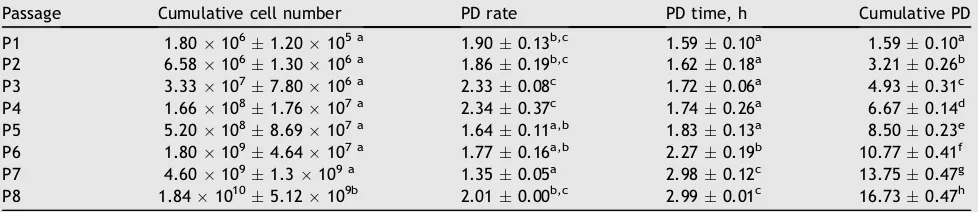
The results of evaluating the effect of oxygen tension (hypoxia, normoxia) on WJ-MSCs proliferation, cumulative cell number, PD, PD time, and cumulative PD for each passage up to P8 are inTables 1e3. Based on these data, PD
time including normoxic, hypoxic (O2 2.5%; O2 5%) P1eP5
had the same PD time and old passage (P6eP8) had higher a
PD time than young passage on normoxic and hypoxic ten-sion. In order to compare the cumulative cell number, PD and PDT among incubation (normoxic and hypoxic tension) at every passage, the data were analyzed using Tukey’s honestly significant differences post hoc test (Tables 4
and5). Hypoxia 2.5% had a higher cumulative cell number compared to normoxia and hypoxia 5% at P4, P5, P6, and P8. The cells were seeded at 480.000 cells for all treatment (normoxia, hypoxia 5%, hypoxia 2.5%) and at all passages (P1eP8).Table 5shows that PD of hypoxia 2.5% at P3, P4,
P7, and P8 was higher than normoxia and hypoxia 5% O2PD
rate. PD time hypoxia 2.5% was lower than normoxia and hypoxia 5% at P7 and P8.
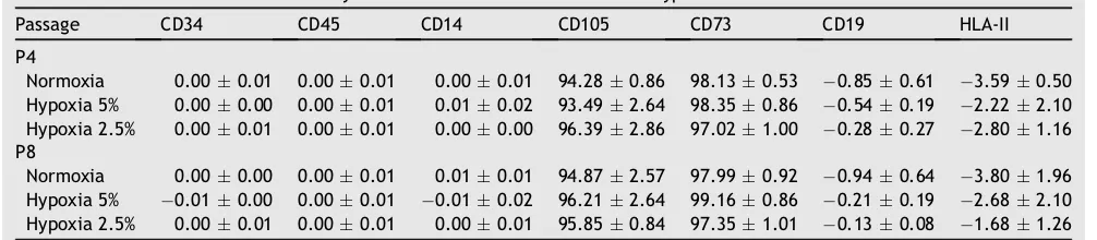
Effect of oxygen tension on phenotype
Human MSCs surface marker are suggested to be positive for CD73 and CD105 and negative for CD14, CD19, CD34, and
Table 1 Cumulative cell number, population doubling (PD), PD time, and cumulative PD of mesenchymal stem cells in nor-moxic tension.
Passage Cumulative cell number PD PD time, h Cumulative PD P1 1.821061.51105 a 1.920.12b 1.570.09a 1.570.09a
P2 6.901064.50105 a 1.940.08b 1.550.06a 3.120.08b P3 3.521071.36106 a 2.340.04c 1.710.03a 4.830.05c
P4 1.711082.85107 a 2.270.19c 1.770.15a 6.600.19d P5 5.651085.78107 a,b 1.730.13b 1.740.14a 8.340.12e
P6 1.951092.97108 b 1.780.08b 2.250.10b 10.590.22f
P7 4.391094.57108 c 1.180.10a 3.410.28c 14.000.21g
P8 1.5310101.37109d 1.800.08b 3.340.14c 17.340.32h
Data are presented as meanstandard deviation. Different letters in the same column (among passage) are significant atp<0.05 (Tukey’s honestly significant differencespost hoctest).
Table 2 Cumulative cell number, population doubling (PD), PD time, and cumulative PD of mesenchymal stem cells in hypoxic tension (5% O2).
Passage Cumulative cell number PD rate PD time, h Cumulative PD P1 1.801061.20105 a 1.900.13b,c 1.590.10a 1.590.10a P2 6.581061.30106 a 1.860.19b,c 1.620.18a 3.210.26b
P3 3.331077.80106 a 2.330.08c 1.720.06a 4.930.31c P4 1.661081.76107 a 2.340.37c 1.740.26a 6.670.14d
P5 5.201088.69107 a 1.640.11a,b 1.830.13a 8.500.23e P6 1.801094.64107 a 1.770.16a,b 2.270.19b 10.770.41f
P7 4.601091.3109 a 1.350.05a 2.980.12c 13.750.47g P8 1.8410105.12109b 2.010.00b,c 2.990.01c 16.730.47h
CD45.26The effect of oxygen tension on the surface marker
of WJ-MSCs can be seen inTable 6.Table 6shows that the surface marker of WJ-MSCs on hypoxia and normoxia, both P4 and P8, were not significantly different (p>0.05).
Discussion
It has been reported that reduced oxygen tension enhances proliferation of some cell types, for example, hypoxia (1e5
% oxygen) enhanced the self-renewal of hematopoietic stem cells and murine embryonic stem cells in several
previous studies. Moreover, when rat BM-MSCs were cultured in reduced oxygen condition, and the proliferation of MSC was increased and had a greater number of colonies.27e31
In the current study, the increased hypoxic (O2 2.5%)
condition was the best microenvironment for stem cells proliferation compared to normoxic and hypoxic (O25%) for
cells at a high passage (P7, P8). This result was consistent with previous reports that MSCs maintain viability when cultured in 2%e5% O2, and increase their proliferation rate
after an initial lag phase.32Hypoxic preconditioning of MSCs
Table 3 Cumulative cell number, population doubling PD, PD time, and cumulative PD of mesenchymal stem cells in hypoxic tension (2.5% O2).
Passage Cumulative cell number PD PD time, h Cumulative PD P1 1.881061.20105 a 1.970.09a 1.530.07a 1.530.07a
P2 7.201063.40105 a 1.940.04a 1.550.04a 3.070.05b P3 4.361073.14106 a,b 2.590.04a 1.540.03a 4.620.07c
P4 2.641082.50107 a,b 2.600.04a 1.540.03a 6.150.10d P5 9.741081.04107 a,b 1.880.07a 1.600.06a 7.750.11e
P6 3.781097.05108 a,b 1.940.16b 2.070.17b 9.820.25f
P7 1.3110101.93109b 1.800.17c 2.230.22b 12.050.19g
P8 6.4810101.21010 c 2.300.09c 2.620.10c 14.670.28h
Data are presented as meanstandard deviation. Different letters in the same column (among passage) are significant atp<0.05 (Tukey’s honestly significant differencespost hoctest).
Table 4 Cumulative cell number among normoxic and hypoxic tension.
Passage Cumulative cell number
Normoxia Hypoxia 5% Hypoxia 2.5%
P1 1.821061.51105 a 1.801061.20105 a 1.881061.20105 a P2 6.901064.50105 a 6.581061.30106 a 7.201063.40105 a
P3 3.521071.36106 a 3.331077.80106 a 4.361073.14106 a,b
P4 1.711082.85107 a 1.661081.76107 a 2.641082.50107 b
P5 5.651085.78107 a 5.201088.69107 a 9.741081.04107 b
P6 1.951092.97108 a 1.801094.64107 a 3.781097.05108 b
P7 4.391094.57108 a 4.601091.3109 a 1.3110101.93109 b
P8 1.5310101.37109a 1.8410105.12109a 6.4810101.21010 b
Data are presented as meanstandard deviation. Different letters in the same row (among normoxic and hypoxic) are significant at p<0.05 (Tukey’s honestly significant differencespost hoctest).
Table 5 Population doubling PD, PD time among normoxic and hypoxic tension.
Passage PD PD time, h
Normoxia Hypoxia 5% Hypoxia 2.5% Normoxia Hypoxia 5% Hypoxia 2.5% P1 1.920.12a 1.900.13a 1.970.09a 1.570.09a 1.590.10a 1.530.07a P2 1.940.08a 1.860.19a 1.940.04a 1.550.06a 1.620.18a 1.550.04a
P3 2.340.04a 2.330.08a 2.590.04b 1.710.03b 1.720.06b 1.540.03a P4 2.270.19a 2.340.37a 2.600.04b 1.770.15a 1.740.26a 1.540.03a
P5 1.730.13a 1.640.11a 1.880.07a 1.740.14a 1.830.13a 1.600.06a P6 1.780.08a 1.770.16a 1.940.16a 2.250.10a 2.270.19a 2.070.17a
P7 1.180.10a 1.350.05a 1.800.17b 3.410.29b 2.980.12b 2.230.22a P8 1.800.08a 2.010.00a 2.300.09b 3.340.14c 2.990.01b 2.620.10a
Data are presented as meanstandard deviation of PD and PD time. Different letters in the same row (among normoxia and hypoxia of PD, PDT) are significant atp<0.05 (Tukey’s honestly significant differencespost hoctest).
in 0.5% oxygen for 24 hours increased expression of pro-survival and proangiogenic factors including hypoxia-inducible factor 1, angiopoietin-1, and vascular endothe-lial growth factor and its receptor. Cell death of hypoxic stem cells and caspase-3 activation in these cells were significantly lower compared with normoxic stem cells both
in vitro and in vivo.33 Hypoxic conditions enhance cell
amplification, and culturing under hypoxia could be an alternative approach without the need for extra additives to stimulate primary culture and further expansion, yielding a sufficient supply of cells and avoiding multiple passages.34Low-oxygen tension is an important component of the stem-cell microenvironment (the stem-cell niche) and provides signals conducive to maintenance of stem-cell function.35Compared with the normoxic condition, hypoxia
enhances proliferation with an approximately six- to seven-fold higher expansion of adipose tissue-derived stromal cells over 6 weeks.36 Hypoxia provides a favorable culture
condition to promote proliferation of MSCs.37The long-term (1 month) effect of human MSC culture in hypoxic tension (2% O2) showed improved survival and increased adipocytic
and osteogenic differentiation capacity.32The MSC culture
under hypoxic conditions was associated with the induction of hypoxia-inducing factor-aand an elevated expression of
energy metabolism-associated genes including glucose transporter 1(GLUT-1),lactate dehydrogenase(LDH), and
pyruvate dehydrogenase kinase 1 (PDK1).38 High
concen-trations of oxygen can cause oxidative stress via production of reactive oxygen speciesdfree radicals that can damage
lipids, proteins, and DNA, altering cell metabolism.39 Mod-erate hypoxia may lower intracellular reactive oxygen species generation and accumulation and thereby increase the metabolic efficiency.40
The flow cytometric analysis (Table 6) showed that ox-ygen level and passage did not affect the MSC’s character. The surface markers expression are positive for CD 105 and CD 73 (more than 95%) and negative for CD 14, CD 19, CD 34, CD 45 and HLA-II (less than 2%). CD45 is a pan-leukocyte marker; CD34 marks primitive hematopoietic progenitors and endothelial cells; CD14 is prominently expressed on monocytes and macrophages, the most likely hematopoi-etic cells to be found in an MSC culture; CD19 is a marker of B cells that may also adhere to MSCs in culture; and HLA-II-DR molecules are not expressed on MSCs.41Table 6 shows
that surface markers in hypoxic condition were not signifi-cantly different when compared to normoxic. However, this
result was not consistent with previous results that showed CD90 expression reduced in BMSCs harvested under hypoxia may be associated with improved chondrogenesis,42 hyp-oxic culture for expansion of adipose tissue-derived stromal cells, and maintenance of their undifferentiated state.36
In conclusion, hypoxic 2.5% O2 yield the highest
prolif-eration, and the lowest PD and PD time. Oxygen level does not affect surface markers of WJ-MSCs at P4 or P8
Conflicts of interest
All contributing authors declare no conflicts of interest.
Acknowledgments
The authors acknowledge gratefully the financial support from the Ministry of National Education, Republic of Indonesia for the research grant of Hibah Unggulan Per-guruan Tinggi 2012e2013. We are thankful to Dwi Agustina
from Stem Cell and Cancer Institute, Jakarta, Indonesia for her valuable assistance. This research was also supported by the Stem Cell and Cancer Institute, Jakarta, Indonesia.
References
1.Ma T, Grayson WL, Fro¨hlich M, et al. Hypoxia and stem cell-based engineering of mesenchymal tissues.Biotechnol Prog. 2009;25:32e42.
2.Anzalone R, Lo Iacono M, Corrao S, et al. New emerging po-tentials for human Wharton’s jelly mesenchymal stem cells: immunological features and hepatocyte-like differentiative capacity.Stem Cells Dev. 2010;19:423e438.
3.Nekanti U, Dastida S, Venugopal P, et al. Increased prolifera-tion and analysis of differential gene expression in human Wharton’s jelly-derived mesenchymal stromal cells under hypoxia.Int J Biol Sci. 2010;6:499e512.
4.Studeny M, Marini FC, Champlin RE, et al. Bone marrow-derived mesenchymal stem cells as vehicles for interferon beta delivery into tumors.Cancer Res. 2002;62:3603e3608. 5.Prockop DJ. Marrow stromal cells as stem cells for
non-hematopoietic tissues.Science. 1997;276:71e74.
6.Stenderup K, Justesen J, Clausen C, et al. Aging is associated with decreased maximal life span and accelerated senescence of bone marrow stromal cells.Bone. 2003;33:919e926. 7.Batsali AK, Kastrinaki MC, Papadaki HA, et al. Mesenchymal
stem cells derived from Wharton’s Jelly of the umbilical cord:
Table 6 Surface markers of mesenchymal stem cells under normoxic and hypoxic tension.
Passage CD34 CD45 CD14 CD105 CD73 CD19 HLA-II P4
biological properties and emerging clinical applications.Curr Stem Cell Res Ther. 2013;8:144e155.
8. Bongso A, Fong CY. The therapeutic potential, challenges and future clinical directions of stem cells from the Wharton’s jelly of the human umbilical cord.Stem Cell Rev. 2013;9:226e240. 9. Ghannam S, Bouffi C, Djouad F, et al. Immunosuppression by mesenchymal stem cells: mechanisms and clinical applica-tions.Stem Cell Res Ther. 2010;1:2.
10. Lindross B. Characterization and Optimization of In Vitro Culture Conditions of Adult Stem Cells for Clinical Therapy. Academic Dissertation. University of Tampere; 2009.
11. Cao C, Dong Y, Dong Y. Study on culture andin vitro osteo-genesis of blood-derived human mesenchymal stem cells. Zhongguo Xiu Fu Chong Jian Wai Ke Za Zhi. 2005;19:642e647 [In Chinese].
12. Kassis I, Zangi L, Rivkin R, et al. Isolation of mesenchymal stem cells from G-CSF-mobilized human peripheral blood using fibrin microbeads.Bone Marrow Transplant. 2006;37:967e976. 13. Locke M, Feisst V, Dunbar PR. Concise review: human adipose
derived stem cells: separating promise from clinical need. Stem Cells. 2011;29:404e411.
14. Griffiths MJ, Bonnet D, Janes SM. Stem cells of the alveolar epithelium.Lancet. 2005;366:249e260.
15. Beltrami AP, Barlucchi L, Torella D, et al. Adult cardiac stem cells are multipotent and support myocardial regeneration. Cell. 2003;114:763e776.
16. Gronthos S, Mankani M, Brahim J, et al. Postnatal human dental pulp stem cells (DPSCs)in vitroandin vivo.Proc Natl Acad Sci USA. 2000;97:13625e13630.
17. Secco M, Zucconi E, Vieira NM, et al. Multipotent stem cells from umbilical cord: cord is richer than blood. Stem Cells. 2008;26:146e150.
18. Can A, Karahuseyinoglu S. Concise review: human umbilical cord stroma with regard to the source of fetus-derived stem cells.Stem Cells. 2007;25:2886e2895.
19. Troyer DL, Weiss ML. Wharton’s jelly-derived cells are a primitive stromal cell population. Stem Cells. 2008;26: 591e599.
20. Ezashi T, Das P, Roberts RM. Low O2tensions and the preven-tion of differentiapreven-tion of hES cells.Proc Natl Acad Sci USA. 2005;102:4783e4788.
21. Obradovic B, Carrier RL, Vunjak-Novakovic G, et al. Gas ex-change is essential for bioreactor cultivation of tissue engi-neered cartilage.Biotechnol Bioeng. 1999;63:197e205. 22. Wang FN, Thirumangalathu S, Loeken MR. Establishment of
new mouse embryonic stem cell lines is improved by physio-logical glucose and oxygen. Cloning Stem Cells. 2006;8: 108e116.
23. Lee A-H, Moon JH, Cho EA, et al. Monoclonal antibody-based screening assay for factor inhibiting hypoxia-inducible factor inhibitors.J Biomol Screen. 2008;13:494e503.
24. Ivanovic¸ Z, Dello Sbarba P, Trimoreau F, et al. Primitive human HPCs are better maintained and expanded in vitro at 1 percent oxygen than at 20 percent.Transfusion. 2000;40: 1482e1488.
25. Fong CY, Richards M, Manasi N, et al. Comparative growth behaviour and characterization of stem cells from human Wharton’s jelly.Reprod Biomed Online. 2007;15:708e718. 26. Pittenger MF, Mackay AM, Beck SC, et al. Multilineage potential
of adult human mesenchymal stem cells.Science. 1999;284: 143e147.
27. Zhang FB, Li L, Fang B, et al. Passage-restricted differentiation potential of mesenchymal stem cells into cardiomyocyte-like cells.Biochem Biophys Res Commun. 2005;28(336):784e792. 28. Sethe S, Scutt A, Stolzing A. Aging of mesenchymal stem cells.
Ageing Res Rev. 2006;5:91e116.
29. Kretlow JD, Jin YQ, Liu W, et al. Donor age and cell passage affects differentiation potential of murine bone marrow-derived stem cells.BMC Cell Biol. 2008;9:60.
30. Bonab MM, Alimoghaddam K, Talebian F, et al. Aging of mesenchymal stem cellin vitro.BMC Cell Biol. 2006;7:14. 31. Nayan M, Paul A, Chen G, et al. Superior therapeutic potential
of young bone marrow mesenchymal stem cells by direct intramyocardial delivery in aged recipients with acute myocardial infarctionin vitroandin vivoinvestigation.J Tis-sue Eng. 2011;2011:741213.
32. Grayson WL, Zhao F, Izadpanah R, et al. Effects of hypoxia on human mesenchymal stem cell expansion and plasticity in 3D constructs.J Cellular Physiol. 2006;207:331e339.
33. Hu X, Yu SP, Fraser JL, et al. Transplantation of hypoxia-preconditioned mesenchymal stem cells improves infarcted heart function via enhanced survival of implanted cells and angiogenesis.J Thorac Cardiovasc Surg. 2008;135:799e808. 34. Grayson WL, Zhao F, Bunnell B, et al. Hypoxia enhances
pro-liferation and tissue formation of human mesenchymal stem cells.Biochem Biophys Res Commun. 2007;358:948e953. 35. Mohyeldin A, Garzo´n-Muvdi T, Quin˜ones-Hinojosa A. Oxygen in
stem cell biology: a critical component of the stem cell niche. Cell. 2010;7:150e161.
36. Yamamoto Y, Fujita M, Tanaka Y, et al. Low oxygen tension en-hances proliferation and maintains stemness of adipose tissue-derived stromal cells.Bio Res Open Access. 2013;2:199e205. 37. Hung SP, Ho JH, Shih YR, et al. Hypoxia promotes proliferation
and osteogenic differentiation potentials of human mesen-chymal stem cells.J Orthop Res. 2012;30:260e266.
38. Lavrentieva A, Majore I, Kasper C, et al. Effects of hypoxic culture conditions on umbilical cord-derived human mesen-chymal stem cells.Cell Com Signaling. 2010;8:1e9.
39. Wiseman H, Halliwell B. Damage to DNA by reactive oxygen and nitrogen species. Role in inflammatory disease and progression to cancer.Biochem J. 1996;313:17e29.
40. Miller WM, Wilke CR, Blanch HW. Effects of dissolved-oxygen concentration on hybridoma growth and metabolism in continuous culture.J Cellular Physiol. 1987;132:524e530. 41. Dominici M, Le Blanc K, Mueller I, et al. Minimal criteria for
defining multipotent mesenchymal stromal cells.Cytotherapy. 2006;8:315e317.
42. Adesida AB, Sierra AM, Jomha NM. Hypoxia mediated isolation and expansion enhances the chondrogenic capacity of bone marrow mesenchymal stromal cells.Stem Cell Res Ther. 2012;3:1e12.