COMPUTER-AIDED DRUG REPURPOSING:
A CYCLOOXYGENASE-2 INHIBITOR CELECOXIB AS A LIGAND
FOR ESTROGEN RECEPTOR ALPHA
Enade Perdana Istyastono
1,4,*, Florentinus Dika Octa Riswanto
2, and Sri Hartati Yuliani
3 1Division of Drug Design and Discovery, Faculty of Pharmacy, Sanata Dharma University Paingan, Maguwohardjo, Depok, Yogyakarta 55282, Indonesia
2
Division of Food and Drug Safety, Quality and Efficacy, Faculty of Pharmacy, Sanata Dharma University Paingan, Maguwohardjo, Depok, Yogyakarta 55282, Indonesia
3
Division of Pharmaceutics and Pharmaceutical Technology, Faculty of Pharmacy, Sanata Dharma University Paingan, Maguwohardjo, Depok, Yogyakarta 55282, Indonesia
4
Center for Environmental Studies Sanata Dharma University (CESSDU) Soropadan, Condongcatur, Depok, Yogyakarta 55283, Indonesia
Received April 17, 2015; Accepted August 6, 2015
ABSTRACT
A cyclooxygenase-2 (COX-2) inhibitor celecoxib has been previously reported to have cytotoxic activities towards gastric, prostate, ovarian, colon and breast cancer cell lines. This article reports that the cytotoxic activities of celecoxib could be resulted from its activity as a potent ligand for estrogen receptor alpha (ERα). Aided by molecular docking simulations, anin silicotest to examine whether celecoxib is a ligand for estrogen receptor alpha (ERα) was performed followed by in vitro test employing cytotoxic assay using 3-[4,5-dimethylthiazol-2-yl]-2,5-diphenyltetrazolium bromide (MTT) method. The compound was extracted from Celebrex®. Measured by using UV spectrophotometric method at 255.5 nm, it was identified that the content of celecoxib was 102.15 mg/271.48 mg capsule content. The in silico test indicated that celecoxib is a potent ligand for ERα. This finding was confirmed experimentally by anin vitro test that celecoxib has a comparable activity as an ERα ligand to tamoxifen, a drug of choice for breast cancer treatment.
Keywords:cyclooxygenase-2; estrogen receptor alpha; molecular docking; celecoxib
ABSTRAK
Obat anti-inflamasi celecoxib, sebuah inhibitor siklooksigenase (COX-2), diketahui memiliki aktivitas sitotoksik pada sel kanker saluran pencernaan, prostat, indung telur dan payudara. Dalam artikel ini disampaikan bahwa aktivitas sitotoksik tersebut diduga berkaitan dengan aktivitas senyawa tersebut sebagai ligan pada reseptor estrogen alfa (REα). Bertulangpunggungkan simulasi penambatan molekuler, uji in silico untuk mengevaluasi aktivitas celecoxib sebagai ligan pada ERα yang diverifikasi secara in vitro melalui uji sitotoksik dengan metode 3-[4,5-dimethylthiazol-2-yl]-2,5-diphenyltetrazolium bromide (MTT). Senyawa uji celecoxib diekstrak dari kapsul Celebrex® yang ditetapkan kadarnya dengan metode spektrofotometri UV pada panjang gelombang 255,5 nm. Diketahui bahwa kadar celecoxib dalam kapsul adalah 102,15 mg tiap 271,48 mg isi kapsul. Hasil uji in silico mengindikasikan bahwa celecoxib merupakan ligan poten pada ERα. Hasil ini dikonfirmasi secara in vitro bahwa celecoxib memiliki aktivitas sebagai ligan pada ERα dengan potensi serupa dengan tamoxifen, obat pilihan untuk kanker payudara.
Kata Kunci:siklooksigenase-2; reseptor estrogen alfa; penambatan molekuler; celecoxib INTRODUCTION
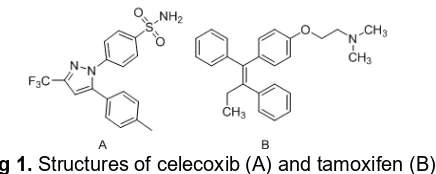
Celecoxib (Fig. 1A) is widely marketed as a selective cyclooxygenase-2 (COX-2) anti-inflammatory drug and has proven to be a blockbuster drug [1-2]. Interestingly, it shows some indications of its role in the cancer pathophysiology, especially in the apoptosis
Fig 1.Structures of celecoxib (A) and tamoxifen (B)
celecoxib shows anticancer properties in some cancer cell lines including colon cancer [8] and prostate cancer [9]. However, this drug suppresses also another COX-2 independent molecular pathway in prostate cancer [9]. Celecoxib is suggested also to have activities against breast cancer since breast is a tissue involved in COX-2 up regulation [10]. Crosstalk of COX-2 with estrogen receptor alpha (ERα) pathway was suggested to play an important role in the activities of celecoxib in the breast cancer pathophysiology [11].
An ERα antagonist tamoxifen (Fig. 1B) is the standard adjuvant for postmenopausal women with hormone-receptor-positive early breast cancer [12]. Notably, tamoxifen is a prodrug since it is metabolized to some active metabolites (e.g., 4-hydroxytamoxifen and N-desmethyl-4-hydroxy-tamoxifen) in the liver [13]. The affinity of the metabolites to ERα is 30-100 fold higher compared to tamoxifen in its original form [13]. In a recent prospective structure-based virtual screening (SBVS) campaign on the ZINC drug database (ZDD) [14] to discover ligands for ERα [15], celecoxib (ZINC02570895 [14]) together with 67 other drugs were identified as potent ligands for ERα [15]. It is therefore of a considerable and timely interest to verify whether celecoxib is a potent ligand for ERα.
In this article, exhaustive SBVS campaigns followed by in vitro tests to verify whether celecoxib a potent ligand for ERα are presented. The research aimed to employ computer-aided drug repurposing strategy [16] to verify the activity of celecoxib as a potent ligand for ERα. The SBVS protocol used to perform in silico tests was initially constructed and retrospectively validated by Anita et al. [17] to identify ERα antagonists [18]. By incorporating PyPLIF [19], subsequently the protocol was re-validated and and reported that the protocol employing PyPLIF has a significantly better quality [19-21]. The in silico screenings followed by in vitro tests employing tamoxifen as the reference compound has confirmed that celecoxib is a potent ligand for ERα.
EXPERIMENTAL SECTION
Materials
Celecoxib in three dimensional (3D) mode in the form of a mol2 file was obtained from the ZINC database
[14]. The ZINC code of celecoxib was ZINC02570895 [14,22]. Configuration files to perform in silico tests to identify celecoxib as ERα ligand by using PLANTS docking software were obtained from Anita et al. [17]: (i) The virtual target protein.mol2, (ii) the conserved water molecule water.mol2, and (iii) the configuration files to run PLANTS docking software plants.config. Configuration files to perform protein-ligand interaction fingerprints (PLIF) identification using PyPLIF were obtained from Radifar et al. [19]: (i) The configuration file to run PyPLIF config.txt, (ii) the virtual reference structure OHT.mol2, and (iii) the virtual binding pocket residuesPLANTSactiveSiteResidues.mol2.
Celecoxib standard was purchased from Sigma-Aldrich. Sample was prepared from Celebrex® (Pfizer) contain 100 mg celecoxib dissolved in dimethyl sulfoxide (DMSO) Merck®. MCF-7 breast cancer cell line was derived from Parasitology Laboratory, Faculty of Medicine, Universitas Gadjah Mada, Yogyakarta, Indonesia. Dulbecco's Modified Eagle Medium (DMEM) (Gibco) as culture medium was purcashed from Gibco. Cytotoxic assay was performed using 3-[4,5-dimethylthiazol-2-yl]-2,5 diphenyl tetrazolium bromide (MTT) (Sigma). Sodium dodecyl sulphate as stopper reagent was obtained from Merck. Tamoxifen as the reference compound in DMEM was provided by Setiawati et al. [23]. This research used US BIO ERα primary antibody [23]. All culture plates used in this study were Iwaki and all tips were purchased from Biologix.
Instrumentation and Computation
Instrumentation used in this study was Shimadzu UV-1800 spectrophotometer, cuvette Hellma, Retsch tipe T460 No V935922013 EY ultrasonicator, Scaltec SBC 22 (max 60/210 g, min 0.001 g, d = 0.01/0.1 mg) analytical balance, and Socorex micropipette. Computational medicinal chemistry applications used in this research were: PLANTS docking software version 1.2 (PLANTS1.2) [24, 25] to perform molecular docking simulations, R computational statistics software version 3.1.2 to perform statistical tests [26] and PyPLIF [19,27] to identify PLIF. All calculations and computational simulations were performed on a Linux (Ubuntu 12.04 LTS) machine with Intel(R) Xeon(R)CPU E31220 (@ 3.10 GHz) as the processors and 8.00 GB of RAM.
Procedure
Virtual screening on celecoxib as ligand for ERα
in the molecular docking simulations were subsequently subjected to PyPLIF to identify their PLIF [19]. Only docking poses whose bitstring of 103 is 1 were selected [19]. From the remaining poses, the best pose of each run was selected as the pose with the best Tanimoto coefficient similarity on the PLIF compared to the PLIF of the co-crystal ligand 4-hydroxytamoxifen (Tc-PLIF) [19,24] of all three independent runs. This procedure was performed 1000 times independently. The Tc-PLIF values of all selected poses from those 1000 independent run were subjected to a relevant statistical test to examine whether the Tc-PLIF values were statistically higher or equal to the Tc-PLIF standard 0.720 [19] in level of confidence of 95% [26].
Determination of celecoxib in Celebrex®
Standard solution preparation: A celecoxib stock solution was obtained by weighing 0.1520 mg of celecoxib standard accurately into a 1 mL vial, dissolved, and diluted to volume with DMSO. The intermediate solution was obtained by transferring 0.8 mL of celecoxib stock solution into a 5 mL volumetric flask, dissolved, and diluted to volume with DMSO. The calibration standard solution of celecoxib was obtained by transferring 200, 300, 400, 500, 600, and 700 µL of intermediate solution of celecoxib into 5 mL volumetric flask and dilute to volume with DMSO. Selection of wavelength: The maximum absorption wavelength of celecoxib was determined at the beginning of the study by scanning the standard solution at 200-400 nm. The maximum absorption wavelength was determined and used for the next determination. Sample preparation: Twenty capsules of Celebrex® were accurately weighed. An equivalent amount of 50 mg of the drug was taken and dissolved in DMSO and filtered; volume was made up to 25 mL. The sample solution was obtained by transferring 10 µL of stock sample solution into a 10 mL volumetric flask, dissolved, and diluted to volume with DMSO.
MTT cytotoxic assay and immunocytostaining of
ERα
MCF-7 cells were cultured until confluent in DMEM then 5 x 10³ cells were seeded into 96-well microplate and incubated in 37 °C and 5% CO2for 24 h. Celecoxib
as stock solution was prepared by dissolving the content of Celebrex® capsules in DMSO. Then, DMEM was removed and a series concentration of celecoxib in DMEM (5.00, 10.00, 25.00, 50.00, 75.00, and 100.00 µM) were added into 96-well plate 100 μL each and incubated in the same as previous condition for 24 h. Each concentration was assayed triplo (n = 3). In the end of incubation time, DMEM was removed from cells and 100 μL DMEM containing 5 mg/mL MTT was added into well plate. The plate was incubated for 4 h,
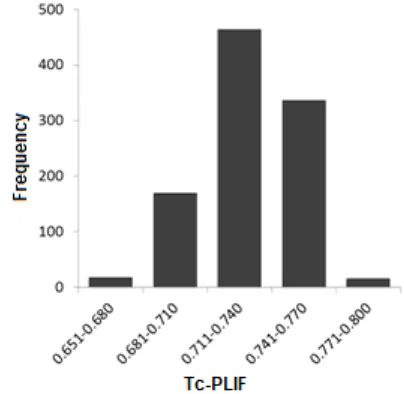
Fig 2. The histogram of all Tc-PLIF values resulted in docking using PLANTS1.2 followed by re-scoring using PyPLIF of celecoxib to ERα
then 100 μL of 10% SDS was added to each well to dissolve crystal formazan. The 96-well microplate was incubated for 24 h in dark room. Formazan crystals were measured by ELISA reader using 595 nm wavelengths. The half-maximal inhibitory concentration (IC50) value was calculated using four parameters
logistic regression by employing R computational statistics software version 3.1.2 [26,28]. Similar procedures were subjected to a series concentration of tamoxifen in DMEM (5.00, 10.00, 25.00, 40.00, 50.00, and 100.00 µM) as the reference compound [23]. By employing the method of ERα immunocytostaining published previously [23], the expression of ERα after adding celecoxib 30 µM was examined compared to the expression of ERα after adding tamoxifen 24 µM.
RESULT AND DISCUSSION
This research aimed to examine whether a widely used and well-known anti-inflammatory compound celecoxib is also a potent ligand for ERα. The identification of celecoxib as a potential ligand for ERα was performed by employing computational chemistry method, which was subsequently confirmed byin vitro tests. This approach can serve as an example of computer-aided drug repurposing in discovery of novel ligands for ERα.
performed instead of one sample t-test to examine whether the Tc-PLIF values are equal or higher than the reference Tc-PLIF value (0.720) [19] (H0) or the Tc-PLIF
values are less than the reference Tc-PLIF value (Hi).
The Wilcoxon test resulted in p-value of 1. This means that at the 95% level of confidences, the Tc-PLIF values are equal or better than 0.720 statistically. This result indicated that celecoxib is a potent ligand for ERα [1,3,17,19,21]. In vitro tests to confirm these in silico results were therefore required [17,19,29-36].
In this study,in vitro tests serve as the verification of the in silico test results [30-34,37-39]. On the other hand,in silicoapproaches could assist in the explanation of in vitro tests [35,40-47]. Very recently, the virtual screening protocol mainly referred in this study [17,19] was retrospectively re-validated [20] using the newest benchmarking dataset provided by Mysinger et al. [36]. The retrospective re-validation resulted in a better parameter that increased the confidence to further examine celecoxib as a ligand for ERα [20]. However, the cutoff value used in this study (Tc-PLIF value of 0.720) was the one suggested by Radifar et al. [19] since the retrospective validation showed a better parameter as a virtual screening protocol to identify potent ERα in an early enrichment (at 1% false positives) compared to the retrospective validation by Setiawati et al. [20]. Unfortunately, the quality of both virtual screening protocols to identify ERα ligands globally (at 100% false positives) could not be compared since Radifar et al. [19] did not provide the value. In fact, combined scoring functions using both scoring functions from PLANTS1.2 (ChemPLP) [24] and PLIF-based scoring functions [7,19,21,48] are highly recommended recently [32,49,50]. On the other hand, both benchmarking datasets [18,36] requires potent ligands with IC50 value ≤ 1 µM, which decreases the predictive
ability of the VS protocols to identify moderate ligands (IC50values of 1 mM to 1 µM) [17,19-21,23].
Since the approach presented in this article was aimed to employ computer-aided drug repurposing strategies, Celebrex® containing 100 mg celecoxib was
employed instead of pure celecoxib. By using UV spectrophotometric method, the amount of celecoxib in Celebrex® was determined [51]. Quantification of drug substance using spectrophotometer was carried out by preparing solution in transparent solvent and measuring its absorbance at suitable wavelength [51]. Different with Revathi et al. [51] which used chloroform as solvent with detection wavelength at 255 nm, the standard solutions of celecoxib (1.46, 1.95, and 2.43 µg/mL) in this study were dissolved in DMSO. The solutions were scanned at the wavelength region of 210-400 nm since the nature of the solvent could alter the position of the spectral peaks [52-53]. The maximum absorption wavelength of 255.5 nm was found. Six points calibration curve were subsequently obtained in a concentration range from 0.97-3.40 µg/mL for celecoxib. The response of the drug was found to be linear in the investigation concentration range and the linear regression equation was y = 0.047x + 0.127 with correlation coefficient 0.9929, which was considered as statistically acceptable since the experimental value of the coefficient of correlation (r) was larger than the
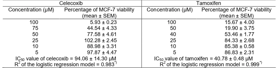
Fig 3. Dose response curves for celecoxib and tamoxifen effect on MCF-7 cell viability
Table 1.Effect of celecoxib and tamoxifen on MCF-7 cell viability after 24 h incubation
Celecoxib Tamoxifen
Concentration (µM) Percentage of MCF-7 viability (mean ± SEM)
Concentration (µM) Percentage of MCF-7 viability (mean ± SEM)
100 5.93 ± 0.23 100 15.67 ± 4.00
75 44.54 ± 4.33 50 19.90 ± 3.75
50 77.58 ± 4.61 40 53.46 ± 1.77
25 102.28 ± 2.45 25 84.33 ± 2.68
10 88.98 ± 3.31 10 85.38 ± 0.58
5 97.87 ± 4.47 5 86.83 ± 2.31
IC50value of celecoxib = 94.06 ± 14.30 µM R2of the logistic regression model = 0.983*)
IC50value of tamoxifen = 40.78 ± 0.48 µM R2of the logistic regression model = 0.999*) *)The R2value of both models show that the models are acceptable to calculate the IC50 value since the values are larger than
Fig 4. The ERα expression using immunocytochemistry method observed under light microscope using 400x magnification [23]. (A) Untreated cells; (B) Untreated cells without antibody; (C) Trated with celecoxib (30 µM); and (D) Treated with tamoxifen (24 µM). Symbol “ ” points the expressed ERα [23]
tabulated one for the given number of determinations (n = 6; r = 0.811) at the 95% level of confidences [54].
Celecoxib concentrations in six determinations were 36.93, 37.32, 38.62, 37.58, 38.2, and 37.09% (w/w), respectively. The mean (standard deviation) of the concentrations was 37.67 (0.67)% (w/w) or 102.15 mg celecoxib in 271.48 mg capsule content. Since the percent relative standard deviation (RSD) obtained on the six determinations of celecoxib was 1.78%, the precision was considered as acceptable (RSD < 7.3%) [52,55].
After determining the celecoxib content in the Celebrex® capsules sample, the cytotoxic activity of celecoxib against MCF-7 cell line was subsequently evaluated. The cell line is one of the established cell line to examine the activity of compounds as ligands for ERα [20,23,56-57]. Celecoxib treatment on MCF-7 cells changed both of cell viability and morphology. The compound showed strong cytotoxic activity towards MCF-7 cell line with IC50(standard error of measurement
(SEM)) value of 94.06 (14.30) µM, while tamoxifen as the reference compound showed IC50 (SEM) value of
40.78 (0.48) µM (Table 1; Fig. 3). Since the Shapiro-Wilk normality test show that the data were normally distributed but the variance were not homogen, Welch two sample t-test were performed to examine whether the IC50 values of celecoxib were different to the IC50
values of tamoxifen (Hi) or not (H0). The Welch two
sample t-test showed that at the 95% level of confidences the IC50 values of celecoxib were
statistically not different to the IC50 values of tamoxifen
(p-value of 0.065). Moreover, the IC50value of celecoxib
is in the same micromolar range with the IC50 value of
tamoxifen. These results are inline with the results from the in silico tests that celecoxib is a potent ligand for ERα. Notably, the immunocytochemistry assay results (Fig. 4) supported these findings by showing decrease of ERα expression in both of celecoxib and tamoxifen treatment.
The activity of celecoxib in the breast cancer pathophysiology was suggested as the results from tight
crosstalk of COX-2 with ERα pathway [11]. Interestingly, our findings presented in this article show that celecoxib plays an important role in ERα pathway, not only because it is a potent COX-2 inhibitor but also celecoxib is a potent ligand for ERα. Since these results emerged from computer-aided drug repurposing strategy using molecular docking simulations, the docking pose of celecoxib in the ERα binding pocket could provide insight to further design its analogs or derivatives with better activity towards ERα [2,20,23,50].
CONCLUSION
Aided by computational chemistry tools, a potent anti-inflammatory drug celecoxib was identified as a potent ligand for ERα. In vitrotests using MTT method subsequently confirmed that celecoxib is a ligand for ERα with IC50 (SEM) value of 94.06 (14.30) µM. It is
comparable with the IC50value of a drug of choice for
breast cancer tamoxifen.
ACKNOWLEDGEMENT
We thank Agustina Setiawati for her technical assistances with the in vitro assay. This research was financially supported by Indonesian Directorate General of Higher Education (Competitive Research Block Grant No. 1320/K5/KM/2014).
REFERENCES
1. Sadée, W., and Bohn, L., 2006,Mol. Intervention, 6 (4), 196–198.
Veenhuizen, A.W., Zhang, Y.Y., and Isakson, P.C., 1997,J. Med. Chem.,40 (9), 1347–1365.
3. Jendrossek, V., 2013, Cancer Lett., 332 (2), 313– 324.
4. Liu, D., Hu, G., Long, G., Qiu, H., Mei, Q., and Hu, G., 2012,Acta Pharmacol. Sin., 33 (5), 682–690. 5. Winfield, L.L., and Payton-Stewart, F., 2012, Future
Med. Chem.,4 (3), 361–383.
6. Rimon, G., Sidhu, R.S., Lauver, D.A., Lee, J.Y., Sharma, N.P., Yuan, C., Frieler, R.A., Trievel, R.C., Lucchesi, B.R., and Smith, W.L., 2010, Proc. Natl. Acad. Sci. U.S.A., 107 (1), 28–33.
7. Chakraborti, A.K., Garg, S.K., Kumar, R., Motiwala, H.F., and Jadhavar, P.S., 2010, Curr. Med. Chem., 17 (15), 1563–1593.
8. Williams, C.S., Watson, A.J.M., and Sheng, H., 2000,Cancer Res., 60 (21), 6045–6051.
9. Patel, M.I., Subbaramaiah, K., Du, B., Chang, M., Yang, P., Newman, R.A., Cardo, C.C., Thaler, H.T., and Dannenberg, A.J., 2005, Clin. Cancer Res., 11 (5), 1999–2007.
10. Turini, M.E., and DuBois, R.N., 2002, Annu. Rev. Med., 53, 35–57.
11. Wang, Y.X., Gao, J.X., Wang, X.Y., Zhang, L., and Liu, C.M., 2012,Tumor Biol., 33 (4), 957–966. 12. Mouridsen, H., Giobbie-Hurder, A., Goldhirsch, A.,
Thürlimann, B., Paridaens, R., Smith, I., Mauriac, L., Forbes, J.F., Price, K.N., Regan, M.M., Gelber, R.D., and Coates, A.S., 2009, N. Engl. J. Med., 361 (8), 766–776.
13. Desta, Z., Ward, B.A., Soukhova, N.V., and Flockhart, D.A., 2004,J. Pharmacol. Exp. Ther., 310 (3), 1062–1075.
14. Irwin, J.J., Sterling, T., Mysinger, M.M., Bolstad, E.S., and Coleman, R.G., 2012, J. Chem. Inf. Model., 52 (7), 1757–1768.
15. Istyastono, E.P., 2013, Virtual Screening on ZINC Drug Database to Discover Novel Antagonists for Estrogen Receptor α, Sanata Dharma University Research Grant Final Report, Universitas Sanata Dharma, Yogyakarta.
16. Bisson, W.H., Cheltsov, A.V., Bruey-Sedano, N., Lin, B., Chen, J., Goldberger, N., May, L.T., Christopoulos, A., Dalton, J.T., Sexton, P.M., Zhang, X., and Abagyan, R. 2007, Proc. Natl. Acad. Sci. U.S.A., 104 (29), 11927–11932.
17. Anita, Y., Radifar, M., Kardono, L., Hanafi, M., and Istyastono, E.P., 2012, Bioinformation, 8 (19), 901– 906.
18. Huang, N., Shoichet, B.K., and Irwin, J.J., 2006, J. Med. Chem., 49 (23), 6789–6801.
19. Radifar, M., Yuniarti, N., and Istyastono, E.P., 2013, Bioinformation, 9 (6), 325–328.
20. Setiawati, A., Riswanto, F.D.O., Yuliani, S.H., and Istyastono, E.P., 2014, Indones. J. Chem., 14 (2), 103–108.
21. Radifar, M., Yuniarti, N., and Istyastono, E.P., 2013,Indones. J. Chem., 13 (3), 283–286.
22. Irwin, J.J., and Shoichet, B.K., 2005,J. Chem. Inf. Model., 45 (1), 177–182.
23. Setiawati, A., Riswanto, F.O.D., Yuliani, S.H., and Istyastono, E.P., 2014, Indonesian J. Pharm., 25 (3), 119–124.
24. Korb, O., Stützle, T., and Exner, T.E., 2009, J. Chem. Inf. Model., 49 (1), 84–96.
25. Korb, O., Stützle, T., and Exner, T.E., 2007, Proc. IEEE Swarm Intell. Symp., 1 (2), 115–134.
26. R Development Core Team, 2008,R: A Language and Environment for Statistical Computing,Vienna. http://www.r-project.org.
27. Salentin, S., Haupt, V.J., Daminelli, S., and Schroeder, M., 2014,Prog. Biophys. Mol. Biol., 116 (2-3), 174–186.
28. Kreidenweiss, A., Kremsner, P.G., and Mordmüller, B., 2008,Malar. J., 7 (187), 1–8.
29. Appiah-Opong, R., Commandeur, J.N.M., van Vugt-Lussenburg, B., and Vermeulen, N.P.E., 2007,Toxicology, 235 (1-2), 83–91.
30. Istyastono, E.P., de Graaf, C., de Esch, I.J.P., and Leurs, R., 2011, Curr. Top. Med. Chem., 11 (6), 661–679.
31. Istyastono, E.P., Nijmeijer, S., Lim, H.D., van de Stolpe, A., Roumen, L., Kooistra, A.J., Vischer, H.F., de Esch, I.J.P., Leurs, R., and de Graaf, C., 2011,J. Med. Chem., 54 (23), 8136–8147.
32. de Graaf, C., Kooistra, A.J., Vischer, H.F., Katritch, V., Kuijer, M., Shiroishi, M., Iwata, S., Shimamura, T., Stevens, R.C., de Esch, I.J.P., Leurs, R., 2011, J. Med. Chem., 54 (23), 8195–8206.
33. Sirci, F., Istyastono, E.P., Vischer, H.F., Kooistra, A.J., Nijmeijer, S., Kuijer, M., Wijtmans, M., Mannhold, R., Leurs, R., de Esch, I.J.P., and de Graaf, C., 2012, J. Chem. Inf. Model., 52 (12), 3308–3324.
34. Kufareva, I., Katritch, V., Stevens, R.C., and Abagyan, R., 2014,Structure, 22 (8), 1120–1139. 35. Schultes, S., Nijmeijer, S., Engelhardt, H., Kooistra,
A.J., Vischer, H.F., de Esch, I.J.P., Haaksma, E.E.J., Leurs, R., and de Graaf, C., 2013, Med. Chem. Commun., 4 (1), 193–204.
36. Mysinger, M.M., Carchia, M., Irwin, J.J., and Shoichet, B.K., 2012, J. Med. Chem., 55 (14), 6582–6594.
38. Kolb, P., Rosenbaum, D.M., Irwin, J.J., Fung, J.J., Kobilka, B.K., and Shoichet, B.K., 2009, Proc. Natl. Acad. Sci. U.S.A., 106 (16), 6843–6848.
39. Carlsson, J., Coleman, R.G., Setola, V., Irwin, J.J., Fan, H., Schlessinger, A., Sali, A., Roth, B.L., and Shoichet, B.K., 2011,Nat. Chem. Biol., 7 (11), 769– 778.
40. Lim, H.D., Istyastono, E.P., van de Stolpe, A., Romeo, G., Gobbi, S., Schepers, M., Lahaye, R., Menge, W.M.B.P., Zuiderveld, O.P., Jongejan, A., Smits, R.A., Bakker, R.A., Haaksma, E.E.J., Leurs, R., and de Esch, I.J.P., 2009, Bioorg. Med. Chem., 17 (11), 3987–3994.
41. Strasser, A., 2009, Expert Opin. Drug Discovery, 4 (10), 1061–1075.
42. Smits, R.A., Adami, M., Istyastono, E.P., Zuiderveld, O.P., van Dam, C.M.E., de Kanter, F.J.J., Jongejan, A., Coruzzi, G., Leurs, R., and de Esch, I.J.P., 2010, J. Med. Chem., 53 (6), 2390–2400.
43. Xiang, Y., Hou, Z., and Zhang, Z., 2012,Chem. Biol. Drug Des., 79 (5), 760–770.
44. Appiah-Opong, R., Commandeur, J.N.M., Istyastono, E.P., Bogaards, J.J., and Vermeulen, N.P.E., 2009,Xenobiotica, 39 (4), 302–311.
45. Schultes, S., Engelhardt, H., Roumen, L., Zuiderveld, O.P., Haaksma, E.E.J., de Esch, I.J.P., Leurs, R., and de Graaf, C., 2013,ChemMedChem, 8 (1), 49–53.
46. Engelhardt, H., Schultes, S., de Graaf, C., Nijmeijer, S., Vischer, H.F., Zuiderveld, O.P., Dobler, J., Stachurski, K., Mayer, M., Arnhof, H., Scharn, D., Haaksma, E.E.J., de Esch, I.J.P., and Leurs, R., 2013,J. Med. Chem., 56 (11), 4264–4276.
47. Lim, H.D., de Graaf, C., Jiang, W., Sadek, P., McGovern, P.M., Istyastono, E.P., Bakker, R. A, de Esch, I.J.P., Thurmond, R.L., and Leurs, R., 2010, Mol. Pharmacol., 77 (5), 734–743.
48. Marcou, G., and Rognan, D., 2007, J. Chem. Inf. Model., 47 (1), 195-207.
49. Schultes, S., Kooistra, A., Vischer, H., Nijmeijer, S., Haaksma, E., Leurs, R., de Esch, I., and de Graaf, C., 2015,J. Chem. Inf. Model., 55 (5), 1030–1044. 50. Istyastono, E.P., Kooistra, A.J., Vischer, H., Kuijer,
M., Roumen, L., Nijmeijer, S., Smits, R., de Esch, I., Leurs, R., and de Graaf, C., 2015, Med. Chem. Commun., 6, 1003–1017.
51. Revathi, R., Perumal, R.V., Sudharshini, S., Ansar, A.M., Thilagalakshmi, A., and Dinesh, A.P., 2011, Int. J. Pharm. Sci. Lett., 1 (2), 49–50.
52. Riswanto, F.O.D., Lukitaningsih, R.R.E., and Martono, S., 2015, Indones. J. Chem., 15 (1), 9– 15.
53. Kealey, D., and Haines, P.J., 2002, Instant Notes Analytical Chemistry, BIOS Scientific Publishers Ltd., Oxford, 225–226.
54. Ermer, J., and Miller, J.H., 2005,Method Validation in Pharmaceutical Analysis: A Guide to Best Practice, Wiley-VCH Verlag GmbH & Co., Weinheim, 87–90.
55. González, A.G., and Herrador, M.A., 2007, TrAC, Trends Anal. Chem., 26 (3), 227–238.
56. Brooks, S.C., Locke, E.R., and Soule, H.D., 1973, J. Biol. Chem., 248 (17), 6251–6253.