www.krspine.org
Adequate Serial Monolayer Passage Number of Human
Intervertebral Disc Cells for Cell Therapy -Growth and
Phenotype of
Cells-Yong Chan Kim, M.D., Ki Bok Kim, M.D., Moon Soo Park, M.D., Seok Woo Kim, M.D.
J Korean Soc Spine Surg 2010 Mar;17(2):57-65. Originally published online June 30, 2010;
doi: 10.4184/jkss.2010.17.2.57
Korean Society of Spine Surgery
Department of Orthopaedic Surgery, Ewha Womans University Collge of Medicine #911-1 Mok-dong, Yangcheon-gu, Seoul, 158-710, Korea Tel: 82-2-2646-6808 Fax: 82-2-2646-6804
©Copyright 2010 Korean Society of Sping Surgery pISSN 2093-4378 eISSN 2093-4386
The online version of this article, along with updated information and services, is
located on the World Wide Web at:
http://www.krspine.org/DOIx.php?id=10.4184/jkss.2010.17.2.57
This is an Open Access article distributed under the terms of the Creative Commons Attribution Non-Commercial License (http:// creativecommons.org/licenses/by-nc/3.0) which permits unrestricted non-commercial use, distribution, and reproduction in any medium, provided the original work is properly cited.
Journal of Korean Society of
Received: January 25, 2010
Revised: April 22, 2010
Accepted: June 1, 2010
Published Online: June 30, 2010
Corresponding author: Seok Woo Kim, M.D.
Department of Orthopaedic Surgery, Hallym University Sacred Heart Hospital 896, Pyeongchon-dong, Dongan-gu, Anyang, Gyeounggi-do 431-070, Korea
TEL: 82-31-380-6000, FAX : 82-31-380-6008
E-mail: [email protected]
“This is an Open Access article distributed under the terms of the Creative Commons Attribution Non-Commercial License (http:// creativecommons.org/licenses/by-nc/3.0/) which permits unrestricted non-commercial use, distribution, and reproduction in any medium, provided the original work is properly cited.”
Adequate Serial Monolayer Passage Number of
Human Intervertebral Disc Cells for Cell Therapy
-Growth and Phenotype of Cells-
Yong Chan Kim, M.D., Ki Bok Kim, M.D., Moon Soo Park, M.D., Seok Woo Kim, M.D.
Department of Orthopaedic Surgery, School of Medicine, Hallym University
Study Design: This is an in-vitro experimental study
Objectives: We wanted to analyze the changes in the growth and phenotype of human degenerative intervertebral disc cells depending on the frequency of subculture in an in vitro monolayer culture system.
Summary of the Literature Review: A subculture of disc cells is needed to obtain an adequate amount of disc cells for cell therapy, tissue engineering and analysis of the biological characteristics of degenerative disc cells
Materials and Methods: The obtained intervertebral discs were divided into the nucleus pulposus (NP) and the annulus fibrosus (AF). The AF and NP cells were cultured in a monolayer manner, respectively. At each subculture time, we analyzed the morphological changes, the adhesion rate, the proliferation rate and the viability. The expressions of types I and II collagen and proteoglycan were analyzed at the mRNA gene level.
Results: Both the AF and NP cells gradually showed a fibroblast-like spindle shape while undergoing subculture. The adhesion rate was higher at the second and third times of subculture. The cell proliferation was the highest at the second subculture time. The viability was markedly lower prior to the subculture. On RT-PCR, the type II collagen expression was gradually decreased in the NP cells. In the AF cells, Type II collagen was not expressed from the second time of subculture. The expression of proteoglycan was gradually decreased in both.
Conclusions: Following the 3rd subculture, the degenerative disc cells had completely changed their original growth and phenotypic characteristics. Therefore, we believe that it is not desirable for us to do passage cultures more than three times for cell therapy.
Key words: Intervertebral disc, Degenerative change, Phenotype, Subculture
서론
만성 요통은 인류가 앓고 있는 가장 흔한 질병으로서 그 중요
한 원인 중의 하나가 추간판의 퇴행성 변화이다.1) 추간판은 노
령화, 비틀림과 같은 비정상적인 역학적 힘과 과도한 체중부하 등에 의하여 퇴행성 변화를 일으키게 된다. 추간판 퇴행성 변화 는 병리학적으로 수핵 세포수의 감소 및 세포 외 기질 분비 능력 의 저하로 수핵 부분의 단백 다당 및 수분 함량이 감소되어 추간
판의 탄력성이 떨어진다.2) 현재, 퇴행성 척추간판 질환의 치료
는 보존적인 치료방법으로 호전이 없을 때, 수술적인 치료방법
을 요하게 되는데 수술적인 치료방법에는 추간판 제거술3,4) 후궁
절제술,5) 척추체간 융합술6,7) 등이 있다. 최근에 정상적인 추간판
의 기능을 대체할 수 있도록 디자인된 인공디스크가 임상적으로 사용되고 있지만 그 적응증이 제한적이고 장기적인 추시 결과가
Yong Chan Kim et al Volume 17 • Number 2 • June 2010 antimycotic (Gibco, New York, NY)을 포함한 phosphate-buffered saline (PBS) 용액으로 충분히 세척하고 멸균된 수술 용 칼로 수핵 조직과 섬유륜 조직을 잘게 잘랐다. 잘게 잘린 섬 유륜 조직은 0.2% 제 1형 및 2형 교원질 분해효소(Collagenase, Worthington Biochemical Co. NJ)가 함유된 �ulbecco�s modi-가 함유된 �ulbecco�s modi-�ulbecco�s modi-fied eagle medium (�MEM, Gibco, New York, NY) 용액, 수 핵 조직은 0.2% 제 2형 교원질 분해효소가 함유된 �MEM 용
액에 넣어서 5% CO2, 37°C 세포 배양기내에서 4시간 동안
충분히 소화시켜 세포를 획득하였다. 이렇게 획득한 세포는
1.5x10⁵cells/cm2 농도로 세포배양 접시에 접종하였다. 세포 배
양액은 10% fetal bovine serum (FBS, Gibco, New York, NY), 1% antibiotic-antimycotic가 포함된 �MEM용액을 사용하였 다. 세포 배양액은 세포가 배양 접시에 부착되기 전 까지 약 2주
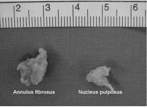
Fig. 1. Digital image of the obtained AF and NP tissue from degenerated
배양 시의 조건은 모두 동일하게 하였으며 총 세 번의 계대 배양
세포의 증식율은 0.05% trypsin-E�TA (Gibco, New York,
NY)로 5% CO2, 37°C 세포 배양기내에서 10분 동안 처리하여 extraction Kit, Gibco-BRL)를 이용하여 총 RNA를 분리하였 다. 얻어진 RNA는 spectrophotometer (Amersham Phamarcia, Ultrospec 21009, USA)를 사용하여 정량하고 동량의 총 RNA를
AMV reverse transcriptase (BMS)를 이용하여 c�NA를 만들었 다. 합성된 c�NA는 유전자 특이 primer 를 이용하여 PCR반응 methylene blue (�MB) colorimetric method22)를 사용하였다. 세 포 배양액 100 ㎕를 �MB 용액 200 ㎕와 반응시킨 후, 525 nm
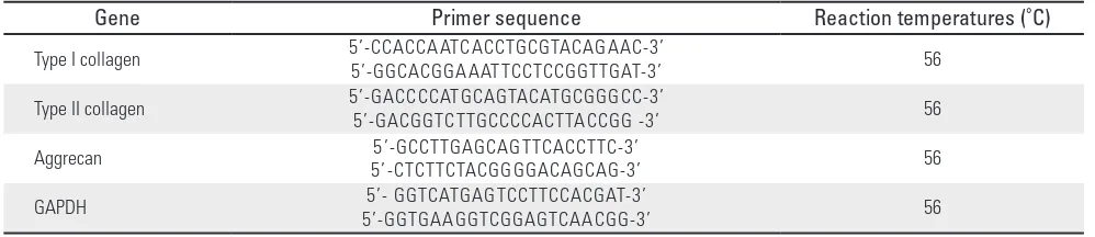
Table 1. The sequence of primers and reaction temperatures used in the RT-PCR
Gene Primer sequence Reaction temperatures (˚C)
Yong Chan Kim et al Volume 17 • Number 2 • June 2010
www.krspine.org 60
결과
1. 형태학적 결과
조직에서 분리한 배양 접시에 부착되기 전 까지 섬유륜 및 수
핵 세포들은 모두 원형을 나타냈으며, 1차 계대 배양을 거치기 전 까지는 불규칙한 다각형의 불규칙한 모양을 나타내고 있었 다. 섬유륜 및 수핵 세포들은 계대 배양을 거치면서 점차적으로 방추형인 섬유모세포 모양으로 변하였으며, 이러한 변화는 섬유 륜 세포에서 더 빨리 진행되었다(Fig. 2 ).
Fig. 2. The morphology of the AF cells and NP cells grown in the monolayer culture system. (x100)
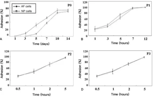
2. 세포의 부착율
1차 계대 배양을 거치기 전에는 매우 낮았으며 1일 째는 0% 로 거의 부착이 되지 않았으며 3일 째 약 8.3±3.2%로 나타났 다. 10일째, 섬유륜과 수핵 세포에서 각각 74.7±2.5%와 70.7 ±2.1%를 나타내었고, 14일째 부착율은 10일째와 비슷하였으 며 더 증가하지는 않았다. 1차 계대 배양을 거치면 세포의 부착 율은 현저히 증가함을 볼 수 있었으며, 2, 3차 계대 배양에서는 5시간 만에 거의 100%의 부착율을 보였다. 부착율은 2, 3차 계 대 배양에서는 통계학적으로 유의한 차이를 보이지 않았으나 (P<0.05), 1차와 2차 계대 배양 사이에는 통계적으로 유의한 차 이를 보였다(P<0.05). 또한 부착율은 섬유륜 세포 및 수핵 세포 지간에 계대 배양 전에는 통계적으로 유의한 차이를 보였으나 (P<0.05), 계대 배양을 거치고 나서는 큰 차이를 보이지 않았다 (P<0.05) (Fig. 3).
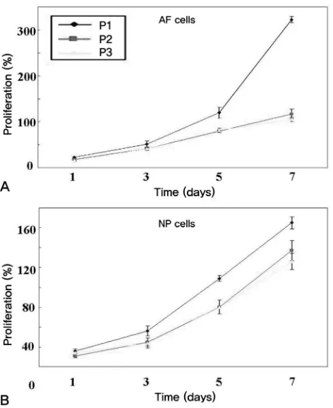
3. 세포의 증식
세포의 증식은 1차 계대 배양에서 제일 높았으며 섬유륜 세포 에서 수핵 세포보다 더 좋은 증식율을 보였으나(P<0.05), 2, 3차 계대 배양에서는 수핵 세포와 섬유륜 세포, 및 계대 배양의 횟수
에 따라서 유의한 차이를 보이지 않았다 (P>0.05)(Fig. 4 ).
4. 세포의 생존율
세포의 생존율은 계대 배양에 따라 큰 차이를 보이지 않았으 며 섬유륜 및 수핵 세포 사이에서도 큰 차이는 없었으며 약 95% 의 높은 생존율을 보였다(P>0.05). 하지만 1차 계대 배양을 거치 기 전 세포의 생존율은 낮았으며 수핵 65.7±4.1%, 섬유륜 세포 는 75.3±6.1%로 나타났다 (P<0.05) (Fig. 5).
5. 유전자 발현
섬유륜 세포는 계대 배양의 횟수에 따라서 제 1형 교원질의 발현은 큰 차이를 보이지 않았으며 제 2형 교원질과 aggrecan 유 전자는 1차 계대 배양에서는 발현이 되었으나 2, 3차 계대 배양 에서는 발현되지 않았다. 수핵 세포에서는 1차 계대 배양에서 제 1형 교원질이 발현되지 않았으나 2, 3차 계대 배양에서 점차 증가하였으며 제 2형교원질과 aggrecan은 제 2차 계대 배양까지 는 발현되었으나 3차 계대 배양에서는 발현되지 않았다(Fig. 6).
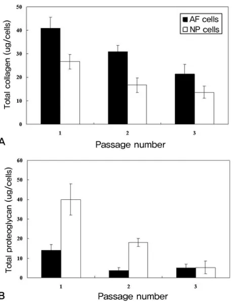
6. 교원질 및 단백다당 함량
총 교원질 함량은 계대 배양의 횟수에 따라서 점차적으로 감 Fig. 4. Proliferation (%) of the AF cells and NP cells at different passage number. (A)
AF cells, (B) NP cells.
Fig. 5. Viability of the AF cells and NP cells at different passage number.
Yong Chan Kim et al Volume 17 • Number 2 • June 2010 Fig 7. Measurement of the total collagen (A) and proteoglycan (B) in the culture
을 증가시키기 위하여 배양접시에 교원질을 코팅하거나 integrin
1. Urban JP, Roberts S. �egeneration of the intervertebral disc. Arthritis Res Ther. 2003;5:120-30.
2. Freemont AJ, Watkins A, Le Maitre C, Jeziorska M, Hoyland JA. Current understanding of cellular and molecular events in intervertebral disc degeneration: implications for therapy. J Pathol. 2002;196:374-9. 3. Hoppenfeld S. Percutaneous removal of herniated lumbar
discs. 50 cases with ten-year follow-up periods. Clin Orthop Relat Res. 1989;238:92-7.
4. Yeung AT, Tsou PM. Posterolateral endoscopic excision for lumbar disc herniation: Surgical technique, outcome, and complications in 307 consecutive cases. Spine. 2002;27:722-31.
5. White AH, von Rogov P, Zucherman J, Heiden �. Lumbar laminectomy for herniated disc: a prospective controlled comparison with internal fixation fusion. Spine. 1987;12:305-7.
6. Eie N. Comparison of the results in patients operated upon for ruptured lumbar discs with and without spinal fusion. Acta Neurochir. 1978;41:107-13.
Yong Chan Kim et al Volume 17 • Number 2 • June 2010
www.krspine.org 64
1983;8:897-900.
8. Freeman BJ, �avenport J. Total disc replacement in the lumbar spine: a systematic review of the literature. Eur Spine J. 2006;15:439-47.
9. An HS, Thonar EJ, Masuda K. Biological repair of intervertebral disc. Spine. 2003;28:86-92.
10. Ganey TM, Meisel HJ. A potential role for cell-based therapeutics in the treatment of intervertebral disc herniation. Eur Spine J. 2002;11:206-14.
11. Gan JC, �ucheyne P, Vresilovic EJ, Swaim W, Shapiro IM. Intervertebral disc tissue engineering I: characterization of the nucleus pulposus. Clin Orthop Relat Res. 2003;411:305-14.
12. Yoon ST. Molecular therapy of the intervertebral disc. Spine J. 2005;5:280-6.
13. Baums MH, Heidrich G, Schultz W, Steckel H, Kahl E, Klinger HM. Autologous chondrocyte transplantation for treating cartilage defects of the talus. J Bone Joint Surg Am. 2006;88:303-8.
14. Brittberg M, Lindahl A, Nilsson A, Ohlsson C, Isaksson O, Peterson L. Treatment of deep cartilage defects in the knee with autologous chondrocyte transplantation. N Engl J Med. 1994;331: 889-95.
15. Benya P�, Shaffer J�. �edifferentiated chondrocytes reexpress the differentiated collagen phenotype when cultured in agarose gels. Cell. 1982;30:215-24.
16. Lemare F, Steimberg N, Le Griel C, �emignot S, Adolphe M. �edifferentiated chondrocytes cultured in alginate beads: restoration of the differentiated phenotype and of the metabolic responses to interleukin-1beta. J Cell Physiol. 1998;176:303-13.
17. Bayliss MT, Johnstone B, O�Brien JP. 1988 Volvo award in basic science. Proteoglycan synthesis in the human intervertebral disc. Variation with age, region and pathology. Spine. 1988;13:972-81.
18. Chou AI, Bansal A, Miller GJ, Nicoll SB. The effect of serial monolayer passaging on the collagen expression profile of outer and inner anulus fibrosus cells. Spine. 2006;31:1875-81.
19. Kluba T, Niemeyer T, Gaissmaier C, Grunder T. Human anulus fibrosus and nucleus pulposus cells of the intervertebral disc: effect of degeneration and culture system on cell phenotype. Spine. 2005;30: 2743-48.
20. Wang JY, Baer AE, Kraus VB, Setton LA. Intervertebral disc cells exhibit differences in gene expression in alginate and
monolayer culture. Spine. 2001; 26:1747-51.
21. Tullberg-Reinert H, Jundt G. In situ measurement of collagen synthesis by human bone cells with a sirius red-based colorimetric microassay: effects of transforming growth factor beta2 and ascorbic acid 2-phosphate. Histochem Cell Biol. 1999;112:271-6.
22. Farndale RW, Buttle �J, Barrett AJ. Improved quantitation and discrimination of sulphated glycosaminoglycans by use of dimethylmethylene blue. Biochim Biophys Acta. 1986;883:173-7.
23. Loeser RF, Sadiev S, Tan L, Goldring MB. Integrin expression by primary and immortalized human chondrocytes: evidence of a differential role for alpha1beta1 and alpha2beta1 integrins in mediating chondrocyte adhesion to types II and VI collagen. Osteoarthritis Cartilage. 2000;8:96-105.
24. Wang H, Kandel RA. Chondrocytes attach to hyaline or calcified cartilage and bone. Osteoarthritis Cartilage. 2004;12:56-64.
25. Hong Y, Gao C, Xie Y, Gong Y, Shen J. Collagen-coated polylactide microspheres as chondrocyte microcarriers. Biomaterials. 2005;26:6305-13.
26. Yashiki S, Umegaki R, Kino-Oka M, Taya M. Evaluation of attachment and growth of anchorage-dependent cells on culture surfaces with type I collagen coating. J Biosci Bioeng. 2001;92:385-8.
27. Eyre �R, Muir H. Quantitative analysis of types I and II collagens in human intervertebral discs at various ages. Biochim Biophys Acta. 1977;492:29-42.
28. Johnstone B, Bayliss MT. The large proteoglycans of the human intervertebral disc. Changes in their biosynthesis and structure with age, topography, and pathology. Spine. 1995;20:674-84.
29. Lee JY, Hall R, Pelinkovic � et al. New use of a three-dimensional pellet culture system for human intervertebral disc cells: initial characterization and potential use for tissue engineering. Spine. 2001;26: 2316-22.
세포 치료를 위한 인간 추간판 세포의 적절한 체외 계대 배양 횟수 -세포의 성장 및 표현형-
김용찬 • 김기복 • 박문수 • 김석우
한림대학교 의과대학 정형외과학교실
연구 계획: 시험관 실험적 연구
목적: 사람의 퇴행성 추간판 세포를 체외에서 배양하면서 계대 배양의 횟수에 따른 성장 및 표현형의 변화를 분석하고자 한다.
선행문헌의 요약: 세포 치료, 조직공학 및 추간판 세포의 생물학적 특성의 분석을 위해서는 퇴행성 추간판 세포의 계대 배양을 거쳐 많은 양의 세포를
획득하여야 한다.
대상 및 방법:. 적출된 추간판 조직을 수핵과 섬유륜 부분으로 나누어서 단층 배양하였다. 계대 배양을 거치면서 세포의 형태 변화, 부착율, 증식율 및 생
존율 분석을 진행하였다. 또한 RT-PCR 검사를 시행하여 제 1, 2형 교원질 (type I, II collagen) 및 단백다당 (proteoglycan) mRNA 발현을 유전자 수준에서 분석하였다.
결과: 세포의 모양은 수핵 및 섬유륜 세포 모두 점차적으로 방추형인 섬유모세포 모양을 나타냈으며, 부착율은 2차 및 3차 계대 배양에서 상대적으로 증가하였고, 증식율은 2차 계대 배양에서 가장 우수했으며, 세포의 생존율은 배양을 거치기 전에는 현저히 낮았다. RT-PCR 분석 상 수핵 세포의 제2형 교원질 발현은 점차적으로 감소하는 양상을 보였으며, 섬유륜 세포의 제 2형 교원질은 2차 계대 배양 시부터 발현이 되지 않았다. 단백 다당의 발현은 수핵 및 섬유륜 세포 모두에서 점차 감소하는 양상을 보였다.
결론: 퇴행성 변화를 일으킨 추간판의 섬유륜 및 수핵 세포는 체외에서 계대 배양의 횟수가 3차 계대 배양에서는 고유의 성장과 표현형을 잃어버렸다. 이에 세포치료에 있어서 체외에서 단층 배양 시 3번 이상의 계대 배양을 거치지 않는 것이 바람직할 것으로 사료된다.