Lung Cancer Nodule Extraction and 3D Modeling
Abstract—The images resulted from CT scan create object’s shape that can be indicated as lung cancer. Pulmonary nodules is one of the indications that are used to diagnose lung cancer. There are some patterns in nodules that identified whether a nodule is indicated as benign tumor or malign tumor or cancer). In this research, the images that will be explored are extracted from CT scan lung images that detected with nodules. The extraction of the images are utilizing bit-plane slicing, thresholding, elimination and outlining. The results of the extracted nodules will be reconstructed in three dimension (3D) modelling nodules. The processes of 3D modeling nodules are done by creating meshes that represent each pixel slice of the extracted image. Each slice of the image then merge into one part. The result of this reconstruction is expected to assist the doctors in diagnosing and analyzing lung nodules based on their shapes and the volume of those shapes. The shape of the nodules and its volume are two of several indicators that can be used to diagnose lung cancer.
Keywords—Bit Plane Slicing, Lung Cancer, Nodule, Outlining I. INTRODUCTION
The resulted image from CT scan can reveals an object that can be identified as lung cancer. Pulmonary nodule is one of many indicators that is used to diagnose lung cancer. There are some patterns on lung nodule that would indicate whether nodule identified with a benign tumour or malignant tumour. If the edges of the nodule is smoother then the nodule can be said to be benign, and vice versa if there are bulges in the nodule and prominent, it can be said to be malignant [1]. A cancerous lung nodule usually the size is double every 25 days. The growth of the nodule of lung cancer is very quick. The morphology characteristic of lung nodules for benign and malignant categories provide the same description as the lung photo, but they have different nodule edges distribution [2]. A nodule that has the same size for two years is likely to be benign. This pattern has some classification which are solid (diffuse), central such as rings, concentric, popcorn or homogeneous with the tendency having smaller nodules, circumscribed, smooth, rounded, and have a fixed size. On the other hand, malignant nodule has eccentric classification pattern and spicules with the tendency of non-demarcated edge nodule, irregular and fast growth [3]. One of the
difficulties in lung nodule detection is the large amount of the objects inside the nodule (e.g., blood vessels, hilum, and mediastinum). To simplify the nodule detection process in the lung, an algorithm model to distinguish the nodules from other objects inside the lung is required [4]. Many researchers were conducted to detect lung nodules. One of the researcher proposing sistem Computer Aided Design (CAD) system for pulmonary nodule detection. The study explain that the first step for lung nodule detection is lung extraction then the next step is nodule segmentation based on its category. The result of this segmentation is classified using Extreme Learning Machine method to detect the assuredness whether an object is nodule positive or not [5]. The research in lung nodule cancer detection are performed by reconstruct the image obtained from the extraction process in three dimension form to determine the volume of the lung nodules in the image automatically. Image reconstruction and extraction in this study is performed with convolution process of input signal based on a continuous interpolation function. The sampling process is implemented on pixel dots of the extracted image. The result of this sampling then stored inside a matrix that has record data structure as a control point for interpolation process. The record data will save the information of each slice of the image position and intensity. The position and intensity of image slice possessed information that can be used as a control point for linear interpolation process. All the points will be reconstructed in a single process. These points (dots) are achieved from the extracted slice nodules without necessary to sort the points of each slice of lung image nodules.
y axis). The techniques of volume rendering can define the thickness of the object [8]. 3D modeling can also be formed from a projection of an image i.e., projection of image volume in two dimension (2D) form as simulation of 3D formation [9].
The study in this paper will extract CT scan lung image that are detected with nodule. The extraction is performed with several techniques: (i) bit-plane slicing, (ii) thresholding, (iii) elimination, and (iv) outlining. The result of nodule extraction then reconstructed in three dimension nodule to have better, clearer and precise image nodule. The reconstruction is perform by creating the meshes which represent each pixel of the image slices. These slices then combined into one piece.
II. METHODSANDMATERIAL
A. Input Image
The images used in this experiment are post-contrast images with the sizes 512 x 512 and 630 x 630 pixel, with the thickness of 3.0mm and 8.0mm. The format of the images are DICOM (.dcm) [10]
.
Basically, the image of DICOM viewer views the image as in Fig. 1. However, since this research is focussed on lung nodules detection then CT lung appearance is preferable since it reveal the images clearly. The objects on the lung are well identified. The resulted CT scan images from these experiments contain hundreds of slice, but not all of the images are used. The images that are used for this experiment are the images that visible have the nodule accordance with HU value [11]. The image input is converted into Bitmap (.bmp) format. This format is readable during the reconstruction 3D modelling.Fig 2. Lung CT scan
B. Lung Cancer Nodule Extraction
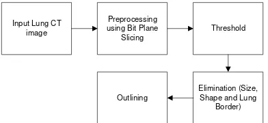
The selected input image will be processed for nodules detection using feature extraction. This extraction separate the selected object with other objects. There are several techniques used for this extraction; (i) bit-plane slicing, (ii) thresholding, (iii) elimination, and (iv) outlining. The techniques can be seen in Fig. 3.
Fig 3. Stages of the processes
The extraction algorithm for lung nodules CT scan detection is divided into several steps:
1. Input lung CT scan image
2. Image preprocessing. This step is carried out using bit-plane slicing process. The purpose of bit-bit-plane slicing is to increase the qualities of nodules area. This process produces few slices of the image that corresponds to the number of bits these slices have. Since the images in this experiment are 8-bit images, the slices of one image contain 8 slices. The first slice of the image is the sharpest one, the value of the sharpness will decrease until the last slice as can be seen in Fig. 4.
Fig 4. Bit Plane Image of Five to Eight Sliced
To obtain more accurate and sharp result on nodules slice detection, this experiment use the fifth until eight sliced. The slice under fifth slice will give a bit rough image thus can be a noise. The comparison of the image before and after bit-plane slicing can be seen in Fig. 5. The processed image looks sharper compare to the original image.
Fig 5. Before and After Bit Plane Slicing Comparison
3. Thresholding. Thresholding is used to change the value of image intensity. This step will transform the images obtained from preprocessing process into binary images or black and white images. Thresholding step will facilitate the elimination process as the noise will be reduced. The noises are slackened due to the intensity enhancement or degradation. Moreover the shape of the nodules becoming clearer (See Fig. 6).
Fig 6. Image after Thresholding Process Input Lung CT
image
Preprocessing using Bit Plane
Slicing
Threshold
Elimination (Size, Shape and Lung
4. Nodules Indication and Elimination
In order to indicate the nodules, it is important to eliminate the objects that then to have round [2]. Beside the shape, the size of the nodules also determines its criteria. The sizes of nodules are less than 0.5 cm2 or 100 pixel. These sizes are determined from the nodules that already identified with cancer. The characteristic of the nodules with cancer are having the size above 0.5cm2 [12]. This experiment use regionprops function with eccentricity properties to get the result close to the circle. The border of the lungs also being eliminated although its size not smaller than the nodule’s size, but it is clear since the location of the nodule is outside the lung. The elimination process can be seen in Fig. 7. The processes start from (a) Circular shape, (b) Indication of the
size, and (c) Lung’s border.
Fig 7. Elimination Process
5. Creating the Outline. Creating the border or outlining serves to facilitate the pixel matching visually. This steps are carried out in modelling process (see Fig. 8).
Fig 8. Outline Process
C. 3D Modelling
The extracted nodules. The collection of the extracted nodules will be combined into a single object with the three dimension modelling using blender. The modelling processes are shown in Fig. 9.
Fig 9. 3D Modeling Process
The following are the 3D Modelling of CT scan lung nodules: 1. The extracted input image. The obtained images are resulted from the extraction of the images that being inputted inside the blender in one folder. These images will be read as an image sequences so that one image represents one frame. This image is used as a background to facilitate the modeling process.
Fig 10. Input Background Image Sequence
For instance, the number of extracted image that has 3.0mm slices contain 23 images. Therefore, the used frame has the same number from 1 until 23. The offset value also being adjusted with the name of the file. Since the first slice is image number 56 then the offset value is 55. The frame starts from 1 (not 0). The value of the offset is the same as the name of the file.
2. Creating the plane. Making a plane or mesh that has minimum length and width that equal to the size of the input image. The forms of the plane or mesh are in top ortho display in order to fit with the image of the CT scan that are used in this experiment. The next step is performing subdivide method to divide the plane and scale them as a result the face has the sized of one pixel of input image.
Fig 11. Creating a Plane
3. The face elimination is compatible with the input image. The face that suits the pixel of the input image will be selected. The next step is performing the inverse process to remove the face that re not suitable with the input image to obtain the result that accordance with the input image. Fig. 12 sows the face elimination process.
Fig 12. Elimination Faces
Input Extraction Lung Nodule
Image
Make a plane Face elimination as the input image
Plane integration Resize to the
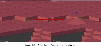
4. The spacing steps between nodules. The created objects nodules are being given the thickness value based on the thickness of the input images, which are 3.0mm and 8.0mm. The thickness of the slices are created using extrude function according to z axis. Once one object nodule is obtained, to connect between one object nodule to the other nodules, we need other object nodule. For that purpose, it is required to move to the next frame to create planes using the next input image (repeating step 2 and 3). Each new created plane is placed in the edges of the previous lower plane as shown in Fig. 13
Fig 13. Spacing between Planes
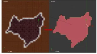
5. Combining the plane. The amalgamations are carried on by join function process. The merging started by selecting both of the planes; the upper planes (first plane) and lower plane (the next plane). Each vertex (that become the edges of both planes) facing each other will be combined using merge function as shown in Fig. 14.
Fig 14. Vertex Amalgamation
The amalgamation of the vertex is performed with two conditions; (i) If the edge of the upper plane faced the lower plane then the lower vertex is combined with the upper vertex with the vertex position still on the upper (ii) and vice versa. These requirements are done in order not to change the starting position of a pixel in the extracted images.
6. Changing the size to original scale. The created nodules do not have the actual size of the original nodule. This is because the size of the face equated with the input image pixel. The size of face is equalized for the references purposes to facilitate the modelling process. Based on radiant DICOM viewer, the size of input images is 512 x 512 pixels with 36.12cm length and 630 x 630 pixel with 37,93cm length. Therefore, for the image with 512 x 512, 1cm = 14.174972314px and 1px = 0.070546875cm. Whereas, the image with 630 x 630, 1cm =
16.60954389665173px and 1px =
0.0602063492063492cm. To obtain face with the size 0.070546875cm or 0.0602063492063492cm each of the edge must has the size 0.2656066170109472cm or 0.2453698213031692, because the shape of the face is square. The standard size of the blender is Blender Unit (BU). The value of one (1) BU equal to one meter, therefore to obtain the real size of each face is 7.055e-007 BU or 6.020e-007 BU.
D. The Calculation of Nodule’s Volume
Not all objects volume inside the blender can be computed. The blender has some conditions to be an algorithm to calculate the volume of the objects inside the blender. The first blender initiate the objects by defining the plane at the origin plane to create the lowest and highest distance from the plane. The calculation of the lowest and highest distance is computed from the number of the vertices of an object.
E. Creating Animation
The animations that are formed tend to loop able (not comparable) in order to continue showing the information from the nodules. The animations nodule are created by displaying the rotation of that nodule. There are two types of animation rotation. This rotation aims to generate variety of nodules viewpoints. For the nodules in left side, they will rotate in the same direction as z axis. The right nodules will rotate in the same direction as x axis. There are 120 frames used in the animation or for five seconds. Key frame for the nodules is 30 frames with 90 degree rotation as shown in Fig. 15
Fig 15. Key Frame Animation
III. RESULTANDDISCUSSION
The experiment of nodules image extraction from CT scan and 3D modelling were conducted on patients with cancer nodules. For instance, a patient with a nodule that has shape tend to be rounded with the slice thickness of 3.0mm.
A. Extraction Experiment
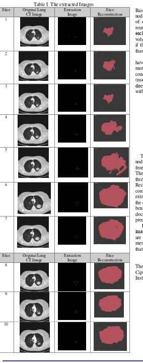
Table I The extracted Images Slice Original Lung
CT Image
Extraction Image
Slice Reconstruction 1
2
3
4
5
6
7
Slice Original Lung CT Image
Extraction Image
Slice Reconstruction 8
9
10
Based on the experiments, the proposed algorithms can detect nodules successfully. The detected nodules meet the criteria of nodule with lung cancer. These nodules tend to have round shape with the wideness above 0.5 cm. The density of
each nodule’s candidate is computed as the ratio of the
volume. Nodules candidate are considered to be non-nodule if the value of their compactness are less than 0.5 or greater than 1.5.
As can be seen from Fig. 16, the resulted nodules have shape that are then to be round with irregular edges. The more irregular geometric shaped of the edges, it can be conclude that the extracted nodules are cancer nodules (malign). The 3D modelling nodules made easy for the
doctors to obtain the data of the nodule’s volume directly
with high level of precision.
Fig 16. 3D Nodule Modelling with Volume = 0.0911109 cm3
IV. CONCLUSION
The conducted research is aims to create pulmonary object nodules in three dimension (3D). The nodules are obtained from CT scan of the extracted lung images reconstruction. The techniques employed in this study are bit-plane slicing, thresholding, elimination and outlining techniques. Reconstruction methods to form a 3D modelling are done by combining the extracted image slices. The result of the extracted images are packed in a simple animation to assist the doctors in diagnosing the nodules whether the nodules are benign or malign. Moreover, these animations can help the doctors to determine the volume of nodules accurately and precisely.
For future work, we plan to develop the algorithm of the
image’s extraction, especially in the case when the nodules
are patched on the lungs. Moreover, the reconstruction methods are expected to run automatically using the method that being proposed in this paper.
ACKNOWLEDGMENT
REFERENCES
[1] M. Gomathi, Dr.P.Thangaraj. A Computer Aided Diagnosis System For Detection Of Lung Cancer Nodules Using Extreme Learning Machine, International Journal of Engineering Science and Technology Vol. 2(10), ISSN: 0975-5462. 2010
[2] Scott Brandman, MD and Jane P. Ko, MD. Pulmonary Nodule
Detection, Characterization, and Management With Multidetector Computed Tomography. J Thorac Imaging Volume 26, Number 2. 2011
[3] Slatore CG , Sullivan DR , Pappas M , Humphrey LL . Patient-centered outcomes among lung cancer screening recipients with computed tomography: a systematic review. J Th orac Oncol. 9(7):927-934. 2014
[4] Rina Noviana, Sarifuddin Madenda, Rodiah and Sigit
Widiyanto. Morphology and Watershed Segmentation for Extracting The Cancer Nodule in Lung CT Image. Advanced Science, Engineering and Medicine.Vol 6, 1-5. 2014
[5] Djojodibroto Darmanto. Respiratory Medicine. Medical Text Book, First Edition. ISBN 978-979-448-980-2. 2007
[6] Rodiah, Sarifuddin Madenda and Fitrianingsih. Three-Dimensional (3D) Reconstruction for Detecting Shape and Volume of Lung Cancer and Nodules. IPTEK Journal of Proceeding Series, Vol.1.eISSN : 2354-6026. 2014
[7] L.Souche, F.Lepage and G.Iskenova. Volume Based Modeling-Automated Construction of Complex Structural Models. EAGE
Conference and Exhibition Incorporating SPE
EUROPEC.London. 2013
[8] Thilakanathan, MR Danan. Blender 3D for Beginners: The Complete Guide: The Complete Beginner's Guide to Getting Started with Navigating, Modeling, Animating, Texturing. ISBN: 152323881X. EAN: 9781523238811. Publisher: CREATESPACE. 2016
[9] A. Padmapriya, S.Vigneshnarthi. Image Processing Operations for 3D Image. International Journal of Scientific and Research Publications, Volume 2. Issue 6, ISSN 2250-3153. 2012 [10] Cipto Mangunkusumo Hospital (RSCM). Lung CT image.
Departement of Radiology, Faculty of Medicine. University of Indonesia.2011
[11] Schreiber Joseph, Paul Anderson, Humberto G. Rosas, Avery L. Buchholz, Anthony G. Au. Hounsfield Units for Assessing Bone Mineral Density and Strength: A Tool for Osteoporosis Management, The Journal of Bone & Joint Surger. 93:1057-1063. 2011