Activity of Dexamethasone Therapy on Pro-Inflammatory Cytokines Profile of Balb/c Mice
with Biliary Atresia
Qonitatul Khasanah1), Muhamin Rifa’i1) 1)
Laboratory of Animal Physiology, Department of Biology, Faculty of Mathematics and Natural Science, Brawijaya University, Malang, Indonesia
ABSTRACT
Biliary atresia is a neonatal obstructive cholangiopathy that progresses to end-stage liver disease. Dexamethasone is one of synthetic glucocorticoid which has function as an anti-inflammatory agent. Here, we investigated whether dexamethasone could modulate the immune activity in mice strain Balb/c with biliary atresia based on the change of quantity of IFN-γ and TNF-α as a pro-inflammatory molecules. This study consists of 2 stages. The first stage is pre-condition which is made the biliary duct become fibrosis by injecting 20 µl of phosphate buffered saline containing 1.5 x 10⁶ fluorescence-forming units Rhesus Rotavirus (RRV) subcutaneously on first day (24 hours) after the mice was born. The second stage is injection with dexamethasone with dose 0,5 mg/kg BW subcutaneously on the 7th-14th day and 14th-21st day. The clinical effect of dexamethasone is investigated on 14th and 21st day by flow cytometry method. Data were analyzed using Kruskal-Wallis test (p<0,05) and Mann-Whitney test using SPSS 16 for Windows. Rotavirus injection subcutaneously was proven to stimulate the production of proinflammatory cytokines, especially in the third week of termination. The result indicated an increasing number of proinflammatory cytokines such as IFN-γ
and TNF-α after RRV injection but after injection of dexamethasone the number of those cytokines is decreased. It can be understood that dexamethasone has a capability to reduce the effect of inflammation regard to the decrease of proinflammatory cytokines.
Keywords : Biliary atresia, Dexamethasone, Inflammation, Obstructive, Rhesus Rotavirus
INTRODUCTION
The evolution of the immune system in mammals have evolved as a defense mechanism against microorganisms that invade the body. This system is important in regulating the body's systemic failure but if it will harm the immune system of the body [1]. Biliary atresia is a condition in which an interruption system of intra and extra-hepatic biliary in the first few months of infant age. Characteristics of biliary atresia is the absence of partial biliary system between the duodenum and liver, causing bile flow resistance and leads to impaired liver function [2]. Reported incidence of biliary atresia between 1:8000 to 1:18000 live births. Biliary atresia is a liver disease causes the terminal which is the main indication of liver transplantation in children. Early symptoms of biliary atresia is often difficult to distinguish from the physiological neonatal jaundice, so that diagnosis and governance becomes too late. Other causes of late diagnosis is the presence of
some differential diagnosis as a cause of direct hyperbilirubinemia that requires time to diagnosis [3].
Portoenterostomi by Kasai operation found in 1959 is a major breakthrough in the management of biliary atresia. The success of the Kasai operation was higher in patients operated on less than 2 months of age. Without surgery, the patient will usually die by age 2 years. Kasai operation is a reliable treatment, but liver damage continues even after Kasai surgery [4].
inflammation. Dexamethasone is a corticosteroid drug. Because fibrosis of the remnant of extrahepatic and intrahepatic bile ducts in BA associated with a strong inflammatory response, steroids would be expected to reduce fibrosis and bile duct blockage to suppress the immune response, so that the flow of bile will remain intact [5]. Therefore, this study is important to determine whether corticosteroids can modulate the activity of inflammation reaction of balb / c mice with biliary atresia based on changes in the quantity of molecules of proinflammatory cytokines IFN-γ and TNF-α.
METHODS
This research was conducted on July 2013 until January 2014 in Laboratory of Animal Physiology, Department of Biology, Faculty of Mathematics and Natural Sciences, Brawijaya University, Malang. The animal experiments were approved by the Animal Care and Use Committee of the Brawijaya University.
Experimental Design. This study uses experimental animals Balb/c mice newborn because in this study biliary atresia is not possible in humans. The sample selection of baby mice population and the placement of the group try and control the allocation is done randomly. This study consists of two phases. The first stage is a pre-condition where the bile ducts are made of baby rats induced fibrosis with 20 mL of phosphate buffered saline containing 1.5 x 10⁶ fluorescence-forming units Rhesus rotavirus (RRV) subcutaneously in the first 24 hours after birth, then the second stage, baby mice were induced dexamethasone 0,5 mg / kg BW subcutaneously on days 7 and 14 after induction of RRV. Further investigated the therapeutic effect of dexamethasone on days 14 and 21 after induction. This study has received Ethical Clearance from the Research Ethics Committee (Animal Care and Use Committee) UB No. 391 / EC / IEC-S3 / 11/2012.
Preparation of Virus Injection. Viruses that used as injection material is Rhesus rotavirus (MMU18006) American Type Culture Collection (ATCC®VR 1739TM). The virus is stored at temperature -700C. In a time of <24 hours after birth, the baby mice were injected
subcutaneously with 20 mL of phosphate buffered saline containing 1.5x10⁶ pfu RRV.
Preparation of Dexamethasone Injection. Injection of dexamethasone 0.5 mg / kg BW subcutaneously or 0.0005 mg / gram BW, with an estimated weight of baby mice aged <24 hours ± 2 grams, then the dexamethasone given is 0,001 mg. Dexamethasone 5 mg / ml was diluted to 125 ml (equivalent to 0.04 mg / ml or 0.004 mg / 0.1 mL or 0.001 mg / 0,025 ml). Injection dose will change according to changes in body weight of mice.
Dexamethasone Therapy on Balb/c Mice with Biliary Atresia. Mice that had been infected with RRV then induced with dexamethasone on day 7 and 14. Mice that had been injected with dexamethasone on day 7 to day 14 was terminated on day 14, and mice that had been injected with dexamethasone on day 14 to day 21 terminated on day 21. Mice were terminated in a manner that has been put on the bottle containing cotton and diethyl ether. Furthermore, the spleen was isolated from the body organs of mice.
Spleen Isolation. Spleen that had been isolated from mice was washed with PBS for 2 times and cleaned from fat tissues. After that, spleen was pushed clockwise using the base of the syringe and filtered with a wire. Homogenates were mixed with PBS included in propylene tube and add 15 ml of PBS until the volume reached 10 ml. Homogenates then were centrifuged at a 2500 rpm, 40C for 5 minutes. Supernatant was discarded while the pellet was taken and resuspended with 1 ml of PBS and homogenized.
Biolegend), pychoerythrien (PE) Rat anti-mouse CD -8a (clone: 53-6.7) and PE antimouse TNF-α (clone: MP6-XT22).
Data Analysis. This study was tested with the Kruskal-Wallis test using a significance level (α) of 0.05. The data used in the form of changes in the quantity of the absolute number of TNF-α and IFN-γ. If the obtained p> 0.05, the results showed no significance between each treatment, otherwise if p <0.05 then shows the significance between treatments were compared. Furthermore, the follow-up Mann-Whitney test with α of 0.05. If the obtained p> 0.05, it did not show any significant difference between the 2 treatments were compared, otherwise if p <0.05 then shows the real difference between the two treatments were compared. Data analysis was performed using SPSS 16.0 for Windows.
RESULT AND DISCUSSION
The Absolute Number of CD4+IFN-γ+ and CD8+IFN-γ+. The results of flow cytometry analysis of the spleen (Figure 1) showed that the number of CD4+IFNγ+ cells increased significantly in the T group (0.91 million or 0.67% of cells) compared to healthy mice (0.21 million or 0.06% of cells) and decreased back to the TND treatment (administration of Rotavirus and Dexamethasone) (0.62 million or 0.56% of million or 0,28% of cells).
Figure 1. The absolute number of CD4+IFNγ+ (K = control, T = Administration of Rotavirus and TND = administration of Rotavirus and Dexamethasone).
Note: Different letters indicate significant difference by Mann-Whitney test
The results of flow cytometry analysis of the spleen (Figure 2) showed that showed that the number of CD8+IFNγ+ increased second week showed that the number of cells in the treatment of T tends to increase when compared to the K (0.07 million or 0.14% of cells to 0.11 million or 0.77% of cells) and decreased in TND group (0.02 million or 0.19% of cells).
Figure 2. The absolute number of CD8+IFNγ+ (K = control, T = Administration of Rotavirus and TND = administration of Rotavirus and Dexamethasone).
Note: Different letters indicate significant difference by Mann-Whitney test
has multiple defense functions. The first is to inhibit viral replication by activating genes which cause damage to cellular mRNA and inhibition of protein translation. The second is to activate Natural Killer cells (NK) that will kill the virus that infected the cells. The third is by inducing the activity of MHC (Major Histocompatibility Complex) class 1 [6].
Both the second and third week, dexamethasone proven to reduce the number of cytokines IFN-γ produced by both CD4 and CD8 cells. Dexamethasone is a synthetic glucocorticoid with immunosuppressant activity and anti-inflammatory. As an immunosuppressant, dexamethasone works by lowering the body's immune response. Anti-inflammatory activity of dexamethasone to suppress or prevent the tissue responses to inflammatory processes and inhibit the accumulation of inflamed cells, including macrophages and leukocytes to sites of inflammation. Mechanism of action of dexamethasone could be the inhibition of arachidonic acid release, modulating substances derived from arachidonic acid metabolism, and a reduction in the amount of 5-HT3 (5-hidroxytryptophan). Dexamethasone has an antiemetic effects, presumably through the mechanism of inhibiting the release of prostaglandins in the central resulting in decreased levels of 5-HT3 in the central nervous system, inhibits the release of serotonin in the gastrointestinal tract so that there is a bond between the serotonin 5-HT3 receptor, endorphin release, and anti-inflammatory strong in the area of surgery and allegedly glucocorticoids have varying effects on the central nervous system and affects the regulation of neurotransmitters, receptor density, signal transduction and configuration of neurons [7].
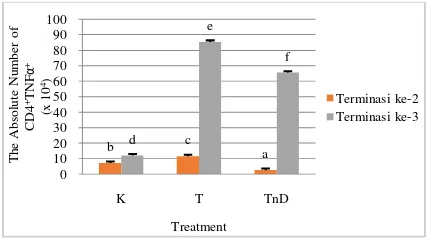
The Absolute Number of CD4+TNF-α+ and second week showed that the number of cells in the treatment of T tends to increase when compared to the K (0.17 million or 0.02% of cells, to 0.10 million or 0.01% of cells) and decreased in TND group (0.07 million or 0.03% of cells).
Figure 3. The absolute number of CD4+TNF-α+ (K = control, T = Administration of Rotavirus and TND = administration of Rotavirus and Dexamethasone).
Note: Different letters indicate significant difference by Mann-Whitney test
The results of flow cytometry analysis of the spleen (Figure 4) showed that showed that the number of CD8+TNF-α+ increased second week showed that the number of cells in the treatment of T tends to increase when compared to the K (0.10 million cells, or 0.05% of cells, to 0.08 million, or 0.08% of cells)and showed the same number of cells in TND group (0.08 million or 0.2% of cells).
Figure 4. The absolute number of CD8+TNF-α+ (K = control, T = Administration of Rotavirus and TND = administration of Rotavirus and Dexamethasone).
Note: Different letters indicate significant difference by Mann-Whitney test
The cytokine is a protein secreted by immune cells that mediate some of the functions of these cells in the regulation of immune system and response to inflammation. Cytokines are also produced in response to microbial and other antigens. This cytokine acts as both a growth factor and stem cell differentiation and activation of immune cells to eliminate antigens [8]. Various types of cytokines include TNF group (Tumor Necrosis Factor), TGF (tumor growth factor), interferon, interleukin, and CSF (Colony Stimulating Factor) [9].
Both the absolute number of CD4+ TNF-α+ and CD8+TNF-α+ mice treated with administration of rotavirus showed significant improvement in the third week when compared to the control treatment. TNF is a main cytokine that played role in acute inflammatory response. The serious infection can trigger the production of large amounts of TNF that causes systemic reaction [10]. TNF-α and-β are structurally related, bind to the same cellular receptor, and produce biological changes that are similar to the various sel. TNF-α is produced by neutrophils, activated lymphocytes, macrophages, NK cells, and some non-lymphoid cells such as astrocytes, endothelial cells and smooth muscle cells, while TNF-β appears to be only produced by T cells [11]. LPS is a potent stimulus to secrete TNF. IFN-γ produced by T cells and NK cells also stimulates macrophage including increased synthesis of TNF. At low levels, TNF work against leukocytes and
endothelium, induces acute inflammation. At the level of being, TNF plays a role in systemic inflammation. At high levels, TNF cause pathological abnormalities of septic shock.
Based on figure 3 and 4, on the termination of third week, dexamethasone proved able to reduce the number of cytokine TNF-α is produced by both CD4 and CD8 cells. Dexamethasone is a synthetic corticosteroid that is one of the most effective. Ability to cope with allergic inflammation and approximately ten times more powerful than prednisone [12]. Dexamethasone able to work through genomic and non-genomic. Through genomic pathway, dexamethasone binds to the glucocorticoid receptor and forms a complex with the transcription factor NFκB to perform blocking the transcriptional activity of proinflammatory cytokines. While on the non-genomic pathway, dexamethasone worked through membrane-associated receptor and second messenger activity to produce an anti-inflammatory protein that can inhibit the production of proinflammatory cytokines (Figure 5).
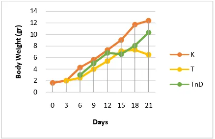
The clinical condition of mice that had cholestasis can be characterized by jaundice (yellow color) in the skin that do not have hair, weight is hard up, and pale feces (akholis). Based on the research, a sign that can be observed in mice after subcutaneously injected Rotavirus is the body weight of mice were less likely to rise (lower) when compared to healthy mice (normal).
The results of the analysis of differences in body weight (Figure 5) showed that there are differences in body weight between the control treatment (K), administration of rotavirus (T), and administration of Rotavirus and Dexamethasone (TND). Weight control mice (K) is the weight of normal mice because mice in this group were not given any treatment. While the T treatment, body weight proved to be lower than the mice in group C on day 0 to day 21. Average body weight of mice in the treatment group TND proved higher than T treatment group but when compared with the treatment group K then the weight of mice in this group were slightly lower. It can be proved that with the administration of dexamethasone can suppress the effects of cholangitis in mice caused by a viral infection.
CONCLUSION
Injection RRV (Rhesus Rotavirus) subcutaneously in Balb / c mice aged <24 hours proved capable of stimulating the production of proinflammatory cytokine, especially at the termination of the third week. This can be evidenced by an increase in proinflammatory cytokines such as IFN-γ and TNF-α. Dexamethasone as a glucocorticoid proved to regulate the activity of immunocompetent cells because dexamethasone had a potential to inhibit the rate of migration of lymphocytes and macrophages into the inflamed tissue. This can be evidenced by the decline in proinflammatory cytokines such as IFN-γ and TNF-α especially in the third week of termination.
ACKNOWLEDGEMENT
The author would like to thank to Mr. Muhaimin Rifa'i S.Si, Ph.D.Med.Sc. as advisor in this study and Pediatric Research Team Dr. Soetomo, Surabaya for funding this research.
REFERENCES
[1] Rifa’I, M., Y. Kawamoto, I. Nakashima, H. Suzuki. 2004. Essential roles of CD8+CD122+ regulatory T cells in the maintenance of T cell homeostasis. The Journal of experimental medicine. 200(9):1123-1124
[2] de Carvalho E., Cláudia A. P I., Jorge A. B. 2007. Extrahepatic biliary atresia:
current concepts and future directions. Jornal de Pediatria. 83(2):105-120 [3] Moyer K, Kaimal V, Pacheco C. 2010.
Staging of biliary atresia at diagnosis by molecular profiling of the liver. Genome Med; 2:33.
[4] Sokol RJ, Mack C. 2001. Etiopathogenesis of biliary atresia. Semin Liver Dis; 21(4):517–24.
[5] Lippi C, Chrousus GP. 1992. Glucocorticoids. In Yaffe SJ, Aranda JV eds.Pediatric Pharmacology, Theurapeutic Principles in Practice.WB Saunders, Philadelphia: 466 – 75
[6] Jiang B., L. Snipes Magaldi,P. Dennehy,H. Keyserling. 2003. Cytokines as Mediators for or Effectors against Rotavirus Disease in Children. Clin Diag Lab Immunol; 10(6): 995-1001.
[7] Elenkov IJ. 2004. Glucocorticoids and the Th1/Th2 balance. Ann N Y Acad Sci; 1024:138-46.
[8] Abbas, A. K., dan Lichtman, A. H. 2005.
Cellular and Molecular
Immunology,Edisi Kelima. Elsevier. Philadelphia.
[9] Dalhouise University. 2008. Cytokines. http://immunology.medicine.dal.ca/educat ion/ Immunity.htm. Tanggal akses 11 November 2008.
[10] Shivakumar P, Campbell KM, Sabla GE. 2004. Obstruction of extrahepatic bile ducts by lymphocytesis regulated by IFN-gamma in experimental biliary atresia. J. Clin. Invest; 114:322–9.
[11] Detrick, B.,Nagineni, C.N., and J.J. Hooks. 2002. Transforming growth factor-beta in human retinal pigment epithelial cell is enhanched by Toxoplasma gondii : a possible role in the
immunopathologenesis of
retinochoroiditis. Clin. Exp. Immun. 128 (2): 372-378.