Atherosclerosis 153 (2000) 9 – 21
High prevalence of peripheral atherosclerosis in a rapidly
developing country
Franc¸ois Perret
a,b, Pascal Bovet
b,c,*, Conrad Shamlaye
c, Fred Paccaud
b,
Lukas Kappenberger
aaDi6ision of Cardiology,Department of Internal Medicine,Uni6ersity Hospital,Lausanne,Switzerland
bGroup for Cardio6ascular Disease and Epidemiologic Transition,Institute for Social and Pre6enti6e Medicine,Bugnon17, Uni6ersity of Lausanne,Lausanne,Switzerland
cUnit for Pre6ention and Control of Cardio6ascular Disease,Ministry of Health,Seychelles
Received 2 June 1998; received in revised form 22 November 1999; accepted 5 January 2000
Abstract
Cardiovascular disease is rapidly increasing in developing countries experiencing epidemiological transition. We investigated the prevalence of peripheral atherosclerosis in a rapidly developing country and compared our findings with data previously reported in Western populations. A cardiovascular risk factor survey was conducted in 1067 individuals aged 25 – 64 randomly selected from the general population of Seychelles. High-resolution ultrasonography of the right and left carotid and femoral arteries was performed in a random subgroup of 503 subjects (245 men and 258 women). In each of the four arteries, arterial wall thickness (in plaque-free segments) and atherosclerotic plaques (i.e. focal wall thickening at least 1.0 mm thick) were measured separately. The prevalence of peripheral atherosclerosis was high in this population. For instance, at least one plaque ]1.0 mm was found in, respectively, 34.9 and 27.5% of men and women aged 25 – 34 and at least one plaque]2.5 mm was found in, respectively, 58.2 and 36.9% of men and women aged 55 – 64. With reference to data found in the literature, the prevalence of carotid atherosclerosis appeared to be significantly higher in Seychelles than in Western populations. This study provides further evidence for the importance of cardiovascular disease in developing countries. Determinants should be identified and relevant prevention and control programs implemented. © 2000 Elsevier Science Ireland Ltd. All rights reserved.
Keywords:Atherosclerosis; Arteries; Carotid arteries; Ultrasonography; Developing countries
www.elsevier.com/locate/atherosclerosis
1. Introduction
Accumulating data support the view of an emerging epidemic of cardiovascular disease (CVD) in developing countries [1,2]. Increasing CVD in developing countries relate, among other factors, to ageing populations, changing lifestyles accompanying industrialisation and urbanisation and nutrition transition [1,3].
As developing countries will certainly not be able to afford the high costs of CVD treatments for large portions of their populations, appropriate preventive strategies should be promptly implemented after spe-cific determinants of CVD epidemic in these countries have been identified [4,5].
In Seychelles, a survey conducted in 1989 demon-strated high levels of several cardiovascular risk factors (CVRF) in the general population [6 – 9]. Subsequently, a national program for the prevention and control of CVD was designed and implemented [10,11]. In 1994, a second independent population-based survey (‘Sey-chelles Heart Study II’) was conducted to assess trends in CVRF levels in the population. The survey also included high-resolution ultrasonography of the carotid and femoral arteries. This new non-invasive technique permits precise quantification of atherosclerotic changes in peripheral arteries and is now frequently used in observational studies or clinical trials on atherosclerosis [12]. Although many reports based on these measure-ments have been published, especially with regard to their relation with CVRF [13 – 28], data from non-se-lected populations are sparse and no such data have
* Corresponding author. Fax: +41-21-3147373.
E-mail address:[email protected] (P. Bovet).
been collected systematically in populations from devel-oping countries so far.
The aim of this study was therefore to determine the prevalence of persons with peripheral atherosclerosis in the general population of Seychelles and to subse-quently compare these findings with data previously reported in Western populations.
2. Methods
2.1. General population
The Republic of Seychelles consists of 115 islands lying in the Indian Ocean, 1800 km east of Kenya and 1800 km north of Mauritius. The islands were first inhabited when French colonials settled in Seychelles in 1770, joined over the next decades by larger number of African slaves and indentured labourers (coming mainly from East and Central Africa and Madagascar) and, later, by small numbers of Indian and Chinese immigrants [29]. Eighty-nine percent of the total popu-lation lives on the largest island, Mahe´. According to a census carried out in 1994, the total population was 73 442 with 49% aged less than 25 years and 45% aged 25 – 64. Although intermarriage has blurred racial dif-ferences in many Seychellois, ethnic descent of the population is considered to be predominantly African in 67%, Caucasian in 8%, Indian or Chinese in 2% and evidently mixed in 23% [6,30]. The opening of an international airport in 1971 was followed by a rapid increase of tourism, fast economic development (the gross domestic product per year and per inhabitant increased from $600 to 6565 from 1970 to 1994) and major changes in lifestyles [9,30]. CVD currently ac-counts for approximately 40% of the total mortality according to the vital statistics (all deaths in the coun-try are certified by a doctor). Stroke mortality is higher in Seychelles than in several industrialised countries while ischemic heart disease mortality is intermediate (Table 1) [8,31,32]. Major tropical scourges such as malaria, leishmaniasis, bilharziasis, yellow fever, and sleeping sickness are unknown in Seychelles. Apart from a small private medical sector, medical care is delivered without fees to all residents within a national health system operated by the Ministry of Health.
2.2. Sur6ey methods
The Seychelles Heart Study II was designed as a cross-sectional survey of the general population and detailed methods have been published elsewhere [30] (full text available on www.seychelles.net/smdj). The study protocol was approved by the review committee of the Ministry of Health. Selection of eligible subjects was performed using computerised population data
from a census carried out in 1987, which were there-after regularly updated by the local Ministry of Admin-istration. A sex- and age-stratified sample of 1280 eligible subjects was randomly drawn from the popula-tion of 28 695 Seychellois aged 25 – 64 years and resid-ing on the main island of Mahe´. Eligible subjects were sent a letter to invite them to participate in the study. The subjects were free to participate and extensive information on the study was largely disseminated through the media and delivered on-site. Fifty-four subjects turned out to be dead or abroad at the time of the survey and were therefore excluded. Out of the remaining 1226 eligible individuals, 1067 (504 men and 563 women) attended the study, hence a participation rate of 87%. Among the 159 eligible persons who did not attend the study, letters were returned unopened by the postal services to the study centre for 38 (24%) suggesting that these persons had moved. The propor-tions of eligible participants who did not attend the survey were similar in all sex- and age-categories with the exception of a lower participation of men aged 25 – 34 years (76% participation). The survey took place from July to December 1994. During this period, 14 persons were invited to report to the study centre every day in a sequence determined by the alphabetical order of their family name. Bus transportation to the study centre, which did not exceed 1 h from any place in the island, was reimbursed.
A face-to-face structured questionnaire was adminis-tered to all participants by a team of nurses who had previous experience in the conduct of surveys. The questionnaire focused on CVD, CVRF and knowledge, attitudes and practices on cardiovascular health. Eth-nicity was assessed according to the phenotypic appear-ance by a single examiner at the time the participants arrived and registered in the survey centre. Blood pres-sure, weight and height were measured according to common standards [33,34]. Several other tests (includ-ing comprehensive echocardiography, 12-lead rest(includ-ing
Table 1
Age standardised death rates (per 100 000) for ischemic heart disease (IHD, code 27 from ICD-9) and cerebrovascular disease (CBVD, code 29 from ICD-9) in selected developing and industrialised coun-tries in the late 1980s according to the WHO data [31]
IHD CBVD
San Tome´a(1984–1985) 54
72 17
Japan (1987) 32 51
105 40
Switzerland (1987) 40 30
86
US (1986) 172 35 31
UK (1987) 218 95 60 53
F.Perret et al./Atherosclerosis153 (2000) 9 – 21 11
electrocardiogram, blood thiamine concentration, ex-tensive serum lipid analyses and urinary dipstick) were also performed and are described in detail elsewhere [30].
2.3. High-resolution arterial ultrasonography
High-resolution B-mode ultrasonography of the right and left carotid (CA) and femoral (FA) arteries was expected to be performed in approximately half of the study participants. This restriction was necessary as only up to 7 of the 14 convoked participants every day could materially be submitted to ultrasonography (vas-cular and cardiac ultrasonography lasted approximately 1 h per subject) [30]. Participants undergoing ultrasono-graphy were selected randomly by matching each par-ticipant’s rank of arrival at the study centre with a list of seven numbers (comprised between 1 and 14) ran-domly generated every day. Finally, 245 men and 258 women (47% of participants) underwent high-resolution arterial ultrasonography.
All scans were performed by the same examiner (FPe), a cardiologist with a long experience in ultra-sonic exploration of arteries [35,36] and who had been previously specifically trained for the present procedure. This cardiologist was kept blind to all CVRF data throughout the study. The echographic system (Vingmed CFM 800C, Horten, Norway) was equipped with a mechanical 7.5 MHz annular phased array probe having a theoretical axial resolution of 0.3 mm. Except for image depth, settings of the system (e.g. image gain and compression) were maintained unchanged through-out the study. Calibration of the ultrasound system was verified several times during the study period and no adjustment was needed.
In this study, we followed the methodological recom-mendations made by Devereux who advised to measure intima – media thickness (IMT) ‘avoiding segments with discrete atheromass’ and to ‘separately identify and measure atherosclerotic plaques’ [37]. The examination protocol had therefore two distinct parts, the first one aimed to measure basal arterial wall thickness in spe-cific arterial segments visually free of plaques (this part will subsequently be referred to as ‘IMT protocol’) while the second part intended to identify and quantify the largest atherosclerotic plaque on the whole range of the examined vessels (this part will subsequently be referred to as the ‘plaque protocol’). All measurements were performed using a computer equipped with a digital frame-grabbing board and a specific software for arterial wall analysis (Eurequa™ 2.5, TSI, Meudon, France). Calibration of this system (along the vertical and horizontal axes) was checked daily with reference to distance markers on the echographic images and remained stable throughout the survey.
2.3.1. IMT protocol
When imaged perpendicularly with high-resolution ultrasonography, longitudinal views of arterial walls typically appear as two parallel lines. This echo-graphic double-line pattern has proved to correspond to the lumen – intima and the media – adventitia histologic interfaces [38] so that the distance between these two lines represents the combined thick-ness of the intima and media layers of the arterial wall [39].
In this study, the best far-wall double-line images were obtained for the right and left common carotid arteries (20 – 40 mm prior to the central edge of flow divider) and the right and left superficial femoral arter-ies (20 – 40 mm after the central edge of flow divider). An end-diastolic frame (snapped at the onset of the QRS on the ECG monitoring) was downloaded on-line on the analysis computer. After further magnification, IMT was precisely measured using the software’s auto-matic line edge detection capability [40]. Three measurements (sampled at the left, middle and right parts of the double-line image, respectively) were recorded on each artery and averaged. The arterial lumen diameter, defined as the distance between the near-wall intima – lumen interface and the far-wall in-tima – lumen interface, was measured using electronic calipers on the same image. From IMT and lumen diameter (LD) values, the walls-to-lumen ratio (WLR=2×IMT/LD) and the cross-sectional wall area (CSWA=[[p×LD/2]+IMT]2
−[p×LD/2]2) were cal-culated.
2.3.2. Plaque protocol
The ‘plaque protocol’ consisted of a thorough scan of the right and left carotid artery (common part from the base of the neck, bifurcation and first 20 mm of the internal and external branches from the central edge of flow divider) and the right and left femoral arteries (common part from the groin, bifurcation and first 20 mm of the superficial and deep branches from the central edge of flow divider). Multiple views were performed to identify all atherosclerotic plaques along the defined artery segments. A plaque was defined as a focal wall thickening of at least 1.0 mm. Plaques were imaged in centred longitudinal view (crossing the most prominent part of the lesion) and in transversal view (at the site of the largest lumen stenosis) and corresponding frozen frames were down-loaded on-line on the computer. When more than one plaque were present on the same arterial tree, only the thickest one (as assessed on the longitudinal view) was recorded.
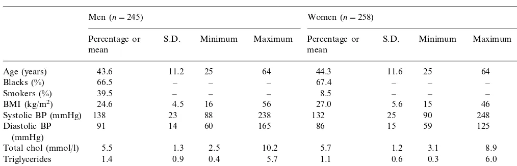
Table 2
Levels of selected cardiovascular risk factors by gendera
Women (n=258)
Men (n=245) P
S.D. Minimum Maximum Percentage or
Percentage or S.D. Minimum Maximum
mean mean
11.2 25 64 44.3 11.6 25 64 0.47
Age (years) 43.6
– – – 67.4
66.5 –
Blacks (%) – – 0.85
39.5
Smokers (%) – – – 8.5 – – – ‡
24.6
BMI (kg/m2) 4.5 16 56 27.0 5.6 15 46 ‡
23 88 238 132
138 25
Systolic BP (mmHg) 90 248 †
91 14 60 165 86 15 59 125
Diastolic BP ‡
(mmHg)
1.3 2.5 10.2 5.7
Total chol (mmol/l) 5.5 1.2 3.1 8.9 *
Triglycerides 1.4 0.9 0.4 5.7 1.1 0.6 0.3 6.0 ‡
(mmol/l)
a*,P50.05; †,P50.01; ‡P50.001.
have been described in detail elsewhere [30]. Among these, maximum plaque thickness (i.e. the distance from the plaque – lumen interface to the media – adventitia interface) was measured on the longitudinal view using electronic calipers. Plaque and total vessel areas were planimetered on the transversal view and the resulting lumen stenosis computed.
2.3.3. Feasibility
Adequate images allowing IMT to be measured could be obtained in all 503 subjects for the common CA. Femoral arteries IMT could not be measured in two subjects (on both sides) because of excessively irregular and poorly defined double-line image. Notice-ably, these two subjects had severe atherosclerosis with large plaques in all four examined sites. There was no missing data for the plaque protocol.
2.3.4. Reproducibility protocols
Reproducibility of IMT and maximum plaque thick-ness (MPT) measurements was tested in 20 randomly chosen participants who were re-examined by the same examiner 2 – 6 weeks after their first session, with the examiner kept blind to the first results and using the same protocol. Mean difference for paired IMT mea-surements was 3.393.0 mm×10−2for carotid arteries and 3.392.9 mm×10−2 for femoral arteries (which represented, respectively, 6.5 and 8.4% of the corre-sponding IMT values). Correlation coefficients for the two sets of measurements were 0.88 and 0.80, respec-tively. The reproducibility of the MPT measurements was examined with thek-statistic after having classified each artery in four lesion categories (i.e. 1, no plaque; 2, plaques measuring 1.00 – 1.49 mm; 3, 1.50 – 2.49 mm; 4, 2.50 mm or more). Inter-rating agreement reached 70.0 and 87.5% (k values of 0.51 and 0.71) for carotid and femoral arteries, respectively.
2.4. Statistical analysis
Statistics were performed using Stata™ 4.0 for Win-dows™ software (Stata Corp., College Station, TX, USA). Differences between two or more means were respectively assessed with unpaired t-test and one-way ANOVA, while differences between two or more pro-portions were respectively assessed with the Fisher’s exact test and the x2-test. P values equal to or lower than 0.05 were considered statistically significant. When not stated otherwise, values mentioned in the text are means9S.D.
3. Results
3.1.1. Study group characteristics
Table 2 displays characteristics of the subjects se-lected for ultrasonography. The distribution of age and black phenotype was similar in both genders. Men had higher prevalence of smoking (]1 cigarette every day), higher systolic and diastolic BP and higher triglycerides concentration. Women had higher BMI and marginally higher total cholesterol concentra-tion. The distribution of these characteristics was not statistically different between the 503 participants selected for ultrasonography and the 564 who were not.
F
.
Perret
et
al
.
/
Atherosclerosis
153
(2000)
9
–
21
13
Table 3
Values (mean9S.D.) of intima–media thickness (IMT), lumen diameter (LD), walls-to-lumen ratio (WLR) and cross-sectional wall area (CSWA) by gender, age and arterya
A B
Men A Women
25–34 35–44 45–54 55–64
25–34 35–44 45–54 55–64
(69) (63) (61) (65)
(55)
(n) (63) (68) (59)
Right carotid 44.097.6 46.398.8 61.8911 ‡ 41.496.6 44.695.4 54.1911 57.2911 ‡ 0.27
IMT (mm×10−2) 50.698.4
52.398.6 63.1913 ‡ 43.895.5 45.996.4 55.7912 57.1911 ‡ 0.12
45.196.4
Left carotid 49.198.5
‡ 33.997.7 38.196.4 45.497.1 49.296.3 ‡
55.1911 ‡
37.996.7 43.396.6 47.798.5 Right femoral
52.3910
Left femoral 38.096.0 42.596.7 46.197.1 ‡ 33.697.9 37.197.4 44.796.0 47.195.0 ‡ ‡
Right carotid 558949 565961 577969 0.14 534955 523941 530958 542959 0.26 ‡
LD (mm×10−2) 553951
* 517945 505942 526956 547948 ‡
572978 ‡
543952 566959
550941 Left carotid
Right femoral 609980 607969 619977 635982 0.19 486958 495961 537967 559963 ‡ ‡
0.55 483958 495963 530972 560957 ‡
632970 ‡
Left femoral 599973 602969 610974
Right carotid 13.692.2 14.192.4 17.792.8 ‡ 13.591.9 14.691.7 17.093.0 17.593.3 ‡ 0.10
WLR (%) 15.592.7
Left carotid 14.191.7 14.892.3 16.292.4 18.193.3 ‡ 14.591.5 15.492.1 17.593.4 17.393.1 ‡ 0.10
‡ 12.392.4 13.492.3 14.592.0 15.191.8 ‡
14.993.0 ‡
13.492.1 12.592.0
11.292.0 Right femoral
‡ 12.292.2 13.192.6 14.592.1 14.591.6 ‡ ‡
Left femoral 11.391.8 12.592.0 13.291.9 14.392.7
‡ 7.591.7 8.091.3 10.092.4 10.892.3
9.691.8 ‡
8.491.8 9.092.4 12.593.0 ‡
CSWA (mm2) Right carotid
Left carotid 8.591.5 9.592.1 9.892.1 12.793.6 ‡ 7.891.4 8.091.4 10.292.6 10.892.5 ‡ ‡
‡
Right femoral 7.891.9 8.991.8 10.192.6 12.093.0 5.691.7 6.491.5 8.491.9 9.592.0 ‡ ‡
‡ 5.591.9 6.291.7 8.191.6 9.091.5 ‡ ‡
11.392.7 Left femoral 7.691.7 8.691.8 9.692.2
ratio and cross-sectional wall area of carotid and femoral arteries were all significantly associated with age (in both genders) while the relation of lumen di-ameter with age was less consistent (especially in men). All values were higher in men than women although carotid IMT and walls-to-lumen ratio were not statisti-cally different between genders.
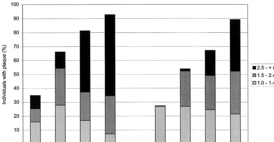
Table 4 lists the proportion of arteries with at least one plaque while Fig. 1 displays the proportions of subjects with at least one plaque (in any of the four examined arteries) by gender, age group and plaque thickness. Carotid and femoral plaque preva-lence were strongly associated with age (in both gen-ders) and with male sex, irrespective of plaque size. At least one plaque ]1.0 mm was found in, respectively, 34.9 and 27.5% of men and women aged 25 – 34 and these numbers reached, respectively, 92.7 and 89.2% in men and women aged 55 – 64. Largest plaques (]2.5 mm) were found in 9.5% of men aged 25 – 34 (none in women) and in, respectively, 58.2 and 36.9% of men and women aged 55 – 64 years. Individuals with only small plaques (1.0 – 1.4 mm) had them more in their carotid than femoral arteries (26.0 vs. 10.4%; PB 0.0005) while individuals with large plaques (2.5 mm or more) had them more in their femoral than carotid arteries (9.9 vs. 16.7%; PB0.002), suggesting a differ-ent time-course of atherosclerosis in these two territo-ries.
3.1.3. Comparison with plaque pre6alence data
originating from industrialised countries
Table 5 compares the prevalence of carotid atherosclerosis in Seychelles and in Western popula-tions. We could identify four studies from Western countries (US, Italy, France and Finland) which were population-based, reached satisfactory participation rates, and used measurement protocols compatible with our study [13,41 – 43]. To permit valid comparisons, prevalence of carotid atherosclerosis in Seychelles were each time calculated using same or similar categories of gender, age and lesion size as in the study taken for comparison.
Carotid atherosclerosis was systematically found to be more frequent in Seychelles than in Western coun-tries within all categories of age, gender and plaque size used to define atherosclerosis. Few exceptions con-cerned categories with small numbers of subjects (hence having poor statistical power to demonstrate a differ-ence) or categories for which Western subjects were significantly older than Seychellois subjects (in the American study). Prevalence differed particularly largely when comparing Seychelles individuals to Ital-ian or Finnish subjects, both for small lesions occurring before age 50 or for large lesions occurring after age 40. In these instances, prevalence of carotid atherosclerosis was often found to be several times higher in Seychelles than in the considered Western populations.
Table 4
Proportions of subjects with at least one plaque by gender, age, plaque thickness and arterya
B
Right or left carotid 23.8 54.1 76.9 ‡ *
† Right or left femoral
Carotid or femoral 34.9 66.2 81.4 92.7 ‡ 27.5 54.0 67.2 89.2 ‡ *
Right femoral 9.5 †
47.5
Left femoral 9.5 20.6 61.8 ‡ 2.9 7.9 23.0 49.2 ‡ ‡
70.9 ‡ 4.4 17.5 27.9 61.5 ‡
Right or left femoral 15.9 30.9 55.9 ‡
* Carotid or femoral 19.1 38.2
2.9
Right or left carotid 4.4 11.9 38.2 ‡ 0.0 0.0
F
.
Perret
et
al
.
/
Atherosclerosis
153
(2000)
9
–
21
15
Table 5
Prevalences of carotid atherosclerosis in Seychelles and in selected Western populationsa
Prevalence (%) A
Definition of plaque
Country (author) Sex and age of subjects Scanning range
Seychelles S/W ratio
Western (n) Seychelles (n) Western
dCCA+BIF+pICA 67.9 81.8 1.2
(55) *
United States (O’Leary) [41] M 65–69 (688) M 55–64 StenosisB25%
M 65–69 (688) M 55–64 (55) Stenosis]25% dCCA+BIF+pICA 28.1 27.3 1.0 n.s.
(65) StenosisB25% dCCA+BIF+pICA 54.2 76.9 1.4 ‡
(1126)
W 65–69 W 55–64
dCCA+BIF+pICA 22.8 24.6 1.1 n.s.
Stenosis]25% W 55–64
(1126) (65)
W 65–69
M 30–39 (101) M 30–39 Thickness]1.0 mm dCCA+BIF+pICA 1.0 34.7 35.0 ‡
Italy (Prati) [42] (75)
M 40–49 (135) M 40–49 (63) Thickness]1.0 mm dCCA+BIF+pICA 7.4 71.4 9.6 ‡
dCCA+BIF+pICA 36.6 64.8 1.8 ‡
Thickness]1.0 mm (54)
M 50–59 (112)
M 50–59
dCCA+BIF+pICA 1.0 27.4 27.4 ‡
W 30–39 (112) W 30–39 (73) Thickness]1.0 mm
dCCA+BIF+pICA 3.9 44.8 11.5 ‡
Thickness]1.0 mm
W 40–49 (127) W 40–49 (58)
Thickness]1.0 mm
W 50–59 (124) W 50–59 (66) dCCA+BIF+pICA 9.7 59.1 6.1 ‡
dCCA+BIF+pICA 2.1 4.7 2.2 n.s.
M 40–69 (337) M 40–64 (143) Stenosis]40%
dCCA+BIF+pICA 0.8 4.3 5.1 *
Stenosis]40%
W 40–69 (355) W 40–64 (157)
W 45–54 (517) W 45–54 Thickness]0.75 mm CCA+BIF 39.1 55.7 1.4 *
France (Bonithon–Kopp) [13] (61)
CCA+BIF 8.7
W 45–54 (517) W 45–54 (61) Thickness]1.75 mm 21.3 2.4 *
CCA+BIF 14.1 48.1 3.4
(52) ‡
(92) M 38–46 Thickness]1.2 mm
Finland (Salonen) [3] M 42
M 42 (92) M 38–46 (52) Stenosis]20% CCA+BIF 0.0 13.5 ‡
CCA+BIF 81.9
M 60 (105) M 56–64 (51) Thickness]1.2 mm 76.5 0.9 n.s.
CCA+BIF 4.8 35.3 7.4 ‡
Stenosis]20% (51)
M 56–64
M 60 (105)
aPrevalence of carotid atherosclerosis in Seychelles were calculated using same categories of gender, age and lesion size (thickness or degree of stenosis) as in the compared populations. M,
Fig. 1. Proportions of men (left) and women (right) with carotid or femoral atherosclerosis (at least one carotid or femoral plaque) by gender, age, plaque thickness and artery.
4. Discussion
This study aimed at quantifying peripheral atherosclerosis in the general population of a develop-ing country. We used high-resolution ultrasonography and measured arterial wall thickness (IMT) and atherosclerotic plaque prevalence in carotid and femoral arteries. The study included fairly large num-bers of subjects of both genders and several age cate-gories, and participation rate was high.
4.1.1. IMT measurement
The IMT measurement protocol used in this study followed the recommendations recently made by De-vereux et al. to measure IMT by avoiding segments with discrete atheromass and to separately identify and measure atherosclerotic plaques [37]. Furthermore, we systematically measured IMT in rectilinear segments, thereby avoiding sites with complex blood rheology such as turns or bifurcations [44]. It is now well known that some lipid deposition often occurs early in such atherosclerosis-prone regions but the true significance of these changes is still debated and could be consid-ered physiological (‘eccentric intimal thickening’) [45]. This methodological approach (used here for the first time in a large population-based study) should permit to better distinguish arterial wall changes due to atherosclerosis or hypertrophy and to investigate the factors associated with each of these pathologic pro-cesses separately.
Our data confirm that IMT is associated with age. In our series, IMT was larger by 30 – 45% in the oldest than youngest subjects (depending on gender and arte-rial territory). Previous histological studies of plaque-free arterial segments have shown that this ageing phenomenon mainly relates to a proliferation of colla-gen fibres and smooth muscle cells in the intima and media layers, together with quantitative and qualitative alterations of the matrix [44,46,47]. Despite arterial wall thickening, lumen diameter (LD) tended to be larger in older than younger subjects and external arterial diame-ter (LD+[2×IMT]) was associated with age for both carotid and femoral arteries and for both genders (PB 0.001). This association of peripheral artery diameter with age, which has already been described by others [48 – 50], was weaker than the association of IMT with age so that both the walls-to-lumen ratio and the cross-sectional wall area were associated with age, with respective proportionate changes of up to 33 and 70% between the 25 – 34 and 55 – 64 age categories.
F.Perret et al./Atherosclerosis153 (2000) 9 – 21 17
basal IMT values might be similar in the Seychelles and Western populations.
4.1.2. High pre6alence of peripheral atherosclerosis
Prevalence of subjects with at least one carotid plaque appeared to be at least twice as high in Sey-chelles as in several Western populations after adjust-ment for age and gender. In selected instances, particularly for young subjects, prevalence of carotid atherosclerosis was up to several times more frequent in Seychelles than in Western populations of same age and gender. Although this descriptive study cannot provide definitive explanations for this discrepancy, areas for discussion include methodological issues, prevalence of classical CVRF in the population and hypothetical conditions likely to have accelerated atherosclerosis in this population.
An overestimation of the prevalence of subjects with plaques in Seychelles is not likely for several reasons. Indeed, the use of a sophisticated echographic system, the protocol requirement to visualise each plaque in two perpendicular planes, the plaque measurements made after study completion based on videotaped im-ages and the frequent controls of the system calibration should all have minimised the risk of an overestimation of the measurements and false-positive findings. On the other hand, an underestimation of the prevalence of carotid atherosclerosis might have occurred in the cited studies from Western countries as these studies were performed several years ago and used potentially less discriminative echographic systems. In this situation, one would nevertheless assume that large lesions would not have been easily missed so that a presumed under-estimation in the plaque prevalence would have oc-curred predominantly for small lesions. This is clearly not the case here, as we found higher prevalence of subjects with both small and large plaques in Seychelles compared to Western countries. These observations support the view that the prevalence of peripheral
atherosclerosis found in Seychelles is valid and truly high.
The high prevalence of atherosclerotic plaques in Seychelles might be secondary to a particularly adverse CVRF profile in this population. The prevalence of the main CVRF in the Seychelles population has been thoroughly investigated in the two population-based surveys conducted in 1989 [6 – 8,32,55] and in 1994 [30] and, as in the WHO-MONICA project, have been age-standardized to the world population for individu-als aged 35 – 64 years. In the 1994 Seychelles Heart Study II, such prevalence in men and women were, respectively, 41.7 and 27.4% for hypertension (BP ] 160/95 mmHg), 41.2 and 8.4% for smoking (at least one cigarette per day), 9.7 and 33.6% for obesity (BMI ]30 kg/m2), 7.8 and 4.6% for diabetes (fasting glyco-suria or history of diabetes), 21.1 and 31.6% for hyper-cholesterolemia (cholesterol ]6.5 mmol/l). These data demonstrate very high prevalence of hypertension and obesity (women), and fairly high prevalence of smoking (men), hypercholesterolemia and diabetes in Seychelles compared to Western countries [56,57] In addition, a larger proportion of the population had high serum lipoprotein (a) levels (\300 mg/l) in Seychelles than, for example, in Switzerland (38 and 25%, respectively) [55], Not unexpectedly in a rapidly developing country, sedentary lifestyle is also being adopted by an increas-ing proportion of the populations. As an example, 36.3% of men and of 7.0% women reported heavy exercise at work in 1989 compared to no more than 13.6 and 2.3% in 1994 (PB0.001 for both). Overall, the markedly adverse CVRF profile documented recently in the Seychelles population is therefore consistent with high prevalence of peripheral atherosclerosis in Seychelles.
Other factors have been linked to high CVD fre-quency in developing countries and might therefore also account for high peripheral atherosclerosis in Seychelles.
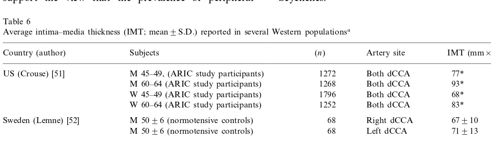
Table 6
Average intima–media thickness (IMT; mean9S.D.) reported in several Western populationsa
Country (author) Subjects (n) Artery site IMT (mm×10−2)
M 45–49, (ARIC study participants) 1272
US (Crouse) [51] Both dCCA 77*
M 60–64 (ARIC study participants) 1268 Both dCCA 93* W 45–49 (ARIC study participants) 1796 Both dCCA 68* 83* Both dCCA
1252 W 60–64 (ARIC study participants)
Sweden (Lemne) [52] M 5096 (normotensive controls) 68 Right dCCA 67910 M 5096 (normotensive controls) 68 Left dCCA 71913
M 42 (population of Kuopio)
Finland (Salonen) [53] 257 Both CCA 73926
M 60 (population of Kuopio) 334 Both CCA 115949
M 42910 (healthy volunteers) 40
France (Gariepy) [54] Right CCA 5096
40 Right CFA 5098
Dietary factors have strong relationship with CVD and special diet characteristics could be involved in the high peripheral atherosclerosis found in Seychelles. Similarly to other developing countries [58,59], Sey-chelles currently experiences rapid dietary transition. It is characterised by a rapid increase in the intake of fat (especially from cheap vegetable hydrogenated oils rich in saturated fats), a decline in complex carbohydrates consumption, an increase in salt intake (partly subse-quent to increased consumption of processed food) and a decrease in dietary fibres and micro-nutrients intake. In Seychelles, the energy intake derived from fats in-creased from 21 to 26% over a decade according to serial household expenditure surveys in 1983/1984 and 1992/1993 [60]. In addition, consumption of fruits and vegetables has been shown to be low in Seychelles [60], which related, e.g. to low thiamin blood levels in the population [61]. It could then be speculated that the current diet in Seychelles provides limited amounts of those micronutrients and antioxidants, which are in-creasingly believed to protect against atherosclerosis. The recently reported inverse relation between birth size and CVD in later life (‘Barker hypothesis’) may have special relevance to developing countries [62]. If this effect can be adequately validated, populations of developing countries would be at enhanced cardiovas-cular risk as substantial numbers of poorly nourished infants born in the past decades would nowadays reach ages at which atherosclerosis becomes apparent. Al-though severe famines are uncommon in tropical coun-tries, some degree of undernutrition has probably occurred in segments of the Seychelles population in the past so that such a mechanism could play a role in the high prevalence of peripheral atherosclerosis found in this study.
Mounting evidence relates infectious organisms (e.g. Chlamydia pneumoniae) to coronary heart disease and, specifically, to atherosclerotic plaques [63,64]. It is how-ever still unknown whether such organisms could have a role at the initiation of the disease, in the progress of the disease or in the complicating events so that specu-lations on a role of such agent in the aetiology of atherosclerosis in developing countries seems prema-ture. This hypothesis could nevertheless have special relevance to developing countries where infectious dis-eases account for a particularly large burden of disease and antibiotics have traditionally been used less exten-sively than in Western countries.
Ethnic factors may also be considered to explain high prevalence of peripheral atherosclerosis in Seychelles, as much of the population is of predominantly African descent and CVD (or CVRF) generally seem to be more frequent in African than Caucasian populations [65 – 67]. Sub-group analysis of our data did not how-ever support substantial difference in peripheral atherosclerosis prevalence between persons of African
or Caucasian descent. For instance, 61.4% of black Africans versus 70.0% of white Caucasian were, respec-tively, found to have at least one plaque (carotid or femoral of at least 1 mm) in this survey (P=0.31). These results have admittedly inherent limited signifi-cance due to low statistical power (small number of Caucasian subjects) and lack of adjustment for con-comitant CVRF. However, carotid atherosclerosis was similarly not found to be more prevalent in African Americans than white Americans in the ARIC study [68].
Some genetic factors, other than those determining ethnicity, could also be involved in the high prevalence of atherosclerosis in Seychelles. Although such consid-erations remain purely speculative due to the lack of data on this aspect in Seychelles, the insular situation of the country has potential for amplification of such phenomena. Moreover, genetic factors that predispose to CVRF (e.g. those modulating lipoprotein (a) levels, central obesity, glucose intolerance or dyslipidemia) have been shown to be particularly detrimental in pop-ulations experiencing rapid environmental changes (e.g. leading to weight gain, rise in plasma cholesterol and blood pressure levels), which may point to detrimental environment – genetic interactions [1,69].
Findings of this study have important public health significance. It has been established that peripheral atherosclerosis detected by ultrasonography closely re-lates to coronary atherosclerosis [16,22,28,70,71], subse-quent coronary [17,24] or cerebral [24,72] ischemic events, cardiovascular mortality [73] and all cause mor-tality [14]. The high prevalence of peripheral atheroscle-rosis (particularly carotid atheroscleatheroscle-rosis) in Seychelles is consistent with high stroke mortality (Table 1). How-ever, ischemic heart disease mortality seemed no higher in Seychelles than in Western countries. Several hy-potheses can be speculated to explain this discrepancy. Under-reporting of ischemic heart disease could occur in Seychelles due to limited availability of sophisticated diagnostic tools. Second, specific protective factors could be present in Seychelles which decrease ischemic heart disease incidence or case-fatality, e.g. by protect-ing the coronary tree from plaque occurrence, decreas-ing the tendency of plaques to rupture or exhibitdecreas-ing antithrombotic properties. Third, prevalence of periph-eral atherosclerosis in Seychelles could have reached high levels only recently and incidence of coronary events would subsequently be expected to increase in a near future.
F.Perret et al./Atherosclerosis153 (2000) 9 – 21 19
countries currently experiencing epidemiological transition.
Acknowledgements
The authors wish to thank L. Chow, G. Madeleine, A. Rwebogora, M. Abel, D. Larue and J. Brioche (Ministry of Health, Seychelles) for their very valuable assistance in the study; Professor R. Darioli, Professor A. Pecoud, Dr M. Depairon and Dr J.P. Gervasoni (University of Lausanne); the Government of Sey-chelles (particularly Health Ministers J. Dugasse and predecessor R. Adam) for their support; and all the participants in the survey for their kind cooperation. The authors also thank the following companies, which provided precious support, Sonotron Ltd. (Switzer-land), Air Seychelles, Skychef Seychelles Ltd., Sey-chelles Marketing Board (SeySey-chelles) and SeySey-chelles Assurance Corporation. Franc¸ois Perret benefited from grants from the ‘Fondation Vaudoise de Cardiologie’ (Switzerland) and the ‘Fondation Emma Muschamp’ (Lausanne, Switzerland). Pascal Bovet benefited from a grant from the Swiss National Science Foundation (No. 3233-038792.93).
References
[1] Reddy KS, Yusuf S. Emerging epidemic of cardiovascular dis-ease in developing countries. Circulation 1998;97:596 – 601. [2] Murray CJL, Lopez AD. Mortality by cause for eight regions in
the world: global burden of disease study. Lancet 1997;349:1269 – 76.
[3] Omran AR. The epidemiological transition. A theory of the epidemiology of population change. Milbank Memorial Fund Q 1971;184:509 – 38.
[4] Manton KG. The global impact of noncommunicable diseases: estimates and projections. World Health Stat Q 1988;41:255 – 66. [5] Shigan EN. Non-transmissible diseases in the world: a health priority problem of increasing importance. World Health Stat Q 1988;41:104 – 6.
[6] Bovet P, Rosalie D, Shamlaye C, Darioli R, Paccaud F. The Seychelles cardiovascular diseases survey 1989 (Methods). Soz-Praventivmed 1991;36(Suppl. 1):S3 – 7.
[7] Bovet P, Rosalie D, Shamlaye C, Darioli R, Paccaud F. The Seychelles cardiovascular diseases survey 1989 (Results). Soz-Praventivmed 1991;36(Suppl. 1):S36 – 87.
[8] Bovet P, Shamlaye C, Kitua A, Riesen WF, Paccaud F, Darioli R. High prevalence of cardiovascular risk factors in the Sey-chelles (Indian Ocean). Arterioscler Thromb 1991;11:1730 – 6. [9] Bovet P. The epidemiologic transition to chronic diseases in
developing countries: cardiovascular mortality, morbidity, and risk factors in Seychelles (Indian Ocean). Investigators of the Seychelles Heart Study. Soz- Praventivmed 1995;40:35 – 43. [10] Gervasoni J, Bovet P, Shamlaye C, Paccaud F. Guidelines for a
collaborative long term program of reduction of cardiovascular risk factors in the population of the Seychelles. Soz- Praven-tivmed 1991;36(Suppl. 1):S30 – 3.
[11] Hungerbuhler P, Bovet P, Shamlaye C. The cardiovascular dis-ease situation in Seychelles. World Health Stat Q 1993;46:108 – 12.
[12] Selzer RH, Hodis HN, Kwong-Fu H, Mack WJ, Lee PL, Liu CR, et al. Evaluation of computerized edge tracking for quan-tifying intima – media thickness of the common carotid artery from B-mode ultrasound images. Atherosclerosis 1994;111:1 – 11. [13] Bonithon-Kopp C, Scarabin PY, Taquet A, Touboul PJ, Malme-jac A, Guize L. Risk factors for early carotid atherosclerosis in middle-aged French women. Arterioscler Thromb 1991;11:966 – 72.
[14] Criqui MH, Coughlin SS, Fronek A. Noninvasively diagnosed peripheral arterial disease as a predictor of mortality: results from a prospective study. Circulation 1985;72:768 – 73. [15] Blankenhorn DH, Azen SP, Crawford DW, Nessim SA,
San-marco ME, Selzer RH, et al. Effects of colestipol – niacin therapy on human femoral atherosclerosis. Circulation 1991;83:438 – 47. [16] Craven TE, Ryu JE, Espeland MA, Kahl FR, McKinney WM,
Toole JF, et al. Evaluation of the associations between carotid artery atherosclerosis and coronary artery stenosis. A case-con-trol study. Circulation 1990;82:1230 – 42.
[17] Salonen JT, Salonen R. Ultrasonographically assessed carotid morphology and the risk of coronary heart disease. Arterioscler Thromb 1991;11:1245 – 9.
[18] Bots ML, Breslau PJ, Briet E, de Bruyn AM, van Vliet HH, van den Ouweland FA, et al. Cardiovascular determinants of carotid artery disease. The Rotterdam elderly study. Hypertension 1992;19:717 – 20.
[19] Salonen JT, Salonen R. Ultrasound B-mode imaging in observa-tional studies of atherosclerotic progression. Circulation 1993;87:II56 – 65.
[20] Demirovic J, Nabulsi A, Folsom AR, Carpenter MA, Szklo M, Sorlie PD, et al. Alcohol consumption and ultrasonographically assessed carotid artery wall thickness and distensibility. The atherosclerosis risk in communities (ARIC) study investigators. Circulation 1993;88:2787 – 93.
[21] Brown SA, Morrisett JD, Boerwinkle E, Hutchinson R, Patsch W. The relation of lipoprotein[a] concentrations and apolipo-protein[a] phenotypes with asymptomatic atherosclerosis in sub-jects of the atherosclerosis risk in communities (ARIC) study. Arterioscler Thromb 1993;13:1558 – 66.
[22] Furberg CD, Adams HP, Jr, Applegate WB, Byington RP, Espeland MA, Hartwell T, et al. Effect of lovastatin on early carotid atherosclerosis and cardiovascular events. Asymptomatic carotid artery progression study (ACAPS) research group. Circu-lation 1994;90:1679 – 87.
[23] Bonithon-Kopp C, Ducimetiere P, Touboul PJ, Feve JM, Bil-laud E, Courbon D, et al. Plasma angiotensin-converting enzyme activity and carotid wall thickening. Circulation 1994;89:952 – 4. [24] Salonen R, Tervahauta M, Salonen JT, Pekkanen J, Nissinen A, Karvonen MJ. Ultrasonographic manifestations of common carotid atherosclerosis in elderly eastern Finnish men. Prevalence and associations with cardiovascular diseases and risk factors. Arterioscler Thromb 1994;14:1631 – 40.
[25] Suurkula M, Agewall S, Fagerberg B, Wendelhag I, Widgren B, Wikstrand J. Ultrasound evaluation of atherosclerotic manifesta-tions in the carotid artery in high-risk hypertensive patients. Risk intervention study (RIS) group. Arterioscler Thromb 1994;14:1297 – 304.
[26] Salomaa V, Stinson V, Kark JD, Folsom AR, Davis CE, Wu KK. Association of fibrinolytic parameters with early atheroscle-rosis. The atherosclerosis risk in communities study. Circulation 1995;91:284 – 90.
[27] Castellano M, Muiesan ML, Rizzoni D, Beschi M, Pasini G, Cinelli A, et al. Angiotensin-converting enzyme I/D polymor-phism and arterial wall thickness in a general population. The Vobarno study. Circulation 1995;91:2721 – 4.
[29] Fauvel AA. Unpublished Documents on the History of the Seychelles Anterior to 1810. Victoria, Seychelles: Government Printing Office, 1909.
[30] Bovet P, Perret F, Shamlaye C, Darioli R, Paccaud F. The Seychelles Heart Study II: methods and selected basic findings. Seychelles Med Dental J 1997;5:8 – 24.
[31] Causes of death by sex and age. In: World Health Statistics Annual, WHO, Geneva, 1988;90 – 399.
[32] Tappy L, Bovet P, Shamlaye C. Prevalence of diabetes and obesity in the adult population of the Seychelles. Diabetic Med 1991;8:448 – 52.
[33] Anonymous MONICA Manual CVD/MNC Version 1.1. World Health Organization, Geneva, 1986.
[34] Rose GA, Blackburn H, Gillum RF, Prineas RJ. Cardiovascular survey methods. World Health Organization Monograph Series 1984.
[35] Perret F, Waeber B, Brunner HR. A new non-invasive method to measure arterial diameter and compliance: development and preliminary studies. Thesis, University of Lausanne, Lausanne, Switzerland, 1994.
[36] Perret F, Mooser V, Hayoz D, Tardy Y, Meister J, Etienne J, et al. Evaluation of arterial compliance – pressure curves: effects of antihypertensive drugs. Hypertension 1991;18:II-77 – 83. [37] Devereux RB, Waeber B, Roman MJ. Conclusions on the
mea-surement of arterial wall thickness: anatomic, physiologic and methodologic considerations. J Hypertens Suppl 1992;10:S119 – 21.
[38] Pignoli P, Tremoli E, Poli A, Oreste P, Paoletti R. Intimal plus medial thickness of the arterial wall: a direct measurement with ultrasound imaging. Circulation 1986;74(6):1399 – 406.
[39] Wikstrand J, Wendelhag I. Methodological considerations of ultrasound investigation of intima – media thickness and lumen diameter. J Intern Med 1994;236:555 – 9.
[40] Touboul PJ, Prati P, Scarabin PY, Adrai V, Thibout E, Ducimetiere P. Use of monitoring software to improve the measurement of carotid wall thickness by B-mode imaging. J Hypertens 1992;10:S37 – 41.
[41] O’Leary DH, Polak JF, Kronmal RA, Kittner SJ, Bond MG, Wolfson SK, Jr, et al. Distribution and correlates of sonograph-ically detected carotid artery disease in the cardiovascular health study. The CHS collaborative research group. Stroke 1992;23:1752 – 60.
[42] Prati P, Vanuzzo D, Casaroli M, Di Chiara A, De Biasi F, Feruglio GA, et al. Prevalence and determinants of carotid atherosclerosis in a general population. Stroke 1992;23:1705 – 11. [43] Salonen R, Seppanen K, Rauramaa R, Salonen JT. Prevalence of carotid atherosclerosis and serum cholesterol levels in eastern Finland. Arteriosclerosis 1988;8:788 – 92.
[44] Stary HC, Blankenhorn DH, Chandler AB, Glagov S, Insull W, Jr, Richardson M, et al. A definition of the intima of human arteries and of its atherosclerosis-prone regions. A report from the committee on vascular lesions of the council on arteriosclero-sis, American Heart Association. Arterioscler Thromb 1992;12:120 – 34.
[45] Stary HC, Chandler AB, Glagov S, Guyton JR, Insull W, Rosenfeld ME, et al. A definition of initial, fatty streak, and intermediate lesions of atherosclerosis. Circulation 1994;89:2462 – 78.
[46] Lie JT. The structure of the normal vascular system and its reactive changes. In: Juergens JL, Spitell JA, Fairbairn JF, editors. Peripheral Vascular Diseases, fifth ed. London: Saun-ders, 1980:51 – 81.
[47] Wilens SL. The nature of diffuse intimal thickening of abdomi-nal aorta in normotensive and hypertensive persons. Am J Pathol 1951;27:825 – 39.
[48] Kawasaki T, Sasayama S, Yagi S, Asakawa T, Hirai T. Non-in-vasive assessment of the age related changes in stiffness of major branches of the human arteries. Cardiovasc Res 1987;21:678 – 87.
[49] Benetos A, Laurent S, Hoeks AP, Boutouyrie PH, Safar ME. Arterial alterations with aging and high blood pressure. A noninvasive study of carotid and femoral arteries. Arterioscler Thromb 1993;13:90 – 7.
[50] Reneman RS, van Merode T, Hick P, Muytjens AM, Hoeks AP. Age-related changes in carotid artery wall properties in men. Ultrasound Med Biol 1986;12:465 – 71.
[51] Crouse JR, Goldbourt U, Evans G, Pinsky J, Sharrett AR, Sorlie P, et al. Arterial enlargement in the atherosclerosis risk in communities (ARIC) cohort. In vivo quantification of carotid arterial enlargement. The ARIC investigators. Stroke 1994;25:1354 – 9.
[52] Lemne C, Jogestrand T, de Faire U. Carotid intima – media thickness and plaque in borderline hypertension. Stroke 1995;26:34 – 9.
[53] Salonen R, Salonen JT. Determinants of carotid intima – media thickness: a population-based ultrasonography study in eastern Finnish men. J Intern Med 1991;229:225 – 31.
[54] Gariepy J, Simon A, Massonneau M, Linhart A, Levenson J. Wall thickening of carotid and femoral arteries in male subjects with isolated hypercholesterolemia. Prevention Cardio-Vascu-laire en Medecine du Travail Group. Atherosclerosis 1995;113:141 – 51.
[55] Bovet P, Rickenbach M, Wietlisbach V, Riesen W, Shamlaye C, Darioli R, et al. Comparison of serum lipoprotein(a) distribution and its correlates among black and white populations. Int J Epidemiol 1994;23:20 – 7.
[56] Pajak A. Geographical variation in the major risk factors of coronary heart disease in men and women aged 35 – 64 years. The WHO MONICA Project. World Health Stat Q 1988;41:115 – 40.
[57] King H, Zimmet P. Trends in the prevalence and incidence of diabetes: non-insulin diabetes mellitus. World Health Stat Q 1988;41:190 – 6.
[58] Drewnowski A, Popkin BM. The nutrition transition: new trends in the global diet. Nutr Rev 1997;55:31 – 43.
[59] Lang T. The public health impact of globalisation of food trade. In: Shetty PS, McPherson K, editors. Diet, Nutrition and Chronic Disease: Lessons from Contrasting Worlds. Chichester: Wiley, 1997:183 – 7.
[60] Larue D. Food consumption patterns in the Seychelles between 1983 and 1993. Seychelles Med Dental J 1996;4:16 – 9. [61] Bovet P, Larue D, Fayol V, Paccaud F. Blood thiamine status
and determinants in the population of Seychelles (Indian Ocean). J Epidemiol Community Health 1998;52:237 – 42.
[62] Barker DJP. Fetal origins of coronary heart disease. Br Med J 1995;311:171 – 4.
[63] Danesh J, Collins R, Peto R. Chronic infections and coronary heart disease: is there a link? Lancet 1997;350:430 – 6.
[64] Kuo CC, Shor A, Campbell LA, Fukushi H, Patton DL, Grayston JT. Demonstration of Chlamydia pneumoniae in atherosclerotic lesions of coronary arteries. J Infect Dis 1993;167:841 – 9.
[65] Muna WFT. Cardiovascular disorders in Africa. World Health Stat Q 1993;46:125 – 33.
[66] Liao Y, Cooper RS. Continued adverse trends in coronary heart disease among blacks. Public Health Rep 1995;110:572 – 9.
[67] Winkleby MA, Kraemer HC, Ahn DK, Varady AN. Ethnic and socioeconomic differences in cardiovascular risk factors. J Am Med Assoc 1998;280:356 – 62.
[68] Li R, Duncan BB, Metcalf PA, Crouse JR, Sharrett AR, Tyroler HA, et al. B-mode-detected carotid artery plaque in a general population. Atherosclerosis risk in communities (ARIC) study investigators. Stroke 1994;25:2377 – 83.
F.Perret et al./Atherosclerosis153 (2000) 9 – 21 21
Indian Subcontinent living in West London and their siblings in India. Lancet 1995;345:404 – 9.
[70] Wofford JL, Kahl FR, Howard G, McKinney WM, Toole JF, Crouse JR. Relation of extent of extracranial carotid artery atherosclerosis as measured by B-mode ultrasound to the extent of coronary atherosclerosis. Arterioscler Thromb 1991;11:1786 – 94.
[71] O’Leary DH, Polak JF, Kronmal RA, Manolio TA, Burke GL, Wolfson SK, et al. Carotid-artery intima and media thickness as
a risk factor for myocardial infarction and stroke in older adults. N Engl J Med 1999;340:14 – 22.
[72] Wolf PA, Kannel WB, Sorlie P, McNamara P. Asymptomatic carotid bruit and risk of stroke. The Framingham study. J Am Med Assoc 1981;245:1442 – 5.
[73] Belcaro G, Barsotti A, Nicolaides AN. ‘Ultrasonic biopsy’ — a non-invasive screening technique to evaluate the cardiovascular risk and to follow up the progression and the regression of arteriosclerosis. Vasa J Vasc Dis 1991;20:40 – 50.