Effects of Estradiol and Progesterone Administration on
Human Serotonin 2A Receptor Binding: A PET Study
Eydie L. Moses, Wayne C. Drevets, Gwenn Smith, Chester A. Mathis,
Brinda N. Kalro, Meryl A. Butters, Mark P. Leondires, Phil J. Greer,
Brian Lopresti, Tammy L. Loucks, and Sarah L. Berga
Background: Preclinical studies demonstrate that 17
b
-estradiol (E
2) increases serotonin-2A receptor (5-HT
2AR)
density in rat frontal cortex.
Methods: We investigated the impact of hormone
replace-ment therapy on 5-HT
2AR binding potential (BP) using
positron emission tomography and [
18F]altanserin in five
postmenopausal women. Subjects were imaged at
base-line, following 8 to 14 weeks of transdermal E
2, 0.1 mg/d,
and following 2 to 6 weeks of E
2plus micronized
proges-terone (P) 100 mg per os twice daily. Regional BPs in the
anterior cingulate cortex, dorsolateral prefrontal cortex,
and lateral orbitofrontal cortex were calculated by Logan
analysis.
Results: There was a main effect of time (p
5
.017) for
5-HT
2AR BP, which increased 21.2%
6
2.6% following
combined E
2and P administration relative to baseline.
This effect was evident in all cerebral cortex regions
examined.
Conclusions: 5-HT
2AR BP increased in widespread areas
of the cerebral cortex following combined E
21
P
admin-istration. Biol Psychiatry 2000;48:854 – 860 © 2000
So-ciety of Biological Psychiatry
Key Words: Estradiol, progesterone, PET, neurotrophin,
behavior, 5-HT
2AIntroduction
O
varian hormones exert prominent effects on the
central serotonergic receptor systems of experimental
animals (Osterlund and Hurd 1998; Pecins-Thompson and
Bethea 1998; Rubinow et al 1998). For example, E
2administration increases serotonin-2A receptor (5-HT
2AR)
density in the anterior cingulate, olfactory, and frontal
cortices of ovariectomized rats (Cyr et al 1998; Sumner
and Fink 1995). Progesterone (P) promotes comparable
5-HT
2AR increases but prevents E
2-related 5-HT
2AR
in-creases when the two hormones are initiated concurrently
(Biegon et al 1983). The 5-HT
2AR system has been
implicated in the pathophysiology of depression (Yates et
al 1990), suicide (Arango et al 1997), schizophrenia
(Jakab and Goldman-Rakic 1998), and Alzheimer’s
dis-ease (Crow et al 1986), so it is conceivable that the effects
of ovarian hormones on cognitive and emotional behavior
(Anderson et al 1987; Coble and Day 1988; Phillips and
Sherwin 1992; Tang et al 1996) may be partly mediated
via indirect effects on 5-HT
2AR binding.
Our study investigates changes in 5-HT
2AR binding
potential (BP) following E
2and P administration, using
the selective 5-HT
2AR radioligand [
18
F]altanserin and
positron emission tomography (PET; Lemaire et al 1991;
Sadzot et al 1995) in healthy postmenopausal women.
Based on the cortical regions where E
2administration was
associated with increased 5-HT
2AR density in rats (Cyr et
al 1998; Sumner and Fink 1995), we tested the hypothesis
that the 5-HT
2AR BP would increase in areas of the frontal
and cingulate cortices during E
2and P administration.
Methods and Materials
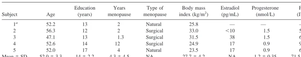
Five postmenopausal women provided informed consent as approved by the University of Pittsburgh Biomedical Institu-tional Review Board. Subject characteristics are listed in Table 1. Inclusion criteria were age over 45 years, laboratory confirma-tion of postmenopausal status (amenorrhea for 12 months unless menopause is surgical and laboratory confirmation of follicle-stimulating hormone [FSH]. 20 IU/L and E2 , 40 pg/mL),
normal gynecologic exam, PAP smear, and mammogram. Ex-clusion criteria were contraindications for magnetic resonance imaging (MRI), presence of psychiatric or medical conditions, and use of psychotropic medications in the past 3 months. Three women who were taking hormone replacement therapy (HRT) at study entry discontinued HRT for 3 months at which time laboratory data confirmed menopause. The MRI scans (Signa 1.5 tesla scanner, GE Medical Systems, Milwaukee) performed before PET scanning ruled out focal abnormalities (e.g. stroke,
From the Departments of Ob/Gyn/ReproSci (BNK, TLL, SLB), Psychiatry (ELM, WCD, GS, MAB, SLB), and Radiology (WCD, CAM, PJG, BL), University of Pittsburgh, Magee-Women’s Research Institute, Pittsburgh, Pennsylvania; NICHD/NIH/ERRB, National Institutes of Health, Bethesda, Maryland (MPL); and Psychiatry Research and Neuroscience Research Center, North Shore– Long Island Jewish Health Care System, Glen Oaks, New York (GS). Address reprint requests to Eydie Moses, M.D., NIMH Clinical Research Fellow,
Magee-Women’s Hospital, Department of OB-GYN/RS, 300 Halket Street, Pittsburgh PA 15213-3180.
Received February 15, 2000; revised June 19, 2000; accepted June 21, 2000.
tumor) and provided an anatomic reference for region-of-interest (ROI) analysis.
For each subject, [15
O]water and [18
F]altanserin PET scans were performed at baseline (PET 1), following 8 to 14 weeks of transdermal 17b-estradiol, Climara 0.1 mg/d (PET 2), and following 2 to 6 weeks of E2plus micronized P, Prometrium 100
mg per os twice daily (PET 3). Laboratory confirmation of E2
and P levels was obtained at each scan to confirm adherence to the protocol and achievement of hormone concentrations within the reproductive range. Serum levels of E2and P were measured
by radioimmunoassay (RIA; Coat-A-Count, DPC, Los Angeles, CA) as previously described (Berga et al 1997). Each specimen was measured in duplicate, and all samples were run in the same assay.
The procedures for [18
F]altanserin arterial blood sampling, image acquisition, and analysis were described previously (Smith et al 1998). Briefly, after arterial and venous cannulation, emission and transmission PET images were acquired using a Siemens/CTI HR1 (63 contiguous slices over 15.2 cm; full width half maximum resolution5560.5 mm transverse and 4.560.5 mm axially; Brix et al 1997). Cerebral blood flow data were obtained in a 3-min dynamic scan following bolus injection of 12-mCi of [15
O]water (Quarles et al 1993). 5-HT2AR BP data
were obtained in a 90-min dynamic scan following bolus injection of 10-mCi high specific activity [18
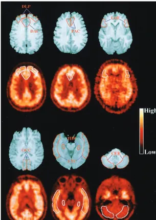
F]altanserin. The PET studies acquired on 3 days were aligned to each other and then to the MRI scan using automated image registration (Woods et al 1993). The ROIs (Figure 1) were defined to approximate prefrontal cortical regions where E2sensitivity was
demonstrated in rodents and where human brain mapping studies showed activation during emotional processing (lateral orbito-frontal cortex and pregenual anterior cingulate cortex) or lan-guage and visuospatial processing (dorsolateral prefrontal cortex and dorsal anterior cingulate gyrus; reviewed in Drevets and Raichle 1998). Control ROIs were defined in hippocampus, because 5-HT2AR density did not significantly change following
E2 administration in the rat dentate gyrus (Sumner and Fink
1995), and the occipital and lateral temporal cortex, which were not examined in preclinical studies.
Decay-corrected time-radioactivity concentrations were ob-tained for each ROI, and the right and left regions were summed. The plasma input function was corrected for the fraction of unmetabolized [18
F]altanserin. The volume of distribution for each ROI (DVROI) was determined by Logan graphical analysis
(Logan et al 1994), as previously described (Smith et al 1998). The DV in the cerebellum (DVcerebellum) represented the
concen-tration of free and nonspecifically bound [18
F]altanserin due to the relatively low concentration of 5-HT2AR in this region (Pazos
et al 1987). The BP was calculated as ([DVROI/DVcerebellum]2
1) to minimize the influence of plasma and tissue nonspecific binding (Mintun et al 1984). The BP relates to the free receptor concentration according to the formula
DVROI where Bmax5 receptor density, L5 concentration of
endoge-nous neurotransmitter bound to receptor, Kd 5 dissociation
constant, and NS 5 ratio of kinetic constants for nonspecific binding.
Cerebral blood flow was assessed using a one tissue compart-mental model (K1, k2, delay), in which K1 (mL/min/100 mL) was used to quantitate cerebral blood flow (Quarles et al 1993). Cognitive, psychiatric, and quality-of-life assessments (Table 2) were administered within one day of each scan. The effects of E2
and P on 5-HT2AR were statistically assessed by
repeated-measures analysis of variance (ANOVA) using the Huynh–Feldt correction. The Tukey honestly significant difference was ap-plied post hoc to examine the significance of specific contrasts in which the ANOVA showed significant differences.
Results
Baseline serum E
2, P, and FSH concentrations confirmed
menopause (Table 1). E
2concentrations (mean
6
SD)
increased to and remained at the targeted postreplacement
levels at PET 2 (73.3
6
36.2 pg/mL) and PET 3 (76.0
6
32.9 pg/mL). (The mean E
2concentration across a normal
menstrual cycle is 110 pg/mL.) The concentrations of P
were negligible at PET 2 (1.3
6
0.46 nmol/L) and
increased to the reproductive range following replacement
at PET 3 (67.7
6
67.7 nmol/L). The wide P and E
2concentration ranges resulted from variable timing of
blood draws with respect to peak and trough levels of the
administered hormone. Whereas the variability was
greater with Prometrium (because of the oral route of
administration and its short half-life), it was narrower for
Subject Age (years) menopause menopause index (kg/m2) (pg/mL) (nmol/L) (IU/L)1a 52.2 13 2 Natural 25.8 — — —
Climara (because transdermal administration provides
uni-form release over 7 days).
Cerebral blood flow data for four subjects (technical
problems precluded use of the cerebral blood flow image
from one subject) showed no significant changes across
scans (Table 3).
There was a main effect of time [F(2,8)
5
9.90, p
5
.017] for 5-HT
2AR BP in the primary ROIs (Figure 2
and Table 4). There was no significant time-by-region
interaction. Post hoc analysis showed that the 21.2%
6
2.6% (mean of all four a priori regions
6
SD) BP
increase between PET 1 and PET 3 was significant (p
5
.0055). The mean increase in BP between PET 1 and
PET 2 and between PET 2 and PET 3 did not reach
significance (mean
6
SD increases in BP were 10.5%
6
3.6% and 10.6%
6
4.2%, respectively; 0.1
,
p
,
.2).
Post hoc, exploratory analysis of control regions also
showed a similar main effect of time [F(2,8)
5
5.28,
p
5
.034] with the 5-HT
2AR BP increasing between
PET 1 and PET 3 (p
5
.029), and no significant time by
region interaction.
Baseline cognitive, psychiatric, and quality-of-life
as-sessments were in the expected range for healthy subjects
(Table 2). There were post-HRT tendencies for
improve-ment in the spatial working memory task, the anxiety
subscale of the Hopkins Symptom Checklist-90, and the
Menopause Specific Quality of Life Questionnaire
(Hilditch et al 1996).
Figure 1. Representative horizontal magnetic resonance imaging sections through each of the regions of interest (ROIs; first and third rows). The coregistered baseline [18
Discussion
These data demonstrate that E
2plus P administration
significantly increases cerebral cortical 5-HT
2AR BP in
humans. These changes were several fold greater than the
test–retest variability for [
18F]-altanserin that was
deter-mined over 2 to 16 days in corresponding regions (Smith
et al 1998). It is unlikely these BP changes were
con-founded by ligand delivery because cerebral blood flow
did not increase between PET 1 and PET 3 (Table 3).
Although the sample size was small, the finding that BP
increased in all five subjects in all ROIs examined is
compelling.
We used BP to quantitate receptor specific binding
because this term presumably excludes the effects of free
and nonspecifically bound [
18F]altanserin from PET
mea-sures of regional 5-HT
2AR concentration. It is nonetheless
noteworthy that although the magnitude of the BP
ROIelevation between PET 1 and PET 2 was similar to that
seen between PET 2 and PET 3, the DV
ROIvalues
increased between PET 1 and PET 2, but not between PET
2 and PET 3 (Table 4). This observation raised the
possibility that E
2administration increased 5-HT
2AR to a
level that plateaued and that P administration did not
further elevate 5-HT
2AR binding. This issue does not
detract from the statistically significant result that HRT
increased 5-HT
2AR BP between PET 1 and PET 3. It does,
however, highlight the inability of our study design to
differentiate the relative contributions of E
2and P to the
change in 5-HT
2AR binding or to delineate the time-course
of E
2and P effects on 5-HT
2AR binding.
The mechanism by which ovarian hormone replacement
leads to the observed 5-HT
2AR BP increases is unclear. A
classic genomic E
2mechanism has been suggested
whereby E
2bound to the intracellular estrogen receptor
translocates into the cell nucleus and directly promotes
DNA expression. Cyr et al (1998; and personal
commu-nication) reported that rodent CNS 5-HT
2AR density
increased in parallel with 5-HT
2AR mRNA expression
following E
2administration in the dorsal raphe nucleus,
frontal cortex, anterior cingulate cortex, and striatum. In
contrast, Sumner and Fink (1995) found that E
2-mediated
5-HT
2AR increases in frontal regions were not associated
with corresponding changes in 5-HT
2AR mRNA,
suggest-ing that alternative mechanisms (such as transynaptic
stimulation of 5-HT
2AR gene transcription,
posttransla-tional 5-HT
2AR processing effects, or putative membrane
Measure Baseline Post-E2 Post-E21P
1. Spatial working memory task (CANTAB)a No. errors 49629 2868.9 2466.2b
2. Rivermead Paragraph Recall Testa Immediate recall 1362.6 1462.9 1363.5
Percent retention 98611 90613 101615 3. California Verbal Learning Testa Immediate recall 6767.5 6467.9 7265.2
Percent retention 9664.3 9766.3 9863.1 4. Brief Visuospatial Memory Testa Immediate recall 2565.4 2765.5 2961.5
Percent retention 9765.6 9864.6 10067.8 5. Beck Depression Inventory Scale 1.561.3 0.561.0 0.861.0 6. Hopkins Symptom Checklist-90 Global severity index 3966.4 3465.2 3565.5 Anxiety subscale 4869 3968.3 3963.5 Depression subscale 3966.0 3766.0 3664.0 7. Medical Outcomes Studyc Physical subscale 5263.9 5462.2 5462.5
Mental subscale 5866.5 6061.9 5863.4 8. Menopause quality of life scaled Total score 3.161.4 1.761.3 1.260.3
n54. One subject underwent a unique battery of tests and so was not included in these results. CANTAB, Cambridge Neuropsychological Automated Test Battery (Sahakian et al 1992).
aAlternate forms of tests were administered at different time points. bn53. Missing data point was a result of technical difficulties. cMedical Outcomes Study Short Form-36 Women’s Health Questionnaire.
dThe Menopause Specific Quality of Life Questionnaire, where score represents total score of vasomotor, psychomotor, physical, and sexual subdomains (Hilditch et al 1996).
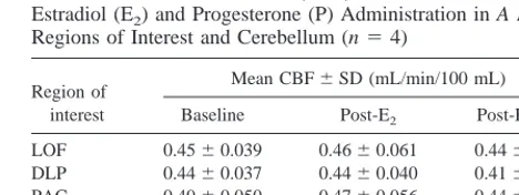
Table 3. Cerebral Blood Flow (CBF) before and after Estradiol (E2) and Progesterone (P) Administration in A Priori
Regions of Interest and Cerebellum (n54)
Region of interest
Mean CBF6SD (mL/min/100 mL)
Baseline Post-E2 Post-E21P
LOF 0.4560.039 0.4660.061 0.4460.030 DLP 0.4460.037 0.4460.040 0.4160.023 PAC 0.4960.050 0.4760.056 0.4460.026 DAC 0.4660.036 0.4560.072 0.4360.029 CER 0.4960.057 0.4760.078 0.4460.042
estrogen-receptor-mediated enhancement of second
mes-senger systems) may also or alternatively account for the
effects of E
2on 5-HT
2AR .
The widespread distribution of 5-HT
2AR BP increases
may also reflect ovarian-hormone-mediated neurotrophic
effects. Administration of E
2promotes neuronal
out-growth (Toran-Allerand 1996), increases expression of
neurotrophin receptor trkA and brain derived neurotrophic
factor (BDNF) mRNA expression, mitogen-activated
pro-tein kinase and cAMP response element binding propro-tein
(Gibbs 1998; McEwen and Alves 1999). Because
5-HT
2ARs are present on nearly all cortical pyramidal cells
and interneurons (Jakab and Goldman-Rakic 1998), the
proliferation of neuritic processes induced by these
neu-rotrophic effects would likely be associated with
upregu-lation of 5-HT
2AR expression.
Nonetheless, the effect of HRT on 5-HT
2AR is not
global in rats. Administration of E
2did not change
5-HT
2AR density in regions including the claustrum,
dentate gyrus, locus coeruleus, and medial preoptic area
(Sumner and Fink 1995). Because of the spatial resolution
limitations of PET, we could not assess the effects of HRT
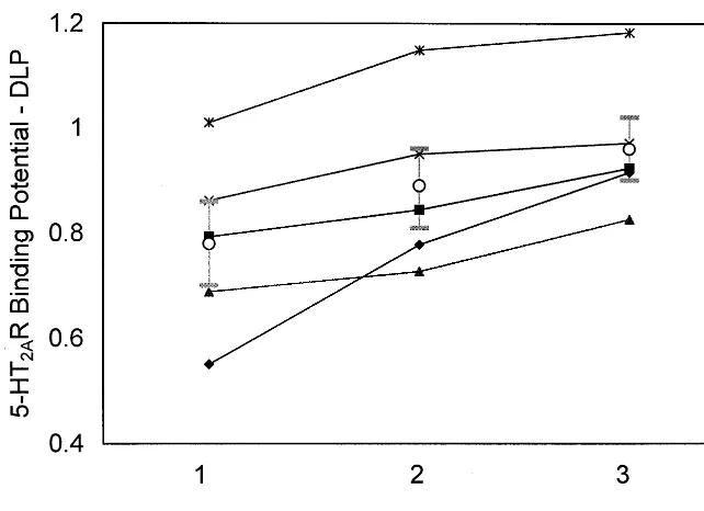
Figure 2. 5-HT2A receptor (5-HT2AR) binding potential (BP) in the dorsolateral prefrontal cortex (DLP) for the five sub-jects at three time points: baseline (positron emission tomography [PET] 1), postestradiol (PET 2), and postestradiol plus progesterone (PET 3) administra-tion. Icons represent the BP value mea-sured in a single PET scan, for each subject, at the designated time point. The mean is shown by the circle with SEM bars.
Table 4. 5-HT2AReceptor Binding Potential (BP) before and after Estradiol (E2) and Progesterone (P) Administration in All Regions of Interest (ROIs)
Mean DV6SD Mean BP6SD Mean %DBP
(E21P vs. baseline)6SD
Baseline Post-E2 Post-E21P Baseline Post-E2 Post-E21P
A Priori ROIs
LOF 2.5160.37 2.7360.21 2.7460.13 0.8460.15 0.8960.23 1.0160.15 20618a
DLP 2.4360.41 2.7560.38 2.6860.15 0.7860.17 0.8960.17 0.9660.13 25624a
PAC 2.6860.64 2.9860.61 2.8960.45 0.9460.24 1.0360.15 1.1060.19 19616a
DAC 2.5560.36 2.8360.37 2.8160.18 0.8760.12 0.9560.15 1.0660.11 22614a
CERb 1.3660.18 1.4660.22 1.3760.13 0 0 0 0
Control ROIs
LTC 2.6460.64 2.8960.56 2.8860.38 0.9260.27 0.9860.23 1.1160.20 26635c
OCC 2.7160.47 2.9560.29 2.9060.11 0.9960.15 1.0460.18 1.1360.15 14618c
HIPd 1.7460.12 1.9760.14 1.7860.13 0.2460.20 0.2960.23 0.2960.20 27623c
DV, volume of distribution; LOF, lateral orbitofrontal cortex; DLP, dorsolateral prefrontal cortex; PAC, pregenual anterior cingulate cortex; DAC, dorsal anterior cingulate cortex; CER, cerebellar cortex; LTC, lateral temporal cortex; OCC, occipital cortex; HIP, hippocampus.
aThere was a main effect of time [F(2,8)59.90, p5.017] for 5-HT
2Areceptor BP in the a priori ROIs. There was no significant effect of region or time-by-region
interaction. Post hoc analysis showed that the increase between baseline and post-E21P scans was significant (p5.0055).
bThe change in cerebellar DV across time was not significant [F(2,8)52.49, p5.14].
cPost hoc analysis of control ROIs showed a similar main effect of time [F(2,8)55.28, p5.034] for 5-HT
2Areceptor BP with a significant increase between baseline
and post-E21P scans (p5.029).
ically resolve the dentate gyrus. Furthermore, our
hip-pocampal measures were affected by spilling-in of
radio-active
counts
from
the
perirhinal,
entorrhinal,
parahippocampal cortices, and amygdala (Links et al
1996).
The cognitive and emotional assessments used to detect
clinical correlations with the imaging data had limited
sensitivity because of the small sample size. In addition,
GABAergic P metabolites may have influenced these
behavioral measures at the third time point. The extent to
which changes in 5-HT
2AR binding may be involved in
mediating the effects of gonadal steroids on behavior, via
either specific serotonin receptor changes or neurotrophic
effects, remains unclear.
This study was supported by National Institute of Health Grant Nos. MH16804, MH01713, MH/HD50748, MH01621, MH49936, MH57078, and MH52247 (Intervention Research Center for Late-Life Mood Disor-ders); National Center for Research Resources Grant No. RR-00056 to the University of Pittsburgh School of Medicine General Clinical Research Center; and the Functional Imaging Research Program of the Department of Radiology, University of Pittsburgh School of Medicine, Pittsburgh, Pennsylvania.
The authors thank Michelle Zmuda, Carolyn Cidis Meltzer, M.D., and the PET facility staff for technical assistance; Florence Hall for assisting with recruitment; and Julie Price, Ph.D., Cristy Matan, B.S., Robert Sembrat, B.S., Tyrone Holliman, and Clara Gautier, Ph.D., for assistance with data analysis.
References
Anderson E, Hamburger S, Liu J, Rebar R (1987): Characteris-tics of menopausal women seeking assistance. Am J Obstet
Gynecol 156:428 – 433.
Arango V, Underwood MD, Mann JJ (1997): Postmortem findings in suicide victims. Implications for in vivo imaging studies. Ann N Y Acad Sci 836:269 –287.
Berga SL, Daniels TL, Giles DE (1997): Women with functional hypothalamic amenorrhea but not other forms of anovulation display amplified cortisol concentrations. Fertil Steril 67: 1024 –1030.
Biegon A, Reches A, Snyder L, McEwen BS (1983): Serotoner-gic and noradrenerSerotoner-gic receptors in the rat brain: Modulation by chronic exposure to ovarian hormones. Life Sci 32:2015– 2021.
Brix G, Zaers J, Adam L-E, Belleman ME, Ostertag H, Trojan H (1997): Performance evaluation of a whole-body PET scanner using the NEMA protocol. J Nucl Med 38:1614 –1623. Coble PA, Day NL (1988): The epidemiology of mental and
emotional disorders during pregnancy and the postpartum period. In: Cohen RL, editor. Psychiatric Consultation in
Childbirth Settings. New York: Plenum Press, 37– 47.
Crow T, Ferrier I, Johnson J (1986): The selectivity of the reduction of serotonin S2 receptors in Alzheimer-type demen-tia. Neurobiol Aging 7:3–7.
Drevets WC, Raichle ME (1998): Reciprocal suppression of regional cerebral blood flow during emotional versus higher cognitive processes: Implication for interaction between emo-tion and cogniemo-tion. Cogn Emoemo-tion 12:353–385.
Gibbs RB (1998): Levels of trkA and BDNF mRNA, but not NGF mRNA, fluctuate across the estrous cycle and increase in response to acute hormone replacement. Brain Res 787: 259 –268.
Hilditch JR, Lewis J, Peter A, van Maris B, Ross A, Franssen E, et al (1996): A menopause-specific quality of life question-naire: Development and psychometric properties. Maturitas 24:161–175.
Jakab RL, Goldman-Rakic PS (1998): 5-Hydroxytryptamine2A serotonin receptors in the primate cerebral cortex: Possible site of action of hallucinogenic and antipsychotic drugs in pyramidal cell apical dendrites. Proc Natl Acad Sci U S A 95:735–740.
Lemaire C, Cantineau, Plenevaux A, Christiaens L (1991): Fluorine-18-Altanserin. A radioligand for the study of sero-tonin receptors with PET: Radiolabeling and in vivo biologic behavior in rats. J Nucl Med 32:2266 –2272.
Links JM, Zubieta JK, Meltzer CC, Stumpf MJ, Frost JJ (1996): Influence of spatially heterogeneous background activity on “hot object” quantification in brain emission computed to-mography. J Comput Assist Tomogr 20:680 – 687.
Logan J, Volkow N, Fowler J, Wang G-J, Dewey SL, MacGregor R, et al (1994): Effects of blood flow on [11C]raclopride binding in the brain: model simulations and kinetic analysis of PET data. J Cereb Blood Flow Metab 14:995–1010. McEwen BS, Alves SE (1999): Estrogen actions in the central
nervous system. Endocr Rev 20:279 –307.
Mintun MA, Raichle ME, Kilbourn MR, Wooten GF, Welch MJ (1984): A quantitative model for the in vivo assessment of drug binding sites with positron emission tomography. Ann
Neurol 15:217–227.
Osterlund MK, Hurd YL (1998): Acute 17b-estradiol treatment down-regulates serotonin 5HT1A receptor mRNA expression in the limbic system of female rats. Mol Brain Res 55:169 – 172.
Pazos A, Probst A, Palacios JM (1987): Serotonin receptors in the human brain. IV. Autographic mapping of serotonin-2 receptors. Neuroscience21:123–139.
Pecins-Thompson M, Bethea CL (1998): Regulation of serotonin re-uptake transporter mRNA expression by ovarian steroids in rhesus macaques. Mol Brain Res 53:120 –129.
Phillips SM, Sherwin B (1992): Effects of estrogen on memory function in surgically menopausal women.
Psychoneuroen-docrinology 17:485– 495.
Quarles RP, Mintun MA, Larson KB, Markham J, MacLeod AM, Raichle ME (1993): Measurement of regional cerebral blood flow with positron emission tomography: A comparison of [15O]water to [11C]butanol with distributed-parameter and compartmental models J Cereb Blood Flow Metab 13:733– 747.
Rubinow DR, Schmidt PJ, Roca CA (1998): Estrogen-serotonin interactions: Implications for affective regulation. Biol
Sadzot B, Lemaire C, Maquet P, Salmon E, Plenevaux A, Degueldre C, et al (1995): Serotonin 5HT2 receptor imaging in the human brain using positron emission tomography and a new radioligand, [18F]altanserin: Results in young normal controls. J Cerebr Blood Flow Metab 15:787–797.
Sahakian BJ, Owen AM (1992): Computerized assessment in neuropsychiatry using CANTAB: Discussion paper. J R Soc
Med 85:399 – 402.
Smith GS, Price JC, Lopresti BJ, Huang Y, Simpson N, Holt D, et al (1998): Test-retest variability of Serotonin 5-HT2A receptor binding measured with positron emission tomogra-phy and [18F]Altanserin in the human brain. Synapse 30: 380 –392.
Sumner BE, Fink G (1995): Estrogen increases the density of 5-HT2A receptors in cerebral cortex and nucleus accumbens in the female rat. J Steroid Biochem Mol Biol 54:15–20.
Talairach J, Tournoux P (1988): Co-Planar Stereotaxic Atlas of
the Human Brain 3-Dimensional Proportional System: An Approach to Cerebral Imaging. New York: Thieme.
Tang MX, Jaocbs D, Stern Y, Marder K, Schofield, Gurland B (1996): Effect of oestrogen during menopause on risk and age at onset of Alzheimer’s disease. Lancet 348:429 – 432. Toran-Allerand CD (1996): Mechanisms of estrogen action
during neural development: Mediation by interactions with neurotrophins and their receptors? J Steroid Biochem Mol
Biol 56:169 –178.
Woods RP, Mazziotta JC, Cherry SR (1993): MRI-PET regis-tration with automated algorithm J Comput Assist Tomogr 17:536 –546.
Yates M, Leake A, Candy JM, Fairbairn AF, McKeith IG, Ferrier IN (1990): 5HT2 receptor changes in major depression. Biol