www.elsevier.com / locate / bres
Research report
Interferon-
a
inhibits long-term potentiation and unmasks a long-term
depression in the rat hippocampus
*
´
´
Victor Mendoza-Fernandez, R. David Andrew, Carlos Barajas-Lopez
Department of Anatomy and Cell Biology, Botterell Hall, Ninth Floor, Queen’s University, Kingston, Ontario K7L 3N6, Canada Accepted 15 August 2000
Abstract
Interferons (IFN) appear to have various neuromodulatory actions. Here, we characterized the actions of IFN-a on the electro-physiological properties of CA1 hippocampal neurons using intracellular recordings. Superfusion of this cytokine did not alter the resting membrane potential, cell input resistance, action potentials, nor GABA-mediated fast synaptic potentials. IFN-a inhibited glutamate-mediated excitatory postsynaptic potentials (gEPSPs) and reversed or prevented long-term potentiation (LTP) induced by high-frequency tetanic stimulation. IFN-areduced gEPSP amplitude far below its control value. Only a short-term potentiation (STP) was observed when either IFN-a or D-2-amino-5-phosphonovalerato (APV; NMDA receptor antagonist) were present during tetanic stimulation. After this
STP in presence of APV, IFN-ahad no effect on gEPSPs. APV had no effect on LTP when applied after tetanic stimulation and did also not prevent IFN-a effect on LTP. Genistein (a tyrosine kinase inhibitor) or heat inactivation prevented IFN-a effects. IFN-a also decreased the depolarization induced by local application of glutamate but did not modify those induced by NMDA. Similarly, IFN-a
reversed the potentiation (induced by tetanic stimulation) of glutamate-induced depolarizations. IFN-adid not affect long-term depression (LTD) induced by low-frequency tetanic stimulation. In conclusion, IFN-a-induced inhibition of LTP is, at least in part, mediated by a postsynaptic effect, by tyrosine kinase activity, and by non-NMDA glutamate receptors. Inhibition of LTP by IFN-aunmasks LTD which is induced by the same high-frequency tetanic stimulation. 2000 Elsevier Science B.V. All rights reserved.
Theme: Excitable membranes and synaptic transmission
Topic: Long-term potentiation: pharmacology
Keywords: Long-term potentiation; Long-term depression; Interferon-a; Hippocampus; Electrophysiology; Synapse
1. Introduction nervous systems indicating a neuromodulatory role for these cytokines [2,5,20].
Interferons are well-known immunomodulators that are The effects of interferon-a (IFN-a) on the hippocampal ´
released from various types of cells during viral infections. electrical activity was first reported by Prieto-Gomez et al. In the brain, these and other cytokines can be synthesized [21], who found that the microiontophoretic application of and released by glial and neuronal cells [18,19,22]. In IFN-a produced an increase of the action potential fre-addition, various neurological side effects are common quency in dorsal hippocampal neurons. This suggests the during IFN-a therapy including: fever, anorexia, fatigue, hypothesis that IFN-a modulates the excitability of hip-behavioral changes, alteration of sleep patterns, mood pocampal neurons. Later D’Arcangelo et al. [4] found that alterations, and impaired learning and memory. These rat IFN-ainhibits long-term potentiation (LTP) in the CA1 observations suggest that these cytokines directly affect the hippocampal region and decreases, at relatively high central nervous system [2,5,20]. In agreement, interferons concentrations, basal synaptic transmission.
affect specific neuronal properties in peripheral and central LTP and long-term depression (LTD) are two well-known experimental models of neural plasticity in the CNS which are thought to play a role in memory [11–13]. The *Corresponding author. Tel.: 11-613-533-2861; fax: 1
1-613-533-biochemical cascades that support these changes in synap-2566.
´
E-mail address: [email protected] (C. Barajas-Lopez). tic strength have been recently reviewed [11,12]. It is
V. Mendoza-Fernandez et al. / Brain Research 885 (2000) 14 –24 15
generally accepted that activation of several kinases are was held at2100 mV by injecting hyperpolarizing current. essential for the development of LTP and this is seen with Cell input resistance and time constant were measured stimuli that produce relatively high concentrations of from electrotonic potentials induced by intracellular in-intracellular calcium. An increase in the activity of several jection of constant current pulses (2100 pA and 100 ms) protein phosphatases, on the other hand, appear to be part applied at the resting membrane potential. After the gEPSP of the biochemical events responsible for LTD, which is amplitude was stable for 20 min, the hyperpolarizing induced with stimuli that produce a low to moderate current was removed and either LTP or LTD was induced. increase in the intracellular calcium concentration. A For LTP two 1-s trains of electrical pulses (100 Hz) were relatively low-frequency stimulation of the glutaminergic applied to the Schaffer collateral-commissural afferents at fibres that innervate CA1 neurons is known to induce LTD, an interval of 10 s. After this tetanic stimulation, the whereas their stimulation at higher frequency induces LTP. membrane potential was again current-clamped to 2100 In the present study, we characterized the effects of mV and gEPSPs recorded over the next 40–120 min. LTD IFN-a on the electrophysiological properties of CA1 was induced by 5 Hz stimulation to the Schaffer collateral-neurons using intracellular recordings. Our evidence indi- commissural afferents for 5 min.
cates that this cytokine inhibits specifically LTP and Unless otherwise stated, drugs were applied by superfu-glutamate excitatory postsynaptic potentials without LTD, sion. Because IFN-a responses did not desensitize, this membrane excitability, or synaptic potentials mediated by cytokine was applied at progressively higher concentration
GABA -receptors.A to obtain the concentration–response curve. In some
experiments, one pipette (tip diameter 3–6mm) was filled with glutamate (5 mM, pH 7.4) or NMDA (10 mM, pH 2. Materials and methods 7.4) and a few nanoliters of these solutions were pressure ejected (typically 160 psi for 10–250 ms) onto CA1 Rat hippocampal slices were prepared as previously stratum radiatum. To avoid leakage effects the pipette tip described [14]. After decapitation, the brain was quickly was always placed |500 mm from the ejection site and removed, and a block of tissue containing the hippocampus advanced to the desired position just prior to ejection. was prepared and placed in an ice-cold artificial cere- Glutamate application was carried out in cells current brospinal fluid (aCSF) with the following composition (in clamped at 2100 mV. NMDA application was performed
21
mM): 124 NaCl, 5 KCl, 1.2 NaH PO , 1.3 MgSO , 2.42 4 4 in extracellular medium without Mg and at resting CaCl , 26 NaHCO , 10 glucose. Coronal slices were cut2 3 membrane potential.
with a vibratome (Campden Instruments) at 400 mm and
were incubated at room temperature in aCSF bubbled with 2.2. Drugs 95% O / 5% CO mixture.2 2
The following drugs were used: human recombinant 2.1. Intracellular recordings IFN-a-2b (IFN-a; Schering-Plough), N-methyl-D-aspartate (NMDA, Sigma), D-2-amino-5-phosphonovalerate (APV; A single slice was placed in the recording chamber and RBI, Natick, MA),L-glutamate (Sigma), picrotoxin (RBI), superfused continuously with heated (34–358C) aCSF at kynurenic acid (Sigma), and genistein (RBI). Stock solu-1.5–2.5 ml / min. Intracellular recordings were made with tion of genistein (50 mM) was prepared with DMSO glass micropipettes filled with 2 to 3 M KCl (resistance whereas stock solutions of all other drugs were prepared in 40–60 mV). Membrane potential was measured with an nanopure water and kept at 48C. NMDA was applied using
21
Axoclamp-2A preamplifier (Axon Instruments, Foster City, a Mg -free aCSF to prevent NMDA channel block. CA). The output of this preamplifier was displayed on an
oscilloscope (TDS 210; Tektronics) and recorded with a 2.3. Statistical analysis PC and Axotape or pClamp software (Axon Instruments).
An intracellular impalement of a CA1 pyramidal cell was Data are expressed as mean6S.E. The paired Student’s judged satisfactory if the resting membrane potential was t-test was used to evaluate differences between mean
$255 mV and action potentials were $60 mV in am- values obtained in the same cell, whereas the unpaired plitude. Glutamate-mediated excitatory postsynaptic po- Student’s t-test was used to compare data collected from tentials (gEPSPs) were evoked by an electrical pulse (20– different cells; two-tailed P values#0.05 were considered 100 ms) applied at 0.1 Hz to the Schaffer collateral- statistically significant.
commissural afferents, using a bipolar electrode made by twisting tungsten wires of a diameter of 20 mm
(Teflon-coated). In the presence of 30 mM picrotoxin, five con- 3. Results secutive gEPSPs were averaged in each experimental
condition and the mean amplitude was calculated. To 3.1. General observations minimize voltage-dependent changes in gEPSP amplitude
Electrophysiological properties of these neurons were antagonist picrotoxin was used to block synaptic potentials similar to those published previously using patch-clamp mediated by these receptors. These potentials were reversal and intracellular recordings [6,13,14]. The mean resting (depolarizing) at 2100 mV due to the high concentration membrane potential was 26162 mV (range,256 to 265 of chloride ions inside the recording electrode (2 M KCl). mV), mean cell input resistance was 5067 MV (range, In 40 analyzed experiments, picrotoxin superfusion di-35–65 MV), and mean time constant was 1360.9 ms minished the amplitude of fast synaptic potentials to an (range, 11–14 ms). The mean action potential amplitude average of 4164% from its control values.
was 7263 mV (range, 64–85 mV) as measured at resting To evaluate IFN-a effects on the GABA-mediated fast
membrane potential. synaptic potential, kynurenic acid (100 mM) was used to
Unless otherwise stated, 30 mM of the GABA receptorA block glutamate ionotropic receptors. Kynurenic acid
V. Mendoza-Fernandez et al. / Brain Research 885 (2000) 14 –24 17 Table 1
diminished the amplitude of the synaptic potentials to an
Parameters (means6S.E., n55) of the action potentials induced by the average of 6263% (n59) of control values.
intracellular application of inward current pulses, in CA1 neurons before
a
(Control) and then in the presence of IFN-a(300 U / ml) 3.2. IFN-a effects on the excitability of CA1 pyramidal
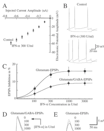
Treatment Control IFN-a effect on the resting membrane potential of pyramidal Rate of rise of the onset (V/ s) 234646 228621 neurons. Control and experimental values were 26361 Rate of rise of the offset (V/ s) 10169 9165 and26261 mV, respectively (n512). IFN-afailed also to a
The observed changes were not significant. alter the voltage-current relationships indicating that cell
input resistance is not altered by this cytokine (Fig. 1A; which component was inhibited by IFN-a. Superfusion of n55). The action potential waveform was also not modify this cytokine had no effect on the GABA-mediated EPSP, by the presence of this cytokine, as shown in Fig. 1B and which was recorded in the presence of 100mM kynurenic
Table 1 (n55). acid and abolished by 30mM picrotoxin. This EPSP was
always completely inhibited by 30mM picrotoxin. 3.3. IFN-a effects on the fast postsynaptic potentials The glutamate mediated EPSP (gEPSP) was inhibited by mediated by glutamate and GABA IFN-ain a concentration-dependent manner with an IC505
148 U / ml and a maximal inhibition of 1662% (Fig. 1C Superfusion of IFN-a (300 U / ml) had a small (963%) and E; n57). The onset of this inhibition was between 6 inhibitory effect on EPSPs mediated by both GABA and and 10 min and reached a maximum about 20–25 min glutamate (see below). Fig. 1C and D show the effect of after starting the superfusion of IFN-a. This was not different cumulative concentrations (IC505151 U / ml) of reversible even after 30–60 min of washing (n56). IFN-aon these EPSPs. This inhibitory effect was present gEPSPs were recorded in the presence of 30mM picrotox-in all tested neurons (n56) and irreversible. in and were abolished by 100 mM kynurenic acid. The Because these EPSPs, as mentioned above, were me- inhibitory effect of IFN-a(300 U / ml; n54) on the gEPSP diated by both glutamate and GABA, we investigated was prevented by pre-treating for 10 min the slices with
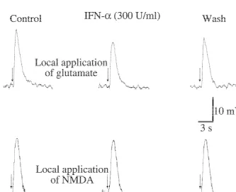
Fig. 2. IFN-ainhibits the depolarization induced by glutamate but did not by NMDA. Pressure ejection of glutamate (5 mM; upper panel) or NMDA (10
mM; lower panel) was at the time indicated by arrows. Depolarization was recorded before (Control), 20 min after adding IFN-a, and after 20 min of IFN-awash-out. Similar observations were obtained in four (upper panel) and six (lower panel) identical experiments. Only one neuron per slice was recorded. Glutamate experiments were carried out in cells current clamped at2100 mV. NMDA experiments were performed in extracellular medium
21
100mM genistein (not shown) which specifically inhibits stimulation (Fig. 3A). After reaching its maximal values, a tyrosine kinase activity in hippocampal tissue [16]. transitory decay in EPSP amplitude was observed during IFN-a (300 U / ml) also reduced the depolarization the first 10–15 min. After that, stable potentiated gEPSPs induced by local application of glutamate (Fig. 2A). (17067% of control values) were observed for as long as Control values were 1663 mV and in presence of IFN-a they were recorded (up to 2 h). In accordance with others these were 1163 mV (P,0.01; n54). IFN-a, however, did [15], we will to the first portion of this response as not modify the depolarization induced by local application short-term potentiation (STP) and to the second portion as of NMDA (n56; Fig. 2B). These observations indicate that the maintenance phase of long-term potentiation (LTP). at least part of the inhibitory effect of IFN-aon the gEPSP This potentiation was prevented by the presence of APV is at the postsynaptic level involving non-NMDA channels (see below) indicating that it is mediated by NMDA and tyrosine kinase activity. channels, as it has been previouly described [11,12].
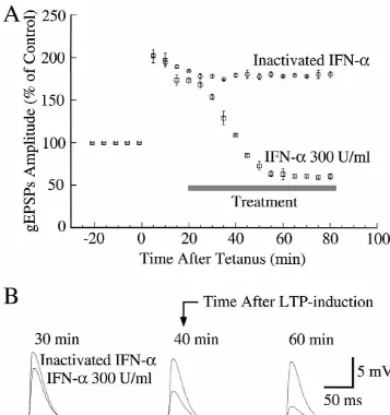
Superfusion of IFN-a (300 U / ml) for 20 min before tetanus prevented the maintenance phase of LTP but a STP 3.4. IFN-a actions on synaptic plasticity in the was still observed (Fig. 3). The average gEPSP amplitude hippocampus reached during the peak of STP was 12464% of control values. After STP, the EPSP amplitude decreased and was Following tetanic stimulation a twofold increase in the maintained at 5665% of control values indicating that gEPSP amplitude (19666% of control values; n55) was IFN-a unmasked a depression of this potential.
observed, with the first recording performed 5 min after Application of IFN-a after STP (Fig. 4; n54) also
V. Mendoza-Fernandez et al. / Brain Research 885 (2000) 14 –24 19
Fig. 4. IFN-ainhibits long-term potentiation in CA1 region. (A) Relative amplitude of glutamate-mediated excitatory postsynaptic potentials (gEPSPs) taken before and after tetanic stimulation (see Section 2) at time 0 (arrow). Both treatments were started during the maintenance phase of LTP, as indicated by bar. Symbols are mean6S.E. of four IFN-a-treated neurons or of four neurons treated with heat-inactivated IFN-asolution. Only one neuron per slice was recorded. (B) Typical gEPSPs from two different neurons treated with either heat-inactivated IFN-asolution or with 300 U / ml of IFN-a, recorded at the indicated times.
inhibited the maintenance LTP. Indeed, in the presence of As expected, inhibition of NMDA receptors with APV this cytokine the gEPSP amplitude reached a new steady- (50mM) during the tetanus stimulation prevented LTP but state value of only|60% of the control values. This effect STP was still present (Fig. 7A; n54). This treatment also was abolished by boiling IFN-a solution for 30 min (Fig. prevented the effects of IFN-aon the gEPSPs. Application
4; n54). of APV after STP neither affected the maintenance phase
Tetanic stimulation also potentiated the depolarization of LTP nor the inhibitory effect of IFN-aon LTP (Fig. 7B; induced by pressure ejection of glutamate (Fig. 5; n54) n54). APV by itself did not alter the EPSP amplitude in but this potentiation was smaller than that observed for control conditions (before tetanus; Fig. 7A) or after STP gEPSPs (Fig. 3A). Application of this cytokine also (Fig. 7B), indicating a null contribution of NMDA chan-reverted this potentiation (Fig. 5; n54), suggesting that the nels for these potentials during such conditions.
IFN-a inhibitory effect on LTP is, at least in part, After applying a low-frequency tetanic stimulation (see
postsynaptic. Section 2) a decrease in gEPSP amplitude was observed,
Fig. 5. IFN-ainhibits the potentiated depolarization induced by local application of glutamate. (A) Relative amplitude of the depolarization induced by local application of glutamate before and after tetanic stimulation at time 0, recorded in CA1 neurons. Symbols are mean6S.E. of four untreated and four IFN-a-treated neurons. IFN-awas applied as indicated by bar. Only one neuron per slice was recorded. (B,C) Typical glutamate-induced depolarizations from two different neurons, one untreated and one treated with IFN-a. These depolarizations were recorded before and after the tetanus, at the indicated times.
4. Discussion high tetanic stimulation widely used by others to induced a NMDA-receptor mediated LTP [1,16]; it lasted for as long In the present study, human recombinant IFN-a spe- as they were recorded (up to 2 h); it was prevented by cifically inhibits LTP and gEPSP in CA1 region neurons of blocking NMDA receptors with APV; and it could not be rat hippocampus slices. This effect was independent of reverted by this antagonist once it had been stablished, as changes in neuronal excitability, NMDA channels or LTD. reported by others [3,8,23]. Finally, IFN-a prevented and Another finding is that inhibition of LTP by IFN-a reverted this LTP but did not affect the gEPSP if APV was unmasked LTD. This indicates that the high-frequency present during the tetanic stimulation.
V. Mendoza-Fernandez et al. / Brain Research 885 (2000) 14 –24 21
Fig. 6. Genistein (100 mM) prevented the IFN-a-inhibitory effects on LTP. (A) Relative amplitude of glutamate-mediated excitatory postsynaptic potentials (gEPSPs) before and after tetanic stimulation at time 0. IFN-awas applied before the tetanic stimulation, as indicated by bar. Symbols are mean6S.E. of five neurons. Only one neuron per slice was recorded. (B) Typical gEPSPs of one neuron taken at the time indicated by letters.
such a depression was seen for as long as our recordings depolarizations were also not modified by IFN-a. This last lasted (.1 h after the tetanic stimulation). Finally, the observation is in agreement with the previous finding that depression induced by IFN-a, at the maintenance phase of rat IFN induces only a small reduction of NMDA-activated LTP, was prevented by the presence of the NMDA receptor currents in cultured embryonic hippocampal neurons [4]. antagonist APV during the tetanic stimulation. This sub- That IFN-a blocks LTP and inhibits gEPSPs through a stance has been previously shown to prevent both the mechanism that involves tyrosine kinase activity is sug-maintenance phase of the LTP and LTD [7,8]. These gested by the fact that receptors to this cytokine are known observations would imply that both biochemical events to couple to a tyrosine kinase domain [17,22]. Here, we responsible for LTP and LTD are activated by the same showed that genistein blocks these IFN-a effects further tetanic stimulation used here to evoked LTP. supporting this hypothesis. A previous study showed that That IFN-a might block LTP by inhibition of NMDA genistein can block tyrosine kinase activity in the CA1 channels was suggested by the fact that opening of NMDA region without affecting the activity of other major protein channels plays a central role in the induction of LTP in the kinases such as the A and C [16]. In the present study, we CA1 hippocampal region [4]. Our observations, however, observed that genistein treatment does not alter the mainte-rule out this hypothesis and demonstrate that the inhibitory nance phase of the LTP, implying that activity of kinases A actions of IFN-a on LTP are independent of NMDA and C is not being inhibited since their activity is known to channels. First, IFN-a also inhibited the maintenance be required for the maintenance phase of LTP [12,16]. phase of the LTP, which is known to be resistant to NMDA Therefore, genistein effects are more likely mediated by channel blockers. Second, neither this LTP phase nor the inhibition of the tyrosine kinase activity.
re-Fig. 7. Inhibition of NMDA channels during the maintenance phase of LTP did not affect the inhibitory effects of IFN-aon LTP. (A) Superfusion of APV (50mM) during the tetanus stimulation prevented LTP but a short-term potentiation (STP) was still noticed. After STP, IFN-afailed to alter the excitatory postsynaptic potentials (gEPSPs). (B) Superfusion of APV (50mM) after LTP induction did not affect IFN-a inhibitory effects on LTP. Symbols are mean6S.E. of four neurons in both A and B; only one neuron per slice was recorded.
ceptors. First, IFN-adoes not modify the NMDA-induced is known to be restricted to inputs that received tetanic depolarization and second, control gEPSPs or the gEPSPs stimulation [10] and this could be the explanation for this recorded during the maintenance phase of LTP are not difference in potentiation. Thus, exogenous glutamate altered by APV, a well known NMDA receptor antagonist. could be mainly mediated by receptors located in mem-gEPSPs are, however, totally abolished by the kynureic brane regions that have not been stimulated during the acid, nonspecific antagonist of ionotropic glutamate re- tetaninc stimulation.
V. Mendoza-Fernandez et al. / Brain Research 885 (2000) 14 –24 23
Fig. 8. IFN-a did not modify LTD. (A) Relative amplitude of glutamate mediated excitatory postsynaptic potentials (gEPSPs) before and after low-frequency tetanic stimulation indicated by the bar to the left (Tetanus). Symbols are mean6S.E. of four neurons. Only one neuron per slice was recorded. The low-frequency tetanic stimulation consisted of a 5-min duration train of electrical pulses at 5 Hz. (B) gEPSPs from a typical experiment recorded at the time indicated by lowercase letters.
cytokines (Review), J. Biol. Regul. Homeostat. Agents 2 (1988) for LTD. This possibility must be considered in studies
107–118. designed to correlate biochemical and synaptic plasticity in
[3] G.L. Collingridge, S.J. Kehl, H. McLennan, Excitatory amino acids the hippocampus. in synaptic transmission in the Schaffer collateral-commissural pathway of the rat hippocampus, J. Physiol. (Lond) 334 (1983) 33–46.
[4] G. D’Arcangelo, F. Grassi, D. Ragozzino, A. Santoni, V. Tancredi, F. Acknowledgements
Eusebi, Interferon inhibits synaptic potentiation in rat hippocampus, Brain Res. 564 (1991) 245–248.
This work was supported by the Medical Research [5] N. Dafny, Is interferon-alpha a neuromodulator?, Brain Res. Rev. 26 Council of Canada and the CCFC. C.B.-L. was partially (1998) 1–15.
supported by the Ontario Ministry of Health (Career [6] F.A. Edwards, A. Konnerth, B. Sakmann, T. Takahashi, A thin slice preparation for patch clamp recordings from neurones of the Scientist Award 04500).
¨
mammalian central nervous system, Pflugers Arch. 414 (1989) 600–612.
[7] P.W. Gean, J.H. Lin,D-2-Amino-5-phosphonovaleate blocks
induc-References tion of long-term depression of the NMDA receptor-mediated synaptic component in rat hippocampus, Neurosci. Lett. 158 (1993) [1] F.P. Bellinger, S. Madamba, G.R. Siggins, Interleukin 1 beta inhibits 170–172.
[9] F. Laezza, J.J. Doherty, R. Dingledine, Long-term depression in [16] T.J. O’Dell, E.R. Kandel, S.G. Grant, Long-term potentiation in the hippocampal interneurons: joint requirement for pre- and postsynap- hippocampus is blocked by tyrosine kinase inhibitors, Nature 353 tic events, Science 285 (1999) 1411–1414. (1991) 558–560.
[10] G.S. Lynch, T. Dunwiddie, V. Gribkoff, Heterosynaptic depression: a [17] P.H. Patterson, H. Nawa, Neuronal differentiation factors / cytokines postsynaptic correlate of long-term potentiation, Nature 266 (1977) and synaptic plasticity, Cell 72 (Suppl.) (1993) 123–137. 737–739. [18] P.H. Patterson, H. Nawa, Neuronal differentiation factors cytokines
and synaptic plasticity, Cell 72 (1993) 123–137. [11] R.C. Malenka, Synaptic plasticity in the hippocampus: LTP and
LTD, Cell 78 (1994) 535–538. [19] V.H. Perry, M.D. Bell, H.C. Brown, M.K. Matyszak, Inflammation in the nervous system, Curr. Opin. Neurobiol. 5 (1995) 636–641. [12] R.C. Malenka, R.A. Nicoll, Long-term potentiation – a decade of
progress?, Science 285 (1999) 1870–1874. [20] C.R. Plata-Salaman, Immunoregulators in the nervous system, Neurosci. Biobehav. Rev. 15 (1991) 185–215.
[13] R. Malinow, J.P. Miller, Postsynaptic hyperpolarization during
conditioning reversibly blocks induction of long-term potentiation, [21] B. Prieto-Gomez, C. Reyes-Vazquez, N. Dafny, Differential effects Nature 320 (1986) 529–530. of IFN-aon ventromedial hypothalamus and dorsal hippocampus, J.
´ Neurosci. Res. 10 (1983) 273–278. [14] V. Mendoza-Fernandez, R.D. Andrew, C. Barajas-Lopez, ATP
inhibits the synaptic release by acting at P2Y receptors in pyramidal [22] N.J. Rothwell, J.K. Relton, Involvement of cytokines in acute neurons of hippocampal slices, J. Pharmacol. Exp. Ther. 293 (2000) neurodegeneration in the CNS, Neurosci. Biobehav. Rev. 17 (1993)
172–179. 217–227.