78
Short Communication:
Morphological Characteristics of The Stomach of Swamp Buffalo (
Bubalus
bubalis
)
Anni Nurliani1*, Teguh Budipitojo2, Dwi Liliek Kusindarta2
1Department of Biology, Faculty of Mathematics and Natural Sciences, Lambung Mangkurat University, Jl. Ahmad Yani Km 35.8, Banjarbaru 70714, South Kalimantan, Indonesia; 2 Department of Veterinary Science, Faculty of
Veterinary Medicine, Gadjah Mada University, Yogyakarta 55284, Indonesia. Corresponding author email: [email protected]
Received : July 19, 2015 Accepted : November 22, 2015 Online : December 31, 2015
Swamp buffaloes (Bubalus bubalis) have been adapted and grown in swampland of South Kalimantan, Indonesia. They have been success to survive in swampland with extreme condition where the feed supply is low on quantity and quality. The ability of swamp buffaloes for adaptation to swampland was suggested to be supported by digestive efficiency. The ability of digestive efficiency on swamp buffalo is often closely related to large rumen volume, slow rumen motility, high cellulolytic activity of microbial population, and slow rate of digestion passage through the reticulo-rumen (Falvey and Chantalakhana, 1999). The efficiency of digestive system in a species maybe also has correlation to the characteristic morphological of stomach. Perez-Barberia, et al. (2004) stated that digestion efficiency can be achieved via stomach adaptation and that this varies between species. To our knowledge, there is no available information concerning morphological adaptation of the stomach of swamp buffaloes. This study was very important to obtain a better understanding of the digestive efficiency of the swamp buffalo by examining the morphology of the stomach of the swamp buffalo with special emphasize on its macroscopic and surface structures.
This study utilized 6 stomachs from 2.5-3 years old healthy male swamp buffaloes. Samples were obtained at the local slaughterhouse, South Kalimantan, Indonesia. The stomach was removed after sectioning the esophagus just cranial to the cardia, and pylorus adjacent to the duodenum. The gross morphology and the interior features were observed and certain portions were measured. Weight of organ was measured by weighing it after it had been opened and contents rinsed with tap water and dried with paper towels. Anatomical measurement was done by spreading the organ. The longest part was measured as length and the widest part was measured as width with soft measuring tape (Perez and Rodolfo, 2012). Pictures were taken with a digital camera.
Stomach filled more less ¾ part of abdomen (Figure 1A). The stomach of the swamp Buffalo composed of the four classic compartments of the ruminants (Figure 1B). The weight mean of the empty rumen was 6370 g. The length mean of the rumen was 108 cm and the width mean was 70 cm. The ruminal papillae were distributed unequally in the rumen. It was more abundant within the ventral rumen than dorsal rumen, whereas the ruminal pillars had no papillae (Figure 2).
79
Figure 1. (A) Topography of swamp buffalo stomach.Rumen (1) filled ½ part of left abdomen. The other part of left abdomen was filled by spleen (5) and some intestine. Reticulum integrated with rumen and it was in front of diaphragm, in this picture it was closed by costae(6). Omasum (2), abomasum (3), and intestine (4) filled right part of
abdomen. (B) Right view of the stomach of the swamp buffalo. Rumen (1), Reticulum (2), Esophagus orifice (3), Omasum (4), and Abomasum (5). Line scale = 10 cm.
Figure 2. Gross anatomical inner views of the rumen in the swamp buffalo. Mucosa surface of the rumen had black color. Surface of ventral rumen (A) was darker than dorsal rumen (B) because ventral rumen had more papillae than
dorsal rumen.Pillar (C) was lighter than others, because it had no papillae.
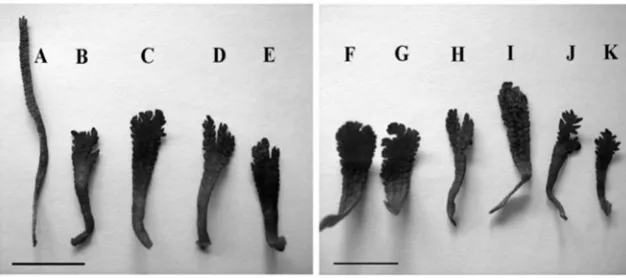
Figure 3. Variation of shape and size ruminal papillae. A = needle-like. B, C, and F = end expansion. D, E, and H = top branching. G and J = lateral branching. I and K = leaf-like. Line scale = 1 cm.
(A) (B) (C)
80
maximum height of the reticular crest (Cristae reticuli) was 1.5 cm (Figure 4C). The reticular cellulae (Cellulae
reticuli) was mostly undivided and contained secondary and tertiary (Figure 4D). They were broader and
deeper near the greater curvature and were becoming smaller toward the lesser curvature.
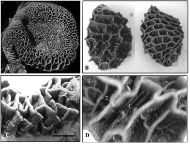
The weight mean of the empty omasum was 630 g. The length mean of the omasum was 36 cm and the width mean was 40 cm. Mucosa surface of the omasum had also black color. Omasum of swamp buffalo had been constructed by longitudinal sheets so called omasal laminae. Surface area of lamina was widened by the papillae. Omasal papillae of swamp buffalo formed dome-like structure, and also indicated long claw-like shape. The mean of length of claw-claw-like papillae was 3 mm (Figure 5).
Morphological observation showed that mucosa surface of rumen swamp buffalo had black color, and pigmentation like this was also showed in the mucosa surface of the reticulum and omasum. This description demonstrates difference with other ruminants. Mucosa surface of cow has brown or dark brown (Chuticul, 1975). Black pigmentation in mucosa surface is caused by increased tissue mass and large blood vessels (vascularization) (Heinrichs and Lesmeister, 2005). Furthermore, Dijkstra et al. (2005) stated that the increasing of tissue mass, surface area and blood flow can increase the absorptive capacity.
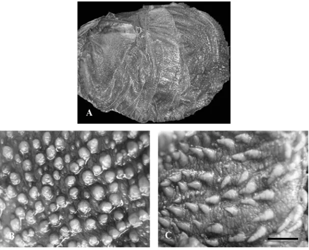
The weight mean of the empty abomasum was 600 g. The length mean of the abomasum was 56 cm and the width mean was 26 cm. Abomasum of swamp buffalo consists of 3 regions (Figure 6). Cardiac region was a small zone that engirdling change orifice between omasum and abomasum. Fundic region had been layered by soft mucous membrane which including high spiral folds. Phyloric region was narrower than other region and it was layered by crinkly mucous containing phyloric glanduler. As in the omasum, the abomasum contained many folds to increase its surface area. These leaves enable the abomasum to be in contact with the large amounts of feed passing through it daily.
Figure 4. Gross anatomical inner views of the reticulum in the swamp buffalo. Primary crest was the highest crest (1), secondary (2) and tertiary (3) crest shorter than primary and intersecting each other to separate reticular cellule
81
Figure 5. Gross anatomical inner views of the omasum in the swamp buffalo. Omasum had longitudinal layers or sheets said laminae (A). Mucosa surface of omasal laminae was widened by dome-like papillae (B) or claw-like papillae
(C). Line scale = 1 cm.
Figure 6. Gross anatomical inner views in the cardiac (A), fundic (B), and phyloric regions (C) of the abomasum of the swamp buffalo. Cardiac region was a small zone that engirdling change orifice between omasum (1) and
abomasum (2).
In conclusion, the stomach of swamp buffalo had morphological peculiarities, such as: mucosa surface of nonglanduler stomach had black color, and there was variation of ruminal papillae of swamp buffalo, including branching. These special characteristic of swamp buffalo stomach morphologically is estimated as supporting factors for increasing digestive efficiency to survive in swampland.
References
Chuticul, K. 1975. The Asiatic water buffalo. Compiled by Food and Fertilizer Technology Center for the Asian and Pasific Region. 116 Huai Ning Street, Taipei, Taiwan, Republic of China.
Dijkstra, J., Forbes, J.M. and France, J. 2005. Quantitative aspects of ruminant digestion and metabolism. CABI Publishing, USA.
Falvey, L. and Chantalakhana, C. 1999. Smallholder dairying in the tropics. International Livestock Research Institute, Nairobi, Kenya.
Heinrichs, A.J. and Lesmeister, K.E. 2005. Rumen development in the dairy calf. In: Calf and heifer rearing. Nottingham University Press, England.
McGavin, M.D. and Morrill, J.L. 1976. Scanning electron microscopy of ruminal papila in calves fed various and form of roughage. American Journal of Veterinary Research, 37: 497-508.
82
Swan, G.E. and Groenewald, H.B. 2000. Morphological changes associated with the development of the rumino-reticulum in growing lambs fed different rations. Journal of Veterinary Research, 67:105-114.