THE PROFILE OF BLOOD TRANSAMINASE ENZYME IN DUCK
(Anas sp.) THAT POLLUTED BY LEAD (Pb) TEXTILE WASTE
Elvia Hernawan and Andi Mushawwir
Laboratory of Animal Physiology and Biochemistry, Animal Science Faculty,
University of Padjadjaran, Jatinangor Sumedang, West Java Indonesia
email : [email protected]
ABSTRACT
Duck which is raised traditionally in around of textile industry have a risk by
lead (Pb) pollution from textile industry
liquid waste, that cause hepatocite to liver.
Decrease in liver function, has the impact to vitellogenesys that is synthesis of
vitellogenin and β
-lipoprotein
as yolk precursor. Transaminase is enzyme which is
indicated decrease in liver function.
Objectives of this research was
to know the profile of blood transaminase
enzyme (SGPT= serum glutamate pyruvate transaminase, SGOT = serum glutamate
oxaloacetate transaminase) in duck that polluted by lead (Pb) from textile waste. This
research has been used survey method with purposive sampling, amount of sample used
was 60 Tegal duck, consisted of 30 duck which was not polluted by Pb and 30 duck
which was polluted by Pb. The data was analysed used statistical analysis of T-students.
Based on research showed that blood SGPT level which was polluted Pb higher
significant different 76,74 ± 1,89
µ
mol/L against which was not polluted 47,93 ± 1,59
µ
mol/L, and so it is with SGOT level was higher significant different 78,73 ± 2,73
µ
mol/L polluted by Pb, against which was not polluted was 46, 52 ± 1,53
µ
mol/L.
Key word : Transaminase, Lead (Pb) and Duck
INTRODUCTION
Duck which is raised traditionally in around of textile industry have a risk by lead
(Pb) pollution from textile industry liquid waste, that cause hepatocite to liver. Decrease
in liver function, has the impact to vitellogenesys that is synthesis of vitellogenin and
β
-lipoprotein as yolk precursor. Transaminase is enzyme which is indicated decrease in
liver function.
Accumulation of excess Pb can potentially cause liver damage that is clinically
characterized by increased SGPT (serum glutamic-pyruvic transaminase) and AST
(serum glutamic-oxaloacetic transaminase). Degree increase in these enzymes correlated
positively with the level of liver damage. Biochemical changes due to liver damage,
manifested by the increase of ALT levels from 20-200 times the normal levels (1-36 M /
L) and AST levels by 10-150 times the normal values (8-40 M / L) (Bergmeyer and
Bernt, 1971).
Pb concentrations in water, soil and air around industrial areas may reach 0.2 ppm
and wastewater regulations limit the conditions 0.05 ppm (Amina, 2006), whereas Pb
concentrations of wastewater based on preliminary research results is 0.207 ppm, there is
also content Other heavy metals such as Cd, Ag and Ni, but the levels in the waste water
is very small.
Pb content of blood contaminated duck waste textiles based on preliminary
research results by using Atomic Absorption Spectrophotometer (AAS type) reached 0.07
ppm, whereas the Pb content of blood is not contaminated duck waste textiles reach
0.0005 ppm. Based on preliminary research results, there is a heavy metal content of Pb
in waste water higher than the content of heavy metals other than Pb content of blood was
contaminated ducks to reach 0.07 ppm indicate the occurrence of heavy metal pollution
Pb in ducks raised in the neighborhood textile industry.
MATERIALS AND METHODS
Animals and Survey
Animals used in this research were 60 Tegal Ducks, 10-12 months age, average body
weight 1.6 kg. Sampling method used was sampling purphosif sampling, consist of 30
ducks polluted by lead and 30 ducks not polluted by lead.
Survey have been made during 30 days and blood sample collected every week
(forth time/ 4 weeks).
Parameters
1.
Glutamate Pyruvat serum transaminase (SGPT/ALT).
2.
Oxaloacetat Glutamate 2 Serum transaminase (SGOT/AST)
This enzyme catalysis the transfer function for alpha-amino group of aspartic acid
to alpha-ketoglutaric acid.
ALT and AST levels are determined by using Clinical Auto Analyzer Cobas Type
C-111. The sample was measured using a photometric system with wavelength
340-659 nm.
Data Analysis
This study uses analysis of T-student population is not paired with:
- Population 1 = duck contaminated textile
- Pollution 2 = duck population is not contaminated textile pollution.
RESULTS AND DISCUSSION
Averages Level of SGPT and SGOT of duck blood polluted and not polluted of Pb
Serum OF Glutamate Pyruvat Transamnase (SGPT) and Serum Glutamate
Oxaloasetat Transaminase (SGOT) were transaminase enzym that referable to evaluation
of liver function. Average level of SGPT and SGOT of duckblood polluted and not
pullted of Pb, showed in Table 1.
Table 1. Averages Level of SGPT and SGOT of duck blood polluted and not
polluted of Pb
Level
Environmental condition
Polluted
Not polluted
Results of Analysis
...µM/L...
SGPT
SGOT
76,74 ± 1,89
78,73 ± 2,73
47,93 ± 1,59
46, 52 ± 1,53
P < 0,01
P < 0,01
Analysis result showed that average of transaminase enzym (SGPT and SGOT)
level was difference significant (P<0.01), between duck polluted and not polluted by Pb.
It was showed that take effect higher Pb accumulation, so much so that caused reduced
of liver function. Increasing SGPT and SGOT level would happened if there were
releasing enzym in accordance with intracellular to into blood that caused hepatocyte, eg
nekrosys hepatoseluler or infark miokardial (Bijanti, 2006).
Serum glutamic-pyruvic transaminase (SGPT/ALT) is an enzyme sitosolik
contained in these organs function as catalyst of removal of the alpha amino acid alanine
to alpha ketoglutrarat. ALT most abundant in the liver. ALT values are considered
normal is 1 to 36 M / L. These levels will rise rapidly and exceed the normal case of
liver cell necrosis, or do not have this enzyme eliminated out (Darmono, 2001). Based on
our research ALT levels average for ducks contaminated with Pb metal reached 76.74 ±
1.89 M / L, this shows an increase of ALT levels reached three times the normal levels
(1-36 M / L), ALT levels whereas the average for the ducks that are not polluted by Pb
metal was 47.93 ± 1.59 M / L.
Serum glutamic-oxaloacetic transaminase
(SGOT/AST) many founded in the heart,
liver, muscle, panckreas, lung, eritrocite, brain cells. Althought this enzime used for lever
testing, its high level founded in the muscle. Contain of SGOT in the blood are 8-40
µM/L. Function of SGPT was transfer catalys of alfa-amino group from aspartate acid to
be alfa ketoglutarat acid (Darmono, 2001).
Based on our research AST levels average for ducks contaminated with Pb metal
reached 76.3 M / L, this shows an increase of AST levels reached twice normal levels
(8-40 M / L), while the average levels of AST average for the ducks that are not
polluted by Pb metal was 46, 52 ± 1.53 M / L.
Increased levels of transaminase enzymes infected duck blood Pb can be explained
due to liver tissue damage occurs through a reduction in its function as a result of ion
exchange of important minerals such as K, Na, P and others into Pb ions and the
formation of the complex formation as Suhendrayatna (2008) suggests that the network
bodies, contamination of Pb
2+ions bind to the cell membranes of two different ways, the
between Pb ions with functional groups like carbonyl, amino, thiol, hydroxyl, phosphate,
and hydroxyl-carboxyl is located on the cell membrane, this phenomenon has led to
decreased cell function until the death of cells (hepatosit).
Toxicity of lead (Pb) in various organs is mediated through several mechanisms
including inactivation of enzymes and other macromolecules through bonds with
sulfhydryl, phosphate, and carboxyl and interaction with cations, especially calcium, zinc
and iron. Pathological processes can occur in the cell membrane and mitochondria,
function and neurotransmitter synthesis, heme synthesis, cellular redox status and
nucleotide metabolism. Adverse effects can occur in nerve, kidney, gastrointestinal tract,
hematopoesis, reproductive and cardiovascular system. Pb metal including metal-metal
bond is more reactive with the ligand in the cell, if the metal binding cells of
(non-essential), it will cause damage to the catalyst capability (detoksikasi) of the cell itself
(Darmono, 1995).
Given that more than 20 amino acids contained in the body, half of it is synthesized
in the liver from other components. The formation of amino acid incorporation requires a
single amino group or nitrogen in the carbon skeleton containing radical group or side
chain that clearly characterize the amino acids are formed. Carbon skeleton is a ketone
acids, such as pyruvic acid or alpha-ketoglutarate which is a product of nitrogen
metabolism of fatty group derived from amino acids found in other large through
transamination processes or deaminase (Piliang, 2000).
All the tissues have the ability to synthesize non-essential amino acids, amino acids
do remodeling, and change the framework of non-carbon amino acids into amino acids
and other derivatives that contain nitrogen. However, the liver is the major site of
nitrogen metabolism. In diet surplus conditions, potentially toxic nitrogen from amino
acids released through transamination, deamination and urea formation. Carbon skeleton
are converted to carbohydrates through gluconeogenesis, or a fatty acid through fatty acid
synthesis. In this regard, amino acids are grouped into 3 categories, namely glukogenik
amino acids, glukogenik and ketogenik and ketogenik.
amine group loosing it toxic. There were two release mechanism of amine groups from
the amino acid group: 1) transamination, moving aminotransferase enzyme amines to
-ketoglutarate, to be glutamate or to oksaloscetate to be aspartate; 2) Deaminasioxidative,
releasing amines from glutamate to be ammonium Ions.
Ketone and acid groups and amino acids found in the form of pairs. In the process
of transamination, the amino group removed from the pair of amino acids
(alanine-pyruvate) into ketone acids from other amino acid pairs (alpha_ketoglutarat_asam
glutamate). Transamination processes need specific enzymes known as enzyme
transaminase (Piliang, 2000).
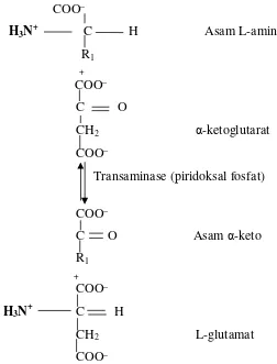
According to Lehninger (1990) and Pilliang (2000) suggested that the removal of
the amino group as proposed earlier enzyme catalyzed by transaminase or amino
transferase, a process called transamination, as illustrated by the following reaction in
Figure 1.
COO
_H
3N
+C H Asam L-amino
R
1 +COO
_C O
CH
2-ketoglutarat
COO
_Transaminase (piridoksal fosfat)
COO
_
C O Asam -keto
R
1+
COO
_H
3N
+C H
CH
2L-glutamat
COO
_Although the main source of amino acids derived from non-essential foods
consumed by ducks, but to maintain the number or the needs of non-essential amino acids
are the endogenous synthesis process (through the transamination process is presented in
the previous paragraph and in Figure 1) will be maintained, then in liver transaminase
levels are maintained within a sufficient level.
Related to this, Alifia and Djawad (2003) and Vodela, et al., (2007) suggests that
the degeneration of parenchymal damage characterized by changes hepatosit or liver cell
death that causes the specific enzymes involved in metabolism of protein migration into
blood vessels. Associated with specific enzyme migration into blood vessels, Kimball
(1983) and Linder (2006) suggested that the transaminase enzymes can be indicators of
liver damage.
CONCLUSION
Based on research inferential that duck polluted by lead significantly undergo
increased level of SGPT and SGOT
REFERENCES
Alifia, F. dan M.I Djawad., 2003. Kondisi Histologi Insang dan Organ Dalam
Ikan
Bandeng Yang Tercemar Logam Timbal Pb
. Jurnal Sains dan Teknologi
. 3(1) :
15-20.
Aminah, N. 2006. Perbandingan Kadar Pb, Hg, Fungsi Hati dan Fungsi Ginjal
Pada Karyawan BBKTL Surabaya. Jurnal Kesehatan Lingkungan. 2(2) :
111-120.
Bergmeyer, H.U., and Bernt. E., 1971.
Methods Of Enzimatis Analysis,
Bergmeyer, H.U., (ed) Vol 2, 755, 760-763, Academic Press Inc, New
York
Bijanti, R. 2006.
Pengaruh Pemberian Perasan Buah Mengkudu (Morinda citrifolia)
Terhadap Kadar SGOT dan SGPT Tikus Putih (Rattus Norcegicus
). Fakultas
Kedokteran Hewan Universitas Airlangga.
Darmono, 1995.
Logam Dalam Sistem Biologi Makhluk Hidup
. Penerbit Universitas
Darmono, 2001.
Lingkungan hidup dan pencemaran
. Universitas Indonesia Press Jakarta
117- 122 : 126.
Hutagalung, H.P, 1984.
Logam Berat Dalam Lingkungan Laut. Pewarta Oceana
Vol IX No.1 LON-LIPI, Jakarta.
Kimball, J.W, 1983.
Biologi
(edisi kelima). Diterjemahkan oleh Siti Soetarmi dan
Nawangsari Sugiri. IPB Press.
Lehninger, L.A. 1990
. Dasar-dasar biokimia
(jilid 2). Diterjemahkan oleh Maggy
Thenawidjaja. IPB Press.
Linder, M.C. 2006.
Biokimia Nutrisi dan Metabolisme
. Diterjemahkan oleh Aminuddin
Prakkasi. UI Press.
Pilliang, W.G. 2000.
Fisiologi Nutrisi
(volume 1). IPB Press.
Suhendrayatna, 2008.
Mekanisme Toksitas Logam Berat
. Institute for Science and
Technology Studies (ISTECS) – Chapter Japan Department of Applied Chemistry
and Chemical Engineering Faculty of Engineering, Kagoshima University